2.1 Preparation of strobilurin X
Strobilurin X was extracted from M. venosolamellata (TUFC 30333, Fungus/Mushroom Resource and Research Center, Faculty of Agriculture, Tottori University). M. venosolamellata was cultured in 5 L of malt extract media for 30–60 days. The culture filtrate was extracted using ethyl acetate (1 L × 3) and fractionated by silica gel column chromatography (acetone-hexane, 10% stepwise from 0 to 30% acetone). The 10% acetone fraction was further fractionated by silica gel column chromatography (ethyl acetate-hexane, 10% stepwise elution from 0 to 30%). Strobilurin X was purified from the 20% ethyl acetate fraction via preparative high-performance liquid chromatography using a Cosmosil 5C18 ARII (10 × 250 mm, Nacalai Tesque, Kyoto, Japan) column and 65% acetonitrile–water as solvent (flow rate: 3 mL/min, ultraviolet detection: 254 nm, [Shimadzu 10A system, Kyoto, Japan]). The identity of the compound was confirmed by nuclear magnetic resonance and mass spectra according to our recent report [16].
2.2 Cell culture
Lung and cervical cancer cell lines (A549 and HeLa, respectively) and normal lung fibroblasts (WI-38) were purchased from Riken Bioresource Center (Tsukuba, Japan) and cultured in Dulbecco’s modified Eagle medium (D6429; Sigma-Aldrich, Tokyo, Japan) supplemented with 10% bovine fatal serum, 100 U/mL streptomycin (Meiji Seika Co. Ltd, Tokyo, Japan) and 100 μg/mL penicillin (Meiji Seika Co. Ltd).
2.3 Detection of cell viability using the WST-8 assay
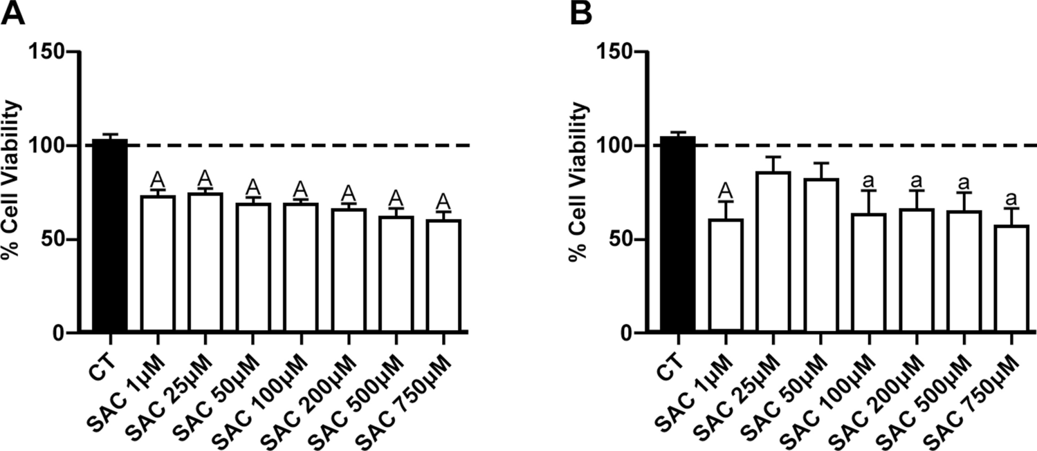
Cell viability was assessed using Cell Counting Kit-8 (WST-8; Dojin, Tokyo, Japan). In brief, the cells (5 × 10³ cells/well) were prepared in a 96-well culture plate the day before the experiment. Various concentrations of strobilurin X were applied for 48 h, and the cell viability was evaluated by measuring the absorbance with a microplate spectrometer (Sunrise™ Fuji film Wako Pure Chemicals, Osaka, Japan) at 450 nm between 1 and 4 h after the addition of the WST-8 reagent to the culture medium in each well. The ratio of each data set to that of the vehicle was calculated.
2.4 Quantitation of lactate production from the cells
The amount of lactate produced through glycolysis was measured using the Lactate Assay Kit-WST (Dojin). The cells (104 cells/well) were prepared in the 96-well culture plate the day before the experiment and incubated with various concentrations of strobilurin X for 48 h. A portion of each conditioned medium was transferred to a new 96-well culture plate and mixed with a working solution, according to the operating instructions. After incubation for 30 min at 37 °C, the absorbance of each sample was measured at 450 nm. The amount of lactate produced by the cells under each condition was calculated using a standard curve.
2.5 Measurement of the mitochondrial respiratory chain complex III activity in cell-free system
The mitochondrial respiratory chain complex III activity was measured using the MitoCheck® Complex II/III Activity Assay Kit (Cayman Chemical, Ann Arbor, MI, USA) according to recent reports [17, 18]. In brief, the reduction of cytochrome c by complex III was detected at 550 nm using a spectrophotometer (Genesys 10S UV–Vis; ThermoFisher Scientific, Tokyo, Japan). A working solution of strobilurin X was mixed with cytochrome c solution and bovine heart mitochondria, and the absorbance was measured every 30 s for 10 min. The activity of complex III was calculated as the ratio of the rate of change in absorbance in each sample to that in the vehicle.
2.6 Detection of the protein synthesis activity
The protein synthesis activity was assessed using the Cayman’s Protein Synthesis Assay Kit (Cayman Chemical). In brief, the cells (3 × 10³ cells/well) were prepared in a 96-well culture plate with a black bottom (Thermo Fisher Scientific) the day before the experiment. The cells were pretreated with strobilurin X for 30 min and labeled with O-Propargyl-puromycin (OPP) for 2 h. The amount of 5-carboxyfluorescein (FAM) bound to OPP was determined by measuring the fluorescence at 535 nm with excitation at 488 nm using a fluorescent microscope (BZ-X810, Keyence, Osaka, Japan).
2.7 Measurement of intracellular reactive oxygen species (ROS) production
After exposure to strobilurin X, the cells harvested by trypsinization were washed with phosphate-buffered saline (PBS) and loaded with chloromethyl-2ʹ,7ʹ—dichlorodihydrofluorescein diacetate (CM-H2DCFDA; 40 μM; C6827, Thermo Fisher Scientific) for 30 min at 37 °C. Subsequently, the cells were washed with PBS and analyzed using a flow cytometer (FACS Aria, BD Japan, Tokyo, Japan).
2.8 Chemicals
Uridine (solvent, stock concentration; PBS, 200 mg/ml), methyl pyruvate (PBS, 110 mg/mL) and N-acetyl cysteine (1 M NaOH, 1 M) are purchased from FUJIFILM Wako Pure Chemical Corp. (Osaka, Japan). PD98059 (dimethylsulfoxide [DMSO], 50 mM), U0126 (DMSO, 20 mM), LY2940001 (DMSO 5 mM), JNK inhibitor II (DMSO, 10 mM) and SB203580 (DMSO, 10 mM) were obtained from Calbiochem (Sigma-Aldrich Japan, Tokyo, Japan).
2.9 Statistical analysis
All analyses were conducted using the SPSS Statistics software (IBM Japan, Tokyo, Japan). Student’s or Welch’s t-test was used for two-group comparisons, and one-way analysis of variance followed by the Games-Hawell test was used for multiple-group comparisons. A p-value of < 0.05 was considered significant.

留言 (0)