2.1 Antibodies, plasmids, and chemicals
Celastrol (purity of > 99%, cat. No. HY-13067) and PI3K activator (purity of > 99%, cat. No. HY-151527) were purchased from MedChem Express (MCE, shanghai, China) and was dissolved in dimethyl sulfoxide (DMSO) to generate a stock concentration of 10 nM, stored at − 80 °C.p-PI3K (cat. No. 4228), PI3K (cat. No. 4257), p-AKT (cat. No. 4060), AKT (cat. No. 4691), p-mTOR (cat. No. 5536), mTOR (cat. No. 2983), and β-actin (cat. No. 4967) were purchased from Cell Signaling Technology (CST, Woburn, USA). rabbit anti-mouse IgG antibody, and the secondary antibody, goat anti-rabbit IgG antibody was purchased from ZSGB-BIO (Beijing, China). BCA protein assay kit and Cytoplasmic Protein Extraction Kits were purchased from Beyotime (Jiangsu, China).
2.2 Cell culture
C57BL/6 J mice (B16-F10) melanoma cell lines were obtained from the Cell Bank of the Chinese Academy of Sciences (Kunming, China). B16-F10 cells were cultured in Dulbecco's modified Eagle’s medium (DMEM, G4511-500ML, Servicebio, Wuhan, China) supplemented with 10% fetal bovine serum (FBS, 10099-141C, GIBCO, California, USA) and a 1% penicillin/streptomycin solution (CM0001-100ML, Sparkjade, Jinan, China). T25 cm2 flasks were used to culture B16-F10 cells at a constant temperature of 37℃ in a moist incubator with 5% CO2. The culture medium wasrefreshed every two days.
2.3 Cell viability assay
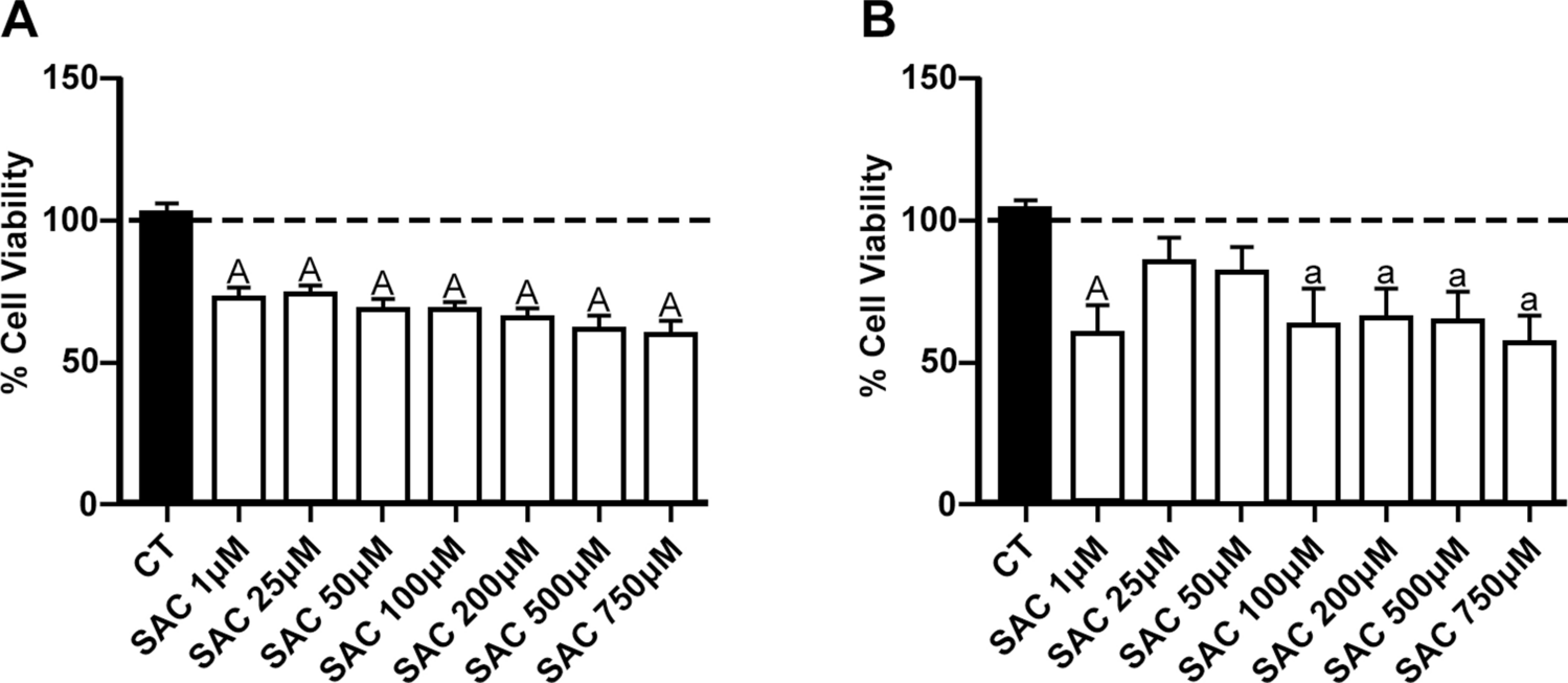
CCK8 (CT0001-B, Sparkjade, Jinan, China) was used to test the inhibitory effect of celastrol on B16-F10 cell proliferation. B16-F10 cells were seeded into 96-well plates at a density of 6 × 10³ cells/well. After overnight incubation, different concentrations (0, 0.03, 0.3, 1, 3, and 10 μM) of celastrol were added to cells for 12, 24, 36, and 48 h, and then cells of each well were treated with CCK-8 solution for 1 h at the end of the incubation period. A wavelength of 450 nm was selected, and an enzyme-linked immunosorbent assay was used to detect the light absorption value of each well.
2.4 Cell clone formation assay
The cells were plated in 6-well plates at a density of 200 cells/well. After 24 h, the cells were treated with different concentrations (0.03, 0.1, 0.3, 1, 3 and 10 μM) of celastrol for 14 days. Subsequently, the medium was discarded, and Cell PBS (1X) was used to rinse the cells three times. Next, colonies were fixed with 4% paraformaldehyde andtreated with 0.1% crystal violet for 15 min, followed by image acquisition using a ZEISS Axio Vert.A1 system.
2.5 Wound healing assay
The effect of various concentrations of celastrol on the migratory ability of B16-F10 cells was evaluated using a wound-healing assay. Approximately 6 × 10⁵ cells were plated in 6-well plates and incubated to 85–95% confluency. A wound was created with a line in the middle using a sterile pipette (200 μl), and cell debris was removed using Cell PBS (1X). Subsequently, various concentrations (0, 0.03, 0.3, 1, 3, and 10 μM) of celastrol were added, and cells were cultured for 0, 12, 24, 36 and 48 h, respectively. Images of wound closure were captured using a ZEISS fluorescence microscope, and subsequent measurements of the wound area were conducted using ImageJ software.
2.6 Flow cytometric analysis of apoptosis
Apoptosis was detected using an Annexin V- FITC Apoptosis Assay Kit (Abs50001, Absin, Shanghai, China). B16-F10 cells were seeded into 24-well plates at a density of 9 × 10⁴ cells/well. The cells were digested using 300 μl of trypsin (CN0004-100ML, Sparkjade, Jinan, China), and then treated with different concentrations of celastrol (0.03, 0.1, 0.3, 1, 3, and 10 μM). After 24 h of treatment, B16-F10 cells were harvested by centrifuging at 1000 rpm for 10 min at 4℃. The cells were then resuspended in 1 ml Cell PBS (1X) and centrifuged repeatedly. Subsequently,300 μl Blinding Buffer (1X) and 5 μl Annexin V-FITC were respectively added to the suspended cells and mixed.The suspended cells were then treated with 5 μl PI for staining after 15 min in the dark. Finally, 200 μl of Blinding Buffer (1X) was added. Assays were conducted using a BD FACSVerse flow cytometer (BD Biosciences).
2.7 Mitochondrial membrane potential assay
A JC-1 Mitochondrial Membrane Potential Assay Kit (C2006, Beyotime, Jiangsu, China) was used for mitochondrial membrane potential (MMP) detection. B16-F10 cells were plated in 6-well plates (6 × 10⁵ cells/well), and various concentrations of celastrol (0, 0.03, 0.3, 1, 3, and 10 μM) were added to respective wells. After 24 h, the cells were harvested, 500 μl of JC-1 staining working solution was added, and the cells were incubated in a CO2 incubator at 37 °C for 20 min. JC-1 staining buffer (1X) was used to rinse the cells twice before adding 2 ml DMEM during the final period. The results were analyzed using a BD FACSVerse flow cytometer.
2.8 Detection of reactive oxygen species
B16-F10 cells were plated in 6-well plates (6 × 10⁵ cells/well). Different concentrations of celastrol (0, 0.03, 0.3, 1, 3, and 10 μM) were added to the B16-F10 cells. After 24 h of treatment, the cells were harvested and cultured with 1 ml DCFH-DA (10 μM/L) in a cell incubator for 20 min at 37 °C (S0033S, Beyotime, Jiangsu, China). Subsequently, the cells were rinsed three times with 1 ml DMEM culture medium. A BD FACSVerse flow cytometer was used to perform all assays.
2.9 Western blot analysis
After being incubated in T25 cm2 flasks and treated with 10 μM concentration of celastrol for 24 h, B16-F10 cells were lysed with cold RIPA solution. According to the standard protocol,proteins were extracted using a BCA protein assay kit (P0012S, Beyotime, Jiangsu, China) to determine protein concentration. A total of 40 μg of protein was separated using 10–12% SDS-PAGE, transferred onto a PVDF membrane, and cultured with a closed buffer (1% BSA in TBST) for 1 h. The primary antibodies, including p-PI3K (1:1000), PI3K (1:1000), p-AKT (1:1000), AKT (1:1000), p-mTOR (1:1000), mTOR (1:1000), and β-actin (1:1500), were respectively incubated overnight at 4℃ after the membranes were cropped based on the expression levels of the target proteins. Subsequently, the membrane was cultured with the primary antibody, rabbit anti-mouse IgG antibody, and the secondary antibody, goat anti-rabbit IgG antibody (1:15000). This incubation was maintained at 37 °C for 1 h. The membranes were then observed with an enhanced chemiluminescence detection system (Fusion FX7, Vilber, France) and quantified using ImageJ software.
2.10 Cell cycle assay
B16-F10 cells were plated at a density of 9 × 10⁵ cells/well in 24-well plates. The cells were digested using 300 μl of trypsin and then treated with different concentrations of celastrol.Next, the cells were fixed overnight in 70% ethanol. Cells were then harvested by centrifugation at 1000 rpm for 10 min at 4 °C and rinsed with 1 ml Cell PBS (1X). Subsequently, cells were incubated with staining buffer, RNase A configuration propidium iodide staining working solution (C1052, Beyotime, Jiangsu, China), and 0.5 ml propidium iodide staining working solution (20X) for 30 min at 37 °C in a cell incubator. Samples were examined using a BD FACSVerse flow cytometer.
2.11 Reverse transcription-quantitative (RT-q) PCR assay
RNA was extracted from B16-F10 cells treated with different concentrations of celastrol using the SPARKeasy Tissue/Cell RNA Rapid Extraction Kit according to the manufacturer’s instructions (AC0202, Sparkjade, Jinan, China). Reverse transcription was conducted using the HiScript II Q Select RT SuperMix for qPCR (R233-01 and Q711-02/03, Vazyme, Nanjing, China). A reaction volume of 20 μl was used for HIF-α analysis, with GAPDH as an internal control to standardize the expression levels of HIF-α. The relative quantitative analysis was conducted using the 2-ΔΔCt method.
The HIF-α primers used were 5'-CAAGTCAGCAACGTGGAAGG-3' (forward) and 5'-ATCAGCACCAAGCACGTCAT-3' (reverse).
The GAPDH primers used were 5'-CCTCGTCCCGTAGACAAAATG-3' (forward) and 5'-TGAGGTCAATGAAGGGGTCGT-3' (reverse).
2.12 Statistical analysis
Data were analyzed by one-way analysis of variance (ANOVA) using GraphPad Prism version 8.0.1 statistical software and were presented as mean ± SD. P < 0.05 was considered statistically significant.

留言 (0)