
記住我


The qualitative analysis of the S. asoca crude methanol extract indicated positive results for the presence of flavonoids, alkaloids, phytosterols, phenols, saponins, tannins and terpenoids. The quantification of polyphenolic content in the S. asoca crude extract, determined from the calibration curve (R2 = 0.998), revealed a concentration of 120 ± 6.82 mg of gallic acid equivalent (GAE) per gram of dry extract. Additionally, the total flavonoid content in the crude extract, estimated from the calibration curve (R2 = 0.999), was found to be 61.54 ± 4.51 mg of quercetin equivalent (QE) per gram of dry extract. These results provide a comprehensive insight into the chemical composition of S. asoca crude methanol extract, highlighting its rich polyphenolic and flavonoid content.
Anticancer propertiesThe cytotoxic effect of S. asoca crude methanol extract was assessed using the trypan blue assay and demonstrated considerable cytotoxic effects on DLA and EAC cells. The concentrations required to achieve 50% cytotoxicity were 42.24 ± 3.65 and 65.44 ± 2.89 μg/mL for DLA and EAC cells, respectively (Fig. 1). In MTT assay, the efficacy of S. asoca extract was evident against triple-negative breast cancer cell lines, MDAMB-231, with an IC50 of 70.22 ± 1.89 μg/mL, and HER-2 positive breast cancer cell line, SKBR3, with an IC50 of 98.41 ± 2.31 μg/mL (Fig. 1). A statistically significant (p < 0.05) decrease in cell numbers was observed in both MDAMB-231 and SKBR3 following extract treatment. Treated cells exhibited a noticeable difference in morphology compared to control cells, characterized by cell shrinkage and shift in morphology from epithelial-like to round in both MDAMB-231 and SKBR3 (Fig. 2). However, the extract did not exhibit any cytotoxicity towards MCF-7, even at a concentration of 300 μg/mL.
Fig. 1
A Cytotoxic effect of S. asoca crude methanol extract on murine tumor cells by trypan blue assay, B Antiproliferative effect of S. asoca on different breast cancer cells by MTT assay. The results are expressed as mean ± SD, with n = 3. Statistical comparisons were conducted using one-way ANOVA, followed by Tukey’s multiple comparison test. Statistically significant probabilities are denoted as *p < 0.05 and **p < 0.01
Fig. 2
Morphology of different breast cancer cells after exposure to varying concentrations of S. asoca crude extract (20× magnification). The black arrow indicates altered morphology from epithelial-like to round
Estrogen-screen assayIn this study, the ERα expressing MCF-7 cells exhibited a proliferative response to 17 β-estradiol (1000 pM), showing a 30% increase in cell count and a 7% increase in response to crude extract within a 72-h timeframe. Along with 17 β-estradiol, the crude extract exhibited a mild estrogenic effect. Conversely, ERβ expressing MDAMB-231 cells demonstrated a 10% decrease in proliferation with 17 β-estradiol treatment, and the cell population was halved when treated with the crude extract at a concentration of 100 μg/mL (Fig. 3). Notably, the crude extract did not induce cytotoxicity in MCF-7 cell lines even at higher concentrations, while they decreased the cell viability of MDAMB-231 cells in a concentration-dependent manner. Consequently, the estrogen-screen assay highlights the estrogenic impact of S. asoca crude extract on MCF-7 and its anti-estrogenic/antiproliferative effect on MDAMB-231 cells.
Fig. 3
Change in cell volume of MCF-7 and MDAMB-231 breast cancer cells following exposure to extract and 17 β-estradiol (1000 pM) in the estrogen-screen assay
Chemical profilingVarious techniques like UV–Vis spectroscopy, fourier-transform infrared spectroscopy (FTIR) and LC–MS were employed to evaluate the chemical profile of S. asoca. The absorption spectrum of UV-Spectrophotometric analysis of S. asoca, showed prominent peaks at 232, 275 and 449 nm which is in good correlation with the reported data [41]. These prominent peaks may have arisen from the phytoestrogens. Henceforth, the UV-spectra of standards (quercetin, kaempferol, β-sitosterol) were cross-checked and found that all of the suspected phytoestrogens have three peaks between 230 and 290 nm in the UV range and a single peak in a visible area (387–390 nm) (Fig. 4A). Accordingly, there is a likelihood of superpositioning of these distinct peaks in the S. asoca crude extract. The biological activity of any molecule is influenced by its functional groups which play a key role in determining the overall physicochemical properties. In FTIR, the results show functional groups such as alcohol, phenol, ester, alkane, aromatic and alkene in the extract. The functional groups identified in the extract are shown in Fig. 4B and Table 1.
Fig. 4
A UV–visible spectrum of S. asoca crude methanol extract and phytoestrogen standards, B FTIR spectrum of S. asoca crude extract
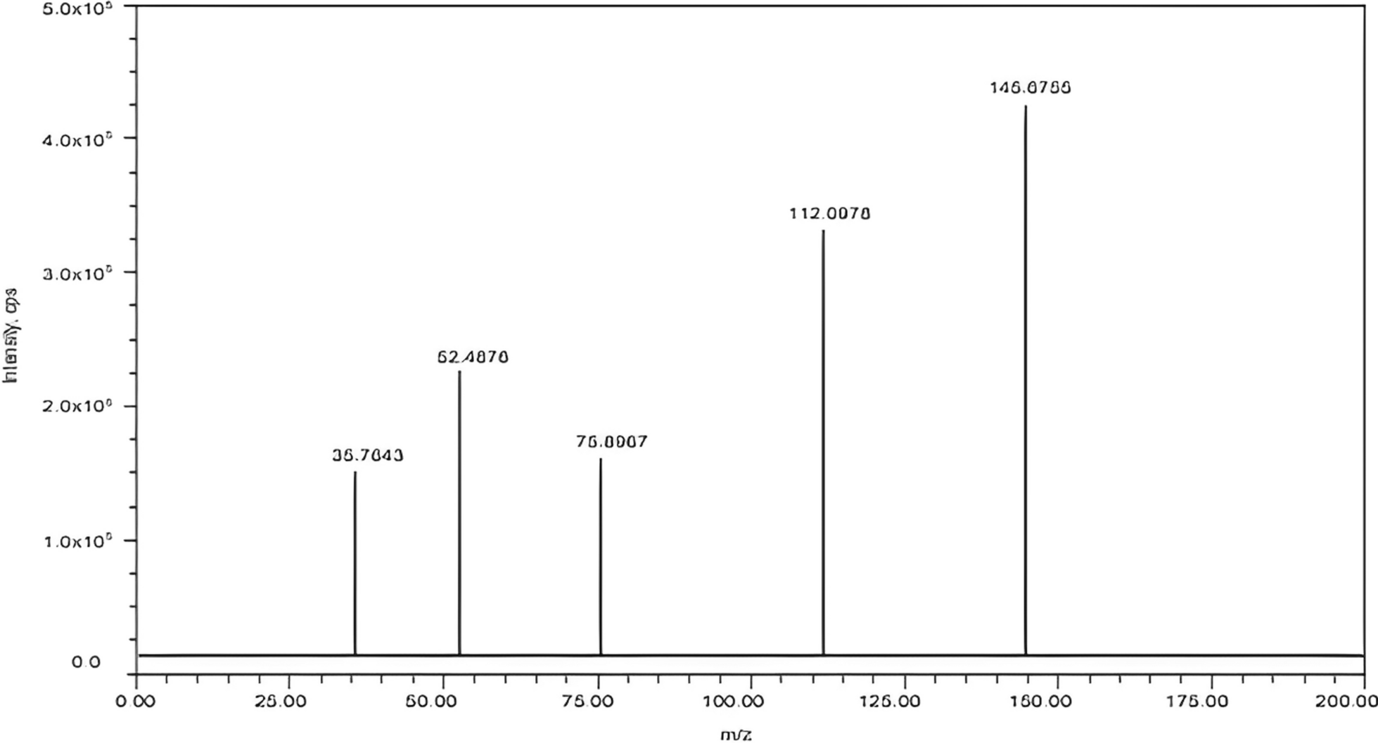
Table 1 FTIR interpretation of compounds of S. asoca crude extractThe LC–MS analysis identified some of the important compounds such as caffeic acid, catechin, quercetin, kaempferol, gallic acid, rutin, β-sitosterol, p-coumaric acid, luteolin etc. (Fig. 5). Phytoestrogenic compounds like β-sitosterol, kaempferol, and quercetin present in the extract are presumed to contribute to the proliferative/antiproliferative effects of the S. asoca crude methanol extract on MCF-7, MDAMB-231, and SKBR3 cancer cell lines.
Fig. 5
LC–MS spectrum of S. asoca crude methanol extract
Molecular dockingThe studies suggest that certain phytoestrogens act as natural agonists for ERβ, making them promising drug candidates for their ability to modulate the cell cycle, influence epigenetic events, and induce apoptosis. Interestingly, in the current investigation, the extract exhibits specific cytotoxicity towards ERβ expressing cells, not affecting ERα. This raises the intriguing possibility that the phytoestrogens in the plant may act as agonists for ERβ. To explore potential interactions between phytoestrogens and ERα/β, molecular docking was performed using Schrodinger Maestro software.
Interaction of phytoestrogens with ERαThe docking of the inbuilt ligand, estradiol into the 3D structure of ERα was done using a glide dock. The amino acid residues in the active site of 3ERT are Trp383, Leu384, Leu387, Met388, Gly390, Lbu391, Val392, Arg394, Met342, Met343, Leu345, Leu346, Thr347, Asn348, Leu349, Ala350, Asp351, Glu353, Leu354, Leu327, Phe404, Leu402, Leu428, Phe425, Ile424, Val422, Met421, Gly420, Glu419, Val418, Met517, Ser518, Lys520, Gly521, Met522, Glu523, Hie524, Leu525, Met528, Lys529, Cys530, Val533, Leu536, Leu539. The estradiol was docked into the active site region and interactions were made with the residues by hydrogen bonding with GLU353 and ARG394 and electrostatic bonding with ASP351. The inbuilt ligand shows a docking score of − 12.17 and binding energy of − 125.19 kcal/mol. The quercetin was docked into the active site region making interactions with the residues by hydrogen bonding with ASP351. The docking score and binding energy were found to be − 6.945 and − 47.026 kcal/mol which was more compared to kaempferol (− 6.93). Tamoxifen shows a docking score of − 10.512 and β-sitosterol did not dock with the binding pocket of ERα. The 3D interaction picture of the study is shown in Fig. 6A and their docking score and binding energy are tabulated in Table 2.
Fig. 6
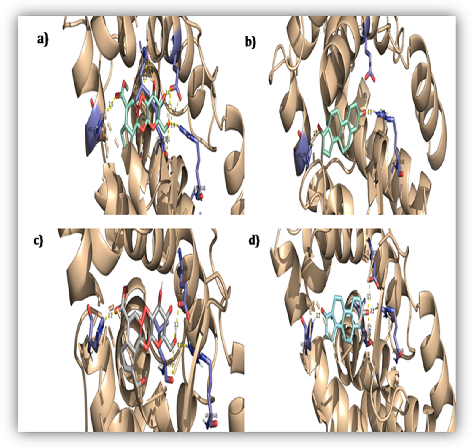
A 2D and 3D image of the interaction between ERα and ligands, B 2D and 3D image of the interaction between ERβ and ligands
Table 2 Docking score and binding energy of estrogen receptors and ligandsThe 2D image (Fig. 6A) reveals the type of interaction between the ligands and amino acids in the active sites of ERα. The inbuilt ligands estradiol, tamoxifen, kaempferol and quercetin form p bonds from their aromatic ring to Phe 404 but the number and nature of hydrogen bonds vary with ligands. Estradiol, kaempferol and quercetin form three hydrogen bonds with Glu353, Hie524 and Arg394, and tamoxifen only once with Asp351.
Interaction of phytoestrogens with ERβThe amino acid residues in the active site of ERβ (PDBID: 3OLL) are Val280, Met295, Ser297, Leu298, Thr299, Leu301, Ala302, Asp303, Glu305, Trp335, Met336, Leu339, Met340, Gly342, Leu343, Met344, Arg346, Leu354, Phe356, Val370, Gly372, Ile373, Ile376, Phe377, Leu380, Ala468, Ser469, Lys471, Gly472, Met473, Hie475, Leu476, Leu477, Met479, Val485, Leu491, Leu495.
The inbuilt ligand was bound deep into the active site area, making hydrogen bonding interactions with Hie475, Arg346, Glu305 and π–π stacking interactions with Phe356. The inbuilt ligand shows a docking score of − 10.5 and binding energy of − 85.248 kcal/mol. The docking of ERβ with quercetin showed the highest docking score of − 9.220 and binding energy of − 66.945 kcal/mol. Quercetin makes hydrogen bond interactions with Arg346, Glu305, Hie475 and π–π stacking with Phe356. Quercetin shows the highest affinity followed by kaempferol (− 8.478) and tamoxifen (− 8.023). Here also, β-sitosterol did not dock with the binding pocket of ER β. The 3D and 2D figures of other ligands are given in Fig. 6B. The docking score and binding energy of other ligands are given in Table 2.
The 2D image (Fig. 6B) reveals the type of interaction between the ligands and amino acids in the active sites of ERβ. The inbuilt ligands estradiol, tamoxifen, kaempferol and quercetin form p bonds from their aromatic ring to Phe 356 but the number and nature of hydrogen bonds vary with ligands. Estradiol, kaempferol and quercetin form three hydrogen bonds with Glu305, Hid475 and Arg346, and tamoxifen only once with Asp351.
留言 (0)