
記住我


The complete genome of PCV3 (2, 000 bp) and PCV4 (1, 770 bp) were synthesized according to the PCV3 and PCV4 genomic sequence (GenBank accession no. KT869077 and MT311852) and cloned into the pUC57 vector by Sangon Biotech Co., Ltd. (Sangon Biotech Co., Ltd., Shanghai, China) to construct standard plasmids for duplex real-time RAA assay, designated as pUC57-PCV3 and pUC57-PCV4. The concentration of the standard plasmids was measured using NanoDrop and Qubit 2.0 fluorometer (Thermo Fisher Scientific, MA, USA). The DNA copy number was calculated using the following formula: DNA copy number/µL = [plasmid concentration (ng/µL)×10− 9 × 6.02 × 1023]/[DNA length (nt)×660]. The gene sequencing was processed by Sangon Biotech Co., Ltd. (Sangon Biotech Co., Ltd., Shanghai, China). The standard plasmids pUC57-PCV3 and pUC57-PCV4 were stored at -20℃ until use.
Design and synthesis of primers and probesThe whole genomic sequences of 30 strains (including genotype 3a, 3b and 3c) of PCV3 and 29 strains (including genotype 4a, 4b and 4c) of PCV4 from GenBank database were aligned respectively using the Lasergene version 7.1 software (DNASTAR Inc., WI, USA) to identify the highly conserved regions. Primer Premier version 5.0 software (PREMIER Biosoft, CA, USA) was used to design RAA primers and probes according to the criteria suggested in the TwistAmp™ amplification guidelines (TwistDx Ltd., Cambridge, UK). An in silico check for the design of the primer pair and probe was conducted utilizing the NCBI Primer-BLAST web tool (https://www.ncbi.nlm.nih.gov/tools/primer-blast). For the development of a duplex real-time RAA assay detecting PCV3 and PCV4, two primer pairs and two RAA probes were designed to target the ORF2 gene of PCV3 (Fig. 1). Concurrently, three primer pairs and two RAA probes were designed to target the ORF1 or ORF2 gene of PCV4 (Figs. 2 and 3). Details of the primers and probes for duplex real-time RAA in this study are shown in Table 1. All the primers and probes were synthesized by Sangon Biotech Co., Ltd. (Sangon Biotech Co., Ltd., Shanghai, China).
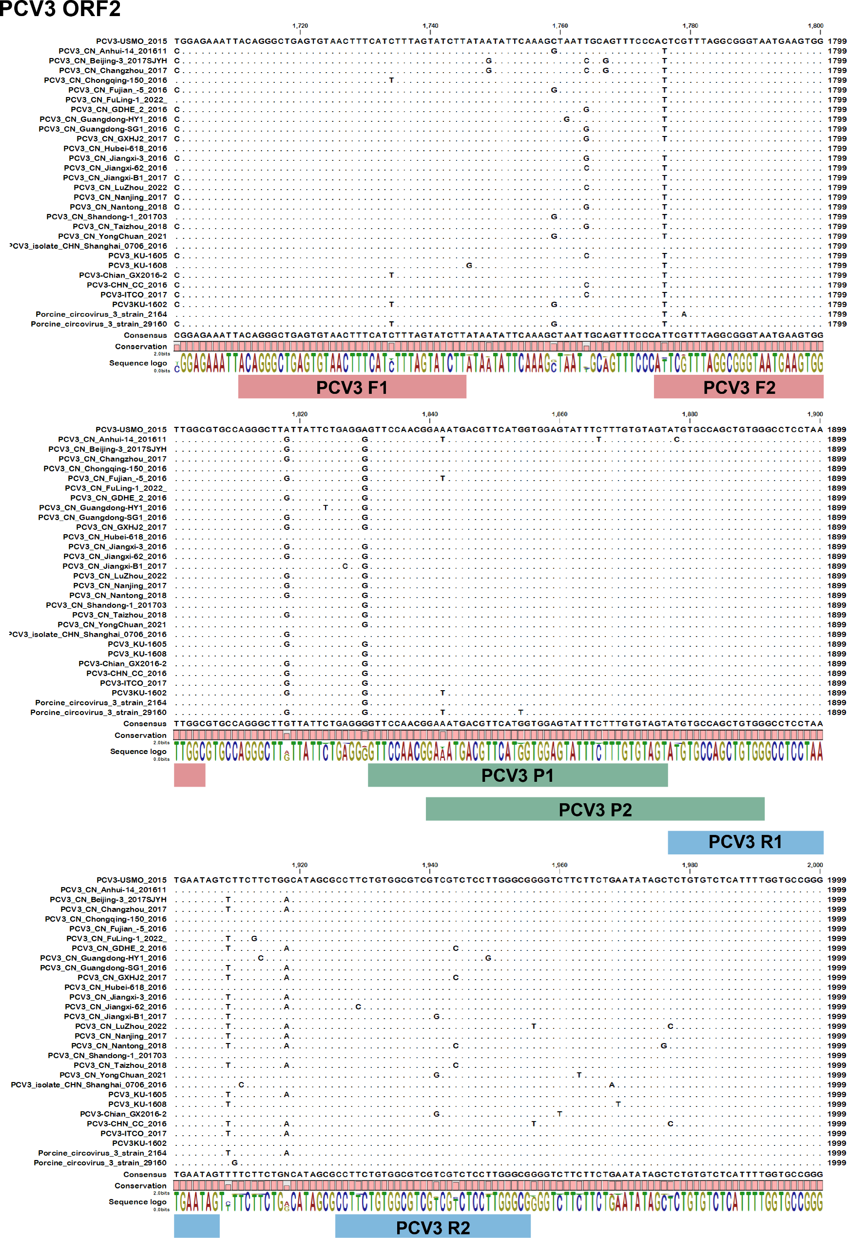
Fig. 1
Positions of the duplex real-time RAA primers and probes in the ORF2 (Cap gene) sequences of different PCV3 strains in the GenBank database. Dots represent nucleotide residues that match the majority. The forward primers are marked with red bars, the reverse primers are marked with blue bars, and probes are marked with green bars
Fig. 2
Positions of the duplex real-time RAA primers and probes in the ORF1 (Rep gene) sequences of different PCV4 strains in the GenBank database. Dots represent nucleotide residues that match the majority. The forward primers are marked with red bars, the reverse primers are marked with blue bars, and probes are marked with green bars
Fig. 3
Positions of the duplex real-time RAA primers and probes in the ORF2 (Cap gene) sequences of different PCV4 strains in the GenBank database. Dots represent nucleotide residues that match the majority. The forward primers are marked with red bars, the reverse primers are marked with blue bars, and probes are marked with green bars
Table 1 Primers and probes used for the duplex RAADuplex real-time RAA assayThe reaction mixture of duplex real-time RAA assay was prepared using the commercial probe-based RAA Kit (Hangzhou Zhongce Biotechnology Co., Ltd., Hangzhou, China). The reaction mixture (50 µL reaction volume) contained Buffer A (25 µL), 2 µL each of PCV3 and PCV4 forward and reverse primers (10 µM), 0.6 µL each of PCV3 and PCV4 probes (10 µM), template DNA (5 µL), nuclease-free water (8.3 µL) and Buffer B (2.5 µL). Briefly, components except template DNA and Buffer B were premixed and transferred into the reaction tube containing RAA enzyme dry powder and overturned to mix thoroughly. Add 5 µL of template DNA to the reaction tube and pipetting 2.5 µL of Buffer B into the tube lid. After a brief vortex mixing and centrifugation, the reaction tube containing the reaction mixture was promptly placed in a ViiA™ 7 real-time PCR instrument (Thermo Fisher Scientific, MA, USA) and incubated at 39 °C for approximately 20 min (1 cycle at 39 °C for 40 s and 40 cycles at 39 °C for 30 s). The fluorescence signal was monitored in real-time, which collected every 30 s. Samples that gave rise to an exponential amplification curve above the negative control threshold within 20 min were judged as positive.
Optimization of reaction systemOptimization experiments were conducted to determine the optimal duplex real-time RAA system. In the optimization experiments, 2.5 µL standard plasmid pUC57-PCV3 (20.0 ng/µL) and 2.5 µL standard plasmid pUC57-PCV4 (20.0 ng/µL) were mixed to serve as templates in the reaction system. 6 sets of primer-probe combinations (PCV3 F1R1 P1/PCV4 F1R1 P1, PCV3 F1R1 P1/PCV4 F2R2 P1, PCV3 F1R1 P1/ PCV4 F3R3 P2, PCV3 F2R2 P2/PCV4 F1R1 P1, PCV3 F2R2 P2/PCV4 F2R2 P1, PCV3 F2R2 P2/PCV4 F3R3 P2) were screened using commercial probe-based RAA Kit under reaction conditions recommended by the kit manual to determine the optimal probe and primer combination.
The concentrations of the best performing primer-probe combination were furtherly optimized by setting the concentration of a single variable component while all other conditions were fixed as recommended by the kit manual. Primer pairs concentration was diluted to 200, 400, and 600 nM, and combinations of concentration were set in Table 2. Probe concentration gradient was set to 60, 90, 120,150 and 180 nM.
Table 2 PCV3 and PCV4 primer pairs concentration ratioNucleic acid extractionViral nucleic acids of clinical samples and artificially spiked samples were extracted from 200 µL of sample lysate with the MagNA Pure LC Total Nucleic Acid Isolation Kit (Roche Diagnostics, Branchburg, NJ, USA), following the manufacturer’s instructions. The extracted nucleic acids were eluted with 50 µL of nuclease-free water and stored at -80℃ until used for duplex real-time RAA assay and qPCR assay. Standard plasmids of PCV3 and PCV4 for duplex real-time RAA were extracted using a Plasmid Mini Kit (Sangon Biotech Co., Ltd., Shanghai, China), according to the manufacturer’s instructions.
Analytical specificityThe specificity of the duplex real-time RAA assay for PCV3 and PCV4 detection was evaluated by using viral DNA or cDNA from other important swine pathogens and standard plasmids (pUC57-PCV3 and pUC57-PCV4) under the optimal reaction system and conditions. The swine pathogens included FMDV (Foot and mouth disease virus), CSFV (Classical swine fever virus), PRV (Pseudorabies virus), PPV (Porcine parvovirus), PCV2 (Porcine circovirus 2), PEDV (Porcine epidemic diarrhea virus), TGEV (Transmissible gastroenteritis virus), PRRSV (Porcine reproductive and respiratory syndrome virus), and PDCoV (Porcine deltacoronavirus). Viral nucleic acids of the above 9 viral pathogens were extracted and maintained in our laboratory. Viral cDNA was obtained using the PrimeScript™ cDNA Synthesis Kit (Takara Biomedical Technology Co., Ltd., Dalian, China), according to the manufacturer’s instructions.
Analytical sensitivityTo evaluate the sensitivity of the duplex real-time RAA assay for PCV3 and PCV4, standard plasmids (pUC57-PCV3 and pUC57-PCV4) was subjected to 10-fold serial dilution ranging from 107 to 100 copies per 2.5 µL. Each dilution was used as a template to carry out the duplex real-time RAA assay according to the optimized system. For a more accurate analysis of the detection limit, 8 independent runs were performed using the above dilution series, and the data were used for the calculation of the 95% limit of detection (LOD) by probit regression analysis using SPSS 24.0 software (SPSS Inc., Chicago, IL, USA).
Analysis of repeatability and reproducibilityThree different dilutions (104, 103, and 102 copies per 2.5 µL) of standard plasmids (pUC57-PCV3 and pUC57-PCV4) were used as templates for the standard plasmids (pUC57-PCV3 and pUC57-PCV4) to assess the intra-assay repeatability and inter-assay reproducibility. In each reaction system, the template consisted of 2.5 µL of diluted pUC57-PCV3 and 2.5 µL of diluted pUC57-PCV4, and both at the same concentration. Each dilution reaction was tested in triplicate in one run or in three independent runs on separate days. The coefficients of variation (CVs) were obtained by calculating the cycle threshold (CT).
Detection of clinical samples and artificially spiked samplesTo evaluate the clinical performance of the duplex real-time RAA assay for PCV3 and PCV4, 60 samples (55 clinical samples and 5 artificially spiked samples) were prepared for detection by the developed assay. 55 clinical samples were screened using the PCV3 qPCR assay (according to local standards in China) and PCV4 qPCR assay reported before respectively [6], and the results showed no qPCR positive samples of PCV4. Therefore, standard plasmid of PCV4 (pUC57-PCV4) was mixed with 3 qPCR positive samples of PCV3 and 5 artificially spiked samples respectively. Specifically, 50 µL of pUC57-PCV4 (205.8 ng/µL) was individually mixed with 1 g, 2 g, and 2.6 g of lymph node samples that tested qPCR positive for PCV3. For the artificially spiked samples, 50 µL of pUC57-PCV4 (205.8 ng/µL) was also individually mixed with 2 g, 3.5 g, and 5 g of pork, as well as 1.5 g and 2 g of lymph node samples. Viral nucleic acid was extracted from each of these mixtures and stored at -20 °C before use. 60 samples were also detected in parallel using the PCV3 and PCV4 qPCR assays respectively for comparison.
Statistical analysisKappa statistics were applied to compare the coincidence rates between the duplex real-time RAA assay and qPCR assays using SPSS 24.0 software (SPSS Inc., Chicago, IL, USA). Statistical analyses and data plotting were performed using the GraphPad Prism software (La Jolla, CA, USA).
留言 (0)