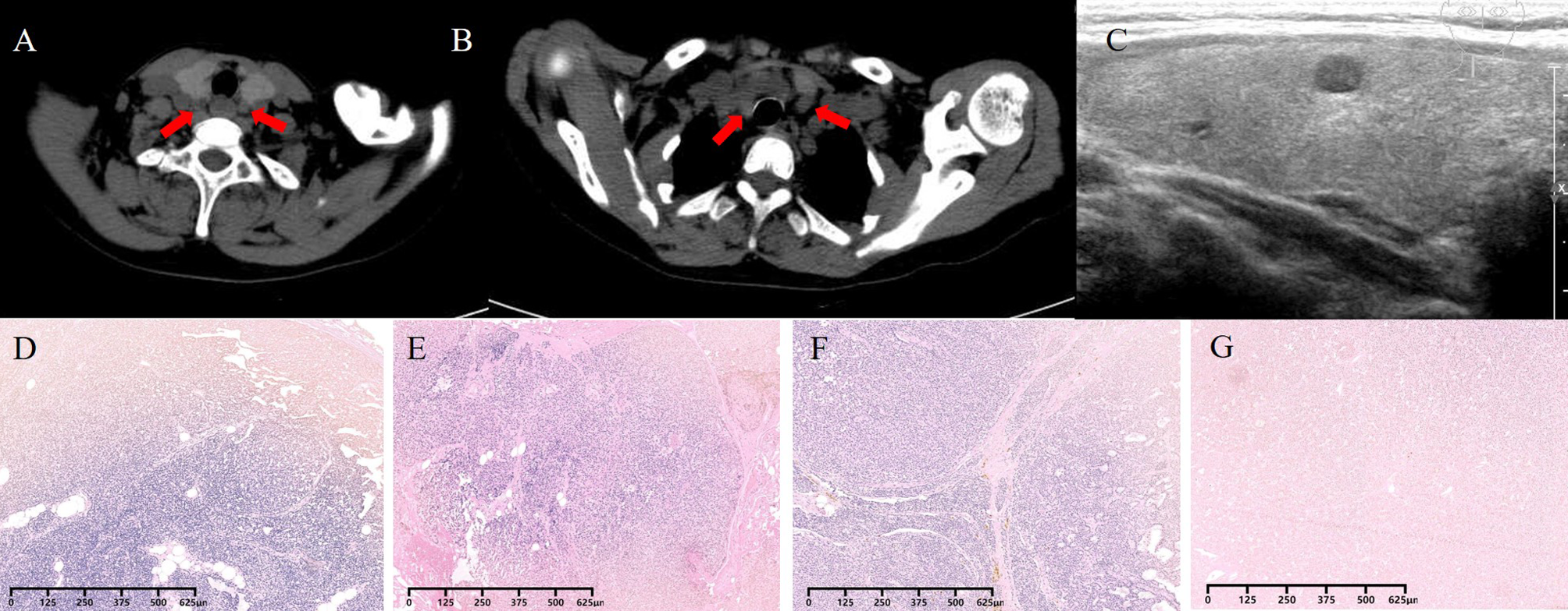
This case clearly shows that bleb formation is observed not only in animal models but also in human ischemic AKI, and bleb formation provides the morphological basis for tubular obstruction mechanism which probably leads to the reduction of urine volume and GFR as this possibility has been previously suggested [7, 9, 12, 13].
According to speculation from animal models, blebs in PT seem to be generated in three ways. (1) A part of membrane-bounded cytoplasm extrudes into the tubular lumen in varied irregular shapes and thereafter separates from the cell. (2) A part of the cytoplasm fluxes into the brush border microvilli, and the microvilli gradually swell and transform into blebs and separate. (3) So-called cytoplasmic bodies are generated intracellularly and extrude into the lumen [6, 14]. Chen et al. also reported two types of bleb formation: at the tips of microvilli and on the apical side of severely damaged cells that had lost their microvilli, with the possibility that alterations in membrane-cytoskeleton linkers may facilitate bleb formation and detachment by weakening membrane-cytoskeleton interactions [8]. Additionally, other previous studies have documented the frequent extrusion of a part of the tubular epithelial cytoplasm into the lumen under ischemic conditions [15, 16]. This time, in electron microscopy, we observed not only apical membrane blebbing but also numerous small structures that seem to be precursors of the blebs, so-called cytoplasmic bodies, in cytoplasm of PTs, and these findings may support tubular obstruction mechanism through bleb formation as a pathogenesis of ischemic AKI in human. Previous studies have highlighted the potential role of blebs forming on the brush border of renal tubules in causing tubular obstruction [7, 9, 12, 13]. Furthermore, several reports have documented the presence of microvesicles in human urine [10, 11], which is likely derived from tubular blebs. This supports the hypothesis that these microvesicles, once shed from the tubules, can persist without degradation until excretion in the urine, thereby suggesting a possible mechanism by which blebs could contribute to tubular obstruction.
Considering the past study reporting that bleb formation was observed within 10 min of onset of ischemia [7], this bleb formation may occur at an earlier stage than cell detachment, potentially contributing to decreased urine output and decreased GFR. This case presented with oliguric AKI along with the sudden onset of nephrotic syndrome, suggesting ischemia directly caused by severe NS seems to trigger bleb formation resulting in oliguric AKI. In addition, electron microscopic finding of apical membrane blebbing with innumerable cytoplasmic bodies in PTs strongly suggests that this bleb formation is literally different from usual debris. While hemorrhagic shock may have further facilitated this bleb formation, given the time course that AKI appeared obviously earlier than hemorrhagic shock, almost simultaneously with the onset of severe NS, it is more likely that blebbing in PTs occurred concurrently with NS because of the intravascular dehydration, ultimately resulting in oliguric AKI as a clinical presentation.
Despite that some reports have demonstrated the bleb formation in animal models as described above [6,7,8], clear evidence of bleb formation, as observed in this case, has not been frequently documented in humans. We speculate that there are two reasons for this. First, kidney biopsy is sometimes clinically avoided in scenarios where systemic deterioration suggests a high risk of post-biopsy complications although a biopsy is desirable for AKI cases without an apparent cause. Studies have shown that AKI patients experience higher rates of post-biopsy complications and blood transfusions compared to non-AKI patients [17,18,19], and despite the KDIGO guidelines recommending kidney biopsy for AKI, its biopsy rate still remains low [18, 19]. For instance, a report from Brigham and Women’s Hospital noted that among 4,903 hospitalized patients who met the laboratory criteria for AKI, only 28 underwent a kidney biopsy [18]. Another study reported that even for early AKI, the biopsy rate was merely 20.5% [20]. Therefore, the histological details of ischemic AKI are still unclear. The second is due to the difficulties of the detection of blebs since this finding is sometimes unclear in usual paraffin sections although it can be easily detected in Epon sections. Thus, we could consider this lesion as cell debris or insignificant artifacts, and they may be removed through the process of pathological preparation in paraffin sections, which could be one of the reasons to be overlooked.
In glomerular pathology, segmental sclerotic lesion was mainly observed. This is the case with pre-existing CKD, hypertension, and diabetes mellitus without diabetic retinopathy, who rapidly developed nephrotic syndrome and oliguric AKI. Although there is the possibility of secondary FSGS, the clinical course could not be fully explained by hypertension- or diabetes-related kidney damage alone and suggested the potential coexistence of MCNS or FSGS, and the histological finding of segmental sclerotic lesions in 22.5% of glomeruli and immunostaining results could not exclude the possibility of FSGS. However, given the further complexity of the case, including the fact that kidney biopsy was performed postmortem, which may have affected the histological findings such as the detachment of podocyte, determining the definitive cause of the nephrotic syndrome based on kidney pathology was challenging. Additionally, because clinical course showed rapid progressive renal dysfunction with nephrotic proteinuria accompanied by hematuria and critical systemic symptoms such as strong generalized fatigue like systematic vasculitis, it was considered different from the usual course of FSGS. Brown et al. previously reported the existence of the treatment-resistant cases of severe NS with microhematuria whose kidney biopsy showed minimal changes with FSGS and suggested the possibility of a different primarily vascular pathogenesis compared to the patients with similar histological appearances [21], and based on light microscopy and immunofluorescence findings, the coexistence of FSGS in this case remains plausible.
Another specific finding in histological analysis was observed in the tubulo-interstitium which corresponds to ATN. This lesion was considered to be caused by ischemia. The kidney of autopsied subjects was also exposed to the ischemia in the agonal stage and the period from death to the autopsy. However, the typical finding of ATN as observed in this case is not commonly observed in autopsy subjects. Therefore, we speculate that the presence of ATN, which appears to be caused by ischemia, occurred at an early stage due to severe NS, rather than changes occurring at the time of death. All this process seems to start with the onset of NS, which led to intravascular dehydration and ischemic AKI. The blebbing might have developed during the course of these processes, causing tubular obstruction and prolonged oliguria.
One of the limitations of our study is the potential influence of hemorrhagic shock on blebbing. Another limitation is that we cannot deny the effect of postmortem changes, which can affect renal histology. However, based on data demonstrating rapid bleb formation following ischemia in animal models and considering the clinical course of this case, we believe that these changes are influenced not only by postmortem alterations but also by ischemic effects caused by severe NS.
In this study, we successfully captured clear bleb formation in proximal tubular epithelial cells in humans. Many aspects of blebbing, including its contribution to tubular obstruction, remain insufficiently elucidated. Further studies are needed to clarify the role of blebbing in ischemic AKI.


留言 (0)