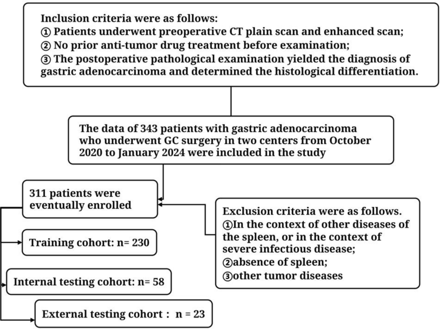
Patient and tumor characteristics
The demographic characteristics of all patients and tumor types in this study are consistent with previous reports. The average age at diagnosis for all patients is 37.6 years, aligning with the previously reported range of 14–52 years (Castellanos et al. 2022; Frič et al. 2023; Ghosh et al. 2023; Guo et al. 2023; Khalid et al. 2022; Wu et al. 2022a, b; Zhao et al. 2021). Among the total patient population, male patients account for 58%, with no observed sex differences, which is in line with former statistics (Bunin et al. 1997; Muller and Merchant et al., 2019; Zhao and Lu et al., 2021). Notably, the average tumor size is15.0 cm3, which is larger than that reported in previous studies (Hu et al. 2021; Iranmehr et al. 2021; Jeswani et al. 2016; Moussazadeh et al. 2016). This finding further validates the effectiveness of the surgical strategy employed and its impact on improved surgical outcomes. The majority of observed tumor types were intrasellar-suprasellar type (27%), suprasellar-third ventricle type I (32%), and suprasellar-third ventricle type II (25%), while occurrences of intrasellar type, third ventricle type, and ectopic CPs were rare. The histological type was mainly aCP, and there was no significant difference between different topological types.
Recurrence and postoperative complications
Although CP is identified as a WHO I grade benign tumor, it has a high recurrence rate ranging from 12 to 52% (Bobeff and Mathios et al., 2023; Lim and Wee et al., 2023; Miao and Fan et al., 2023; Ordóñez-Rubiano and Forbes et al. 2018). The impact of GTR on recurrence is not controversial, is the major predictive factor. But GTR does not warrant survival free of recurrence, it still occurs in 10–20% of cases depending on series (Prieto and Pascual et al., 2013). In our experience, if relapse occurs after radiotherapy, the tumor becomes tightly adhered to surrounding structures, making subsequent manipulation more challenging and increasing the risk of complications. Therefore, the first surgery is vital and always precious for neurosurgeons. Rational strategy and meticulous operation is the key for the successful treatment. This case series achieved 94.5% GTR and an average recurrence rate of 13.56% after 4 years of follow-up. Notably, the recurrence rate for patients undergo first surgery is as low as 8.33%. The higher rate of recurrent cases may be attributed to further radiotherapy after initial incomplete tumor resection, which increases the difficulty of subsequent resection, or to the high proliferation activity of recurrent tumor cells. The recurrence rate among patients varies significantly based on their topographical types, Specifically, patients with intrasellar-suprasellar type and third ventricle type have a lower recurrence rate, indicating the potential of our classification for predicting postoperative recurrence. There is no notable difference with regard to the degree of PS preservation, so the value of sacrificing resection extent to preserve the PS during surgery may be questionable. Sometimes the goal of PS preservation might be contrary to GTR (Ordóñez-Rubiano et al. 2019).
CPs located in the central region of the skull base and surrounded by structures with important physiological functions, complications associated with CPs themselves include tumor stroke, decreased vision, visual field defects, endocrine dysfunction, and diabetes insipidus, among others. There may be various postoperative complications, such as sodium salt disorders, secondary epilepsy, cerebral infarction, cerebral hemorrhage, cerebrospinal fluid leakage, intracranial infection, pulmonary infection, coma, and postoperative unexplained death. This article focuses on long-term complications, such as postoperative obesity, endocrine dysfunction, and diabetes insipidus, which can significantly impact patients’ long-term quality of life. It has been observed that patients’ BMI after surgery is significantly higher than before, which is consistent with previous studies (Beckhaus et al. 2023; Castle-Kirszbaum et al. 2022; Li et al. 2023; Wu et al. 2022a, b) and may be related to damage to the infundibularis nucleus of the hypothalamus (Yang et al. 2020). This change is observed in all six topographical types, but only in the intrasellar-suprasellar type, suprasellar-third ventricle type I, and suprasellar-third ventricle type II is the difference statistically significant. This suggests that these types of CPs are more correlated to the hypothalamus-pituitary axis so the risk of hypothalamic infundibularis nucleus damage is higher.
Regarding endocrine function, there was no statistically significant difference in preoperative, postoperative short-term, and long-term performance among patients with different topographical types. Similarly, no difference was observed in preoperative and postoperative short-term endocrine function between patients with different degrees of PS preservation. However, the long-term endocrine status did differ significantly based on the degree of PS preservation. Notably, patients with a higher degree of pituitary stalk preservation demonstrated a lower risk of long-term endocrine deficiency. This finding was further corroborated through multivariate analysis (Table 4). Other than that, there is a noteworthy variation in the incidence of partial hypopituitarism, with a lower proportion observed in patients with third ventricle type and ectopic type. The proportion of patients experiencing pituitary deficiency after surgery is significantly higher compared to the preoperative period, specifically in the hypothalamic-pituitary-thyroid axis. Nonetheless, no statistically significant differences are observed among the different topographical types concerning the types of pituitary dysfunction. These findings suggest that the etiology of postoperative endocrine dysfunction in CP patients may not be associated with the topographical type, the tumor’s location, and origin. Notably, post-surgical visual disturbances and psychiatric-hypothalamic alterations also significantly influence patients’ quality of life (Pascual et al. 2018, 2021). While our study focuses on endocrine function changes, this represents just one of several crucial factors to consider when developing surgical strategies with patients. In certain cases, clinicians may need to carefully balance these various factors, sometimes necessitating strategic compromises to achieve optimal overall outcomes.
Pituitary stalk preservation
As previously suggested, there was no significant difference in the occurrence of various types and degrees of endocrine dysfunction in patients with different degrees of post-surgery PS preservation. This suggests that the development of endocrine dysfunction in these patients may not be dependent on the degree of PS preservation. Interestingly, there was a significant difference in the proportion of patients with long-term endocrine dysfunction between the groups with varying levels of PS preservation, indicating that patients with complete PS disconnection are more likely to experience long-term endocrine function defects, whereas those with complete PS preservation have a higher chance of recovery. This difference may also be attributed to factors such as the patients’ age, gender, tumor volume, and the surgeon’s experience. However, these indicators did not reveal statistically significant differences among the groups, and all patients were operated on by the same surgeon. Univariate and multivariate analyses (Table 4) were conducted to assess the long-term endocrine function of the patients. The results indicated that only the preoperative BMI and PS preservation were associated with long-term endocrine deficiency, thereby reinforcing the aforementioned conclusions. Therefore, the long-term endocrine status of patients after surgery can be predicted based on PS preservation during the operation.
It is crucial to emphasize that preserving the PS remains an important objective during surgery, aiming for gross total resection (GTR). The ability to retain the PS largely depends on the surgeon’s proficiency and their familiarity with anatomy. This study’s findings reveal that normal preoperative endocrine function serves as a protective factor for the PS, suggesting that the likelihood of intraoperative PS preservation can be predicted based on preoperative endocrine function, thereby guiding the surgical procedure. The choice of surgical approach is typically determined by preoperative imaging data, the patient’s overall condition, and the surgeon’s preferences.
Classification
In 1991, Charles Raybaud et al. firstly proposed a classification scheme based on the anatomical structure displayed by preoperative magnetic resonance imaging in patients with craniopharyngioma, which is divided into the following four types: sellar, infundibulo-tuberal, intraventricular and global type (Raybaud et al. 1991). In 2004, according to the relationship between the tumor and the third ventricle, Jose M. Pascual et al. further divided the intraventricular craniopharyngioma into strictly and non-strictly intraventricular craniopharyngioma. Strictly intraventricular craniopharyngioma only grows in the third ventricle, and has no damage to the third ventricle floor. Non-strictly craniopharyngioma destroys the floor of the third ventricle and grows into the third ventricle. The latter adhesion is tighter, wider, and the prognosis is worse (Pascual et al. 2004). In order to make better surgical decisions, Christian Sainte-Rose et al. divided craniopharyngioma into three types according to the relationship between craniopharyngioma and hypothalamus shown by magnetic resonance imaging: Type0 (no hypothalamus involvement), Type1 (hypothalamus compression elevation or deformation, but still visible), Type2 (hypothalamus involvement, and invisible). Radical resection is the first choice for Type0, and Type1 can still choose complete resection according to the proficiency of the surgeon, while for Type 2, subtotal resection with hypothalamus protection combined with radiotherapy may be the best option (Sainte-Rose et al. 2005). The adhesion of the tumor also affects the outcome of the operation. In order to predict the risk of postoperative hypothalamic injury and the degree of planned tumor resection, Ruth Prieto et al. divided the tumor into five levels: mild, moderate, serious, severe, and critical according to the location, morphology and adhesion strength of the tumor (Prieto et al. 2016).
Tao Hong et al. proposed a new topographical classification for CPs (Tang and Xie et al., 2018), categorizing them based on the origin of the tumors and the hypothalamus observed during endoscopy. This classification divides CPs into two main types: central and peripheral. The central type primarily develops in the pituitary stalk, with the tumor strictly localized in the midline and a low pituitary stalk preservation rate. Conversely, the peripheral type is further categorized into subtypes, namely hypothalamic stalk, suprasellar stalk, and intrasellar stalk CPs, based on their different origins. Suprasellar stalk CPs pose a higher risk of hypothalamus injury, while Intrasellar stalk CPs carry a lower risk. To better understand the growth pattern of tumors, Songtao Qi et al. (Qi and Lu et al., 2011) divided them into four basic growth models according to their relationship with the meninges: subphrenic, extramembranous, intramembranous, and submembranous. This classification serves as a supplement to the existing classification (Prieto and Rosdolsky et al., 2020). Additionally, based on the origin and location of the tumor, the team divided CPs into three types: Q, S, and T, aiming to enhance neurosurgeons’ understanding of the anatomical characteristics of the tumor (Liu et al. 2021). However, Qi’s scheme does not consider the 3 V type, which is the most frequent topography among the papillary type(Prieto et al. 2024).
Yaşargil et al. divided craniopharyngioma into six types according to the relationship between the tumor and the diaphragma sellae and the ventricle (Yaşargil et al. 1990), namely, (a) Purely intrasellar-infradiaphragmatic, (b) intra- and suprasellar, infra- and supradiaphragmatic, (c) supradiaphragmatic, parachiasmatic, extraventricular, (d) intra- and extraventricular, (e) paraventricular in respect to the third ventricle, (f) purely intraventricular. The suitable surgical approaches for each type of CPs were pointed out. For example, it is suggested that the transsphenoidal approach is the most suitable for the small and cystic intrasellar infrasellar craniopharyngioma. For parachiasmatic craniopharyngiomas with large tumors, unilateral pterional approach should be selected. Based on the relationship between craniopharyngioma and infundibulum, Kassam et al. divided CPs into four types (Kassam et al. 2008): pre-infundibulum (type I), trans-infundibulum (type II), post-infundibulum (type III), and all located in the third ventricle (type IV). It is pointed out that type I ∼ III can be treated by endoscopic extended transnasal approach. However, so far, there is no any type of classification that can accurately indicate the origin of the tumor and guide clinical diagnosis and treatment, and reports on the long-term endocrine status of patients with different types are very rare. In this study, CPs were classified according to the different origins from the hypothalamus-pituitary stalk-pituitary axis and the location of tumor growth, and the postoperative endocrine status of each type of patients was described and compared in detail. It has certain value for predicting postoperative endocrine status and recurrence of patients. Preoperatively, the patients’ topographical classification is used to inform the patient of the possible endocrine function after surgery, and the patients’ gain and loss are weighed, which is conducive to the realization of individualized treatment of the patient.
Surgical considerations based on the classification systemStage1 (topographical types)Type1. Intrasellar type
Most tumors are commonly located below the pituitary gland, exerting pressure on it, which results in its upward displacement. In such situations, transsphenoidal surgery using a microscope or endoscopy can be utilized, facilitating the preservation of the hypothalamus-pituitary axis with a relatively low occurrence rate of postoperative endocrine dysfunction. Additionally, the occurrence of cerebrospinal fluid leakage is rare due to the integrity of the sellar diaphragm.
Type2. Intrasellar-suprasellar type
The tumor originates from the lower portion of the pituitary stalk. It exhibits a tendency to invade downwards along the pituitary stalk, causing compression and downward displacement of the pituitary gland. Subfrontal trans-sella approach is most often selected for such type by us. However, this type shows the highest rate of reccurence among all types due to the potential residue tumors in the sella. Therefore, we choose endoscopic transsphenoidal approach for intrasellar-suprasellar type unless the suprasellar portion of tumor reaches huge volume. It is often challenging to ascertain whether preservation of the pituitary stalk is possible, based on the extent of tumor involvement. However, if it becomes apparent that the tumor originates solely from the outer layer of the tubular structure of the PS, known as the peri-stalk pattern, preservation of the pituitary stalk can be achieved. Conversely, if the pituitary stalk demonstrates clear signs of swelling and tumorization, referred to as the intra-stalk pattern, there is a high risk of tumor recurrence if preservation of the pituitary stalk is attempted during surgery.
Type3. Suprasellar-third ventricle type I
The tumor originates in the upper pituitary stalk-infundibular nodule and primarily progresses in two directions: upwards towards the third ventricle and downwards towards the sella. However, unlike the intrasellar type, this tumor tends to exert pressure on the pituitary stalk against the sellar diaphragm. Furthermore, it differs from the intrasellar-suprasellar type as it typically exhibits compression upwards to the third ventricle floor. However, no penetration into the third ventricle is the sign of this type. We commonly utilize the subfrontal approach for this type of tumor. Drilling off the crista galli can widen the visualization and operational corridor to the third ventricle floor. Hypothalamus damage is mild if the care is taken when identifying the interface between tumor capsule and tissue of the third ventricle floor. However, preserving the pituitary stalk can pose challenges, largely depending on whether the tumor follows a peri-stalk or intra-stalk pattern.
Type4. Suprasellar-third ventricle type II
This particular type of CP typically originates from the infundibular nodule-grey nodule. It commonly infiltrates the third ventricle by entering through the ventricular floor. In some cases, it can also spread downward along the pituitary stalk, invading the suprasellar cistern area. However, invasion within the pituitary gland itself is rare. Subfrontal tans-terminal lamina approach is preferenrially chosen for this type If this tumor is detected and treated early, there is a high likelihood of successful preservation of the PS with gross total GTR. Nevertheless, due to the invasion of the hypothalamic structure, even with the complete pituitary stalk preservation, there may still be complications such as endocrine and internal environment disorders following surgery.
Type5. Third ventricle type
This type originates from the internal face of the third ventricle floor, leading to its growth being primarily confined to this area. It also tends to invade the upper and bilateral structures of the third ventricle. However, extension beyond the third ventricle floor into the suprasellar region is uncommon for this tumor. When considering surgical approaches, the longitudinal fissure tanscorpus callosum-third ventricle approach is typically proposed for this type. But we do not propose this surgical corridor as the prefericial approach due to its shortage of managing blood supply and tumor origin at early stage of the operation.
Type6. Ectopic type
The tumor typically originates not in the hypothalamus-pituitary axis but rather in other areas such as the clivus, cerebellopontine angle, and fourth ventricle. These locations are more frequently associated with recurrent cases. Depending on the specific site, a corresponding surgical approach is chosen.
Stage2 (CP growing patterns with PS)
These distinct subtypes of growth patterns not only affect the preservation of the PS but also impact postoperative hypothalamus function. As shown in Fig. 7, patients with intra-stalk have a lower degree of PS preservation. Therefore, for intra-stalk pattern CPs, achieving fine preservation of the PS while getting GTR is challenging. Partial PS preservation or complete PS disconnection is typically opted for intra-stalk pattern CPs. Conversely, most or even complete PS preservation is achievable for peri-stalk pattern CPs. Furthermore, for the same surgeon, predicting the extent of PS preservation can be associated with comprehensive preoperative clinical information, including the patient’s endocrine function status.
Trade-offs between total and subtotal resections
An ongoing area of discussion revolves around the debate between total and subtotal resections. While there no grade I or II evidence to conclusively support either approach, a retrospective review of the literature suggests that gross total resection (GTR) is associated with a higher rate of progression-free survival (Gautier et al. 2012; Hofmann et al. 2012; Mehta et al. 2016; Yaşargil and Curcic et al., 1990), while cases of tumor invasion of the hypothalamus need to be excluded (Elowe-Gruau et al. 2013; Mehta and Jane et al., 2016). The most effective treatment is to achieve GTR while protecting the function of the hypothalamic-pituitary axis, because the hypothalamic-pituitary axis is essential for preventing severe neurological dysfunction (Li and Xiao et al., 2023).
Accurate classification of patients is crucial in guiding the appropriate degree of tumor resection during surgery. For example, in the case of an intrasellar-suprasellar CP with intra-stalk growing ppattern, dissection of the outer layer of the pituitary stalk is necessary to fully expose and remove the intracavitary tumor. Retaining any portion of the tumorous pituitary stalk carries a risk of tumor recurrence. Therefore, despite the potential for postoperative endocrine deficiency, it is necessary to completely electrocoagulate PS at the tumor’s origin site in order to pursue GTR and reduce the risk of short-term recurrence, thereby improving long-term prognosis for this case.
Study limitations
The proposed new classification in this study categorizes CPs into six topographical types, including some rare types. To ensure higher test efficiency, a larger sample size is required for data analysis. Unfortunately, the limitation of sample size prevented the detection of statistical significance for many surgeons’ valuable surgical experience. In addition, visual disturbances and psychiatric-hypothalamic alterations were not included in this study, which limited the more comprehensive application of the research results.What’s more, while the craniotomy surgical approach has a long history in neurosurgery, the development of transnasal endoscopic technology is relatively recent. To accurately compare the effects of different surgical approaches, it is crucial to include a larger number of transnasal endoscopic cases in our institution. We also have to acknowledge the limitations of the classification presented for stalk preservation because it is difficult to anatomically define the degree of stalk preservation when using transcranial approaches (either pterional or subfrontal) compared to endoscopic endonasal approaches.


留言 (0)