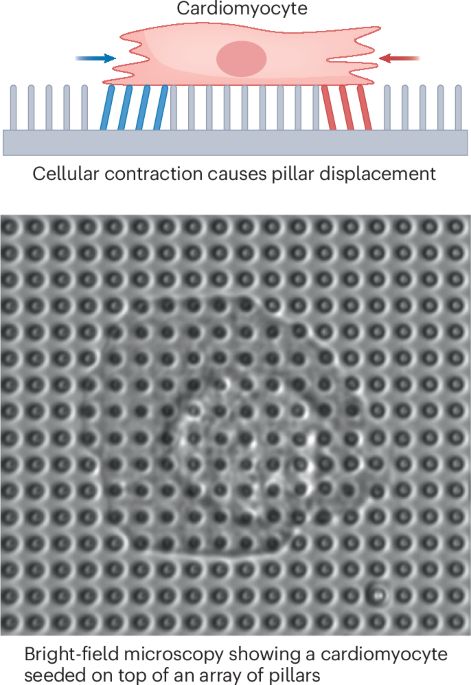
Traction force microscopy of cardiomyocytes
Cardiomyocytes work as the driving force in the contraction and relaxation of the heart. Consequently, these cells are used in in vitro models to study cardiac electrophysiology and biomechanics. The dynamic nature of cardiomyocytes means that their contractile function is a crucial metric to assess experimentally. Novel methods of traction force microscopy (TFM) offer quantitative assessment of single-cell cardiomyocyte contractile function, with the option to alter the cellular environment, either mechanically or chemically, to mimic disease states.
The development, function and pathology of cardiac tissue are highly influenced by microenvironmental factors in both in vivo and in vitro systems. The stiffness of cardiac tissue has a direct effect on cardiac output. For example, after myocardial infarction, many cardiomyocytes die, and remaining healthy cardiomyocytes are left in contact with an extracellular matrix that is stiffer as a result of an increase in collagen. Cardiomyocytes respond to this external stimulus of the stiffer extracellular matrix with abnormal contractile behaviour. Therefore, mimicking this stiffened extracellular matrix in vitro under TFM is a valuable experimental tool. Changes in pillar stiffness can be achieved by modifying the composition or dimensions of pillars. Cardiomyocytes cultured on TFM micropillar arrays of stiffer composition show decreased paxillin displacement and reduced contractile function compared with cardiomyocytes cultured on softer substrates.


留言 (0)