
記住我


Biological information analysis was performed using the RNA-seq data from DEX-stimulated BMSCs and the control group, resulting in the identification of 2133 upregulated and 1552 downregulated genes. Figure 1A shows the intragroup repeatability and intergroup differences of the sequencing samples, Fig. 1B shows a volcano plot, and Fig. 1C displays a heatmap of the DEGs. The GO enrichment analysis of the upregulated and downregulated DEGs revealed that DEX stimulation respectively promoted adipocyte differentiation (Fig. 1D) and inhibited osteoblast differentiation (Fig. 1E). A heatmap was generated for genes enriched in fat cell differentiation among the upregulated DEGs and genes enriched in osteoblast differentiation among the downregulated DEGs (Fig. 1F).
Fig. 1
Bioinformatics analysis of RNA-seq data from DEX-stimulated BMSCs and control BMSCs. A. PCA sample distribution of 4 control group samples and 4 hormone group samples. B. Volcano plot using the criteria |logFC| > 1 and adjusted p value < 0.05 (red dots represent upregulated genes, green dots represent downregulated genes). C. Heatmap showing the clustering relationship of DEGs between the control group and the DEX treatment group. D. GO enrichment analysis of upregulated DEGs. E. GO enrichment analysis of downregulated DEGs. F. Heatmap of genes enriched in fat cell differentiation among the upregulated DEGs and genes enriched in osteoblast differentiation among the downregulated DEGs
Gene expression changes during dexamethasone-induced adipogenic differentiation of BMSCsP5 rat BMSCs were induced to undergo adipogenic differentiation with dexamethasone, simulating the hormonal effects on human BMSCs in vivo. The cells cultured for different durations were divided into 0-, 4-, and 8-day groups for subsequent experiments. The results from Oil Red O staining revealed that the cells in the 0-day group presented very few lipid droplets, whereas those in the 4-day group presented relatively small and lightly stained lipid droplets, and those in the 8-day group presented numerous lipid droplets, some of which were moderately fused and had larger volumes (Fig. 2A).
RNA from the three groups of cells was extracted for RT‒qPCR experiments to validate the effect of dexamethasone on the expression of adipogenic and osteogenic genes in rat BMSCs (Fig. 2B, C). The relative mRNA expression levels of adipogenic genes (PPARγ and C/EBPα) gradually increased in the 0-, 4-, and 8-day groups (P < 0.05), whereas the relative mRNA expression levels of osteogenic genes (RUNX2 and COL1A1) gradually decreased (P < 0.05).
Western blot analysis revealed that the expression levels of the adipogenic proteins PPARγ, C/EBPα, and FABP4 gradually increased (P < 0.05) in the 0-, 4-, and 8-day groups, whereas the expression levels of the osteogenic proteins RUNX2, COL1A1, and OCN gradually decreased (P < 0.05) (Fig. 2D, E, F, G).
Fig. 2
Changes in gene expression during dexamethasone-induced adipogenic differentiation of BMSCs. A. Different time points of dexamethasone-induced adipogenic differentiation of BMSCs: 0 days, 4 days, and 8 days (100x). Scale bar: 100 μm. B: qPCR detection of adipogenic gene mRNA expression in BMSCs. C: qPCR detection of osteogenic gene mRNA expression in BMSCs. D, E: Protein expression of adipogenic genes in BMSCs; F, G: protein expression of osteogenic genes in BMSCs. (ns p > 0.05, * p < 0.05, ** p < 0.01, *** p < 0.001, compared with the 0-day group)
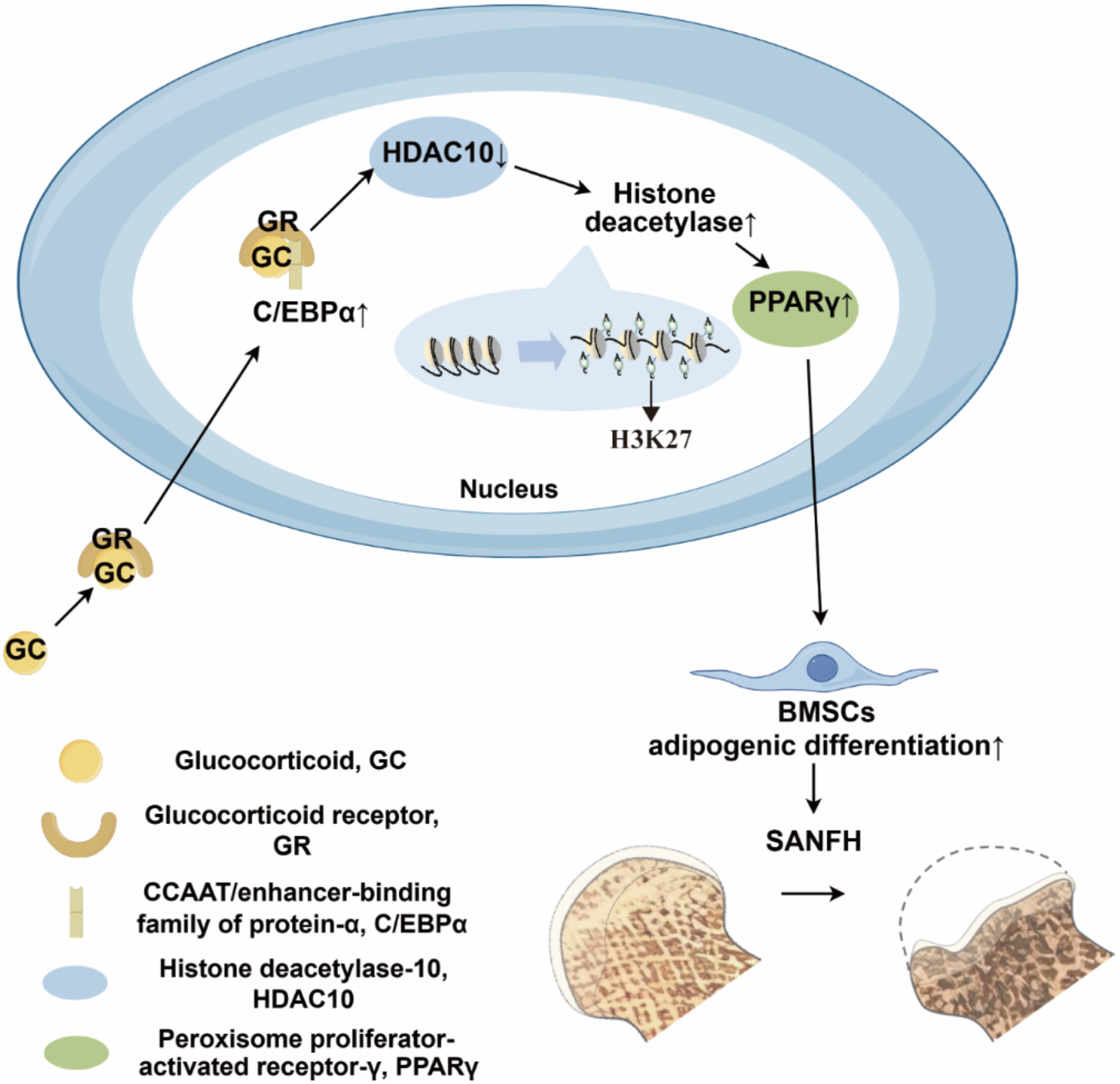
Changes in HDAC expression in the adipogenic differentiation model of BMSCsAfter the successful establishment of an in vitro adipogenic differentiation model of BMSCs, RNA from the three groups of cells was extracted for RT‒qPCR analysis (Fig. 3A). The results revealed a significant increase in the relative expression levels of HDAC 2, HDAC 3, and HDAC 8 mRNAs (P < 0.05), whereas the relative expression levels of HDAC6 and HDAC10 decreased, with HDAC10 showing a significant decrease (P < 0.05).
On the basis of the above experimental results, we selected HDAC10 as the target gene for this study. Western blot analysis revealed that under dexamethasone induction, the expression level of HDAC10 gradually decreased (P < 0.05) (Fig. 3B, C).
Next, we investigated the potential mechanism by which the transcription factor HDAC10 regulated PPARγ expression. Seven potential binding sites of HDAC10 were predicted within 2 kb upstream of the PPARγ transcription start site. ChIP experiments in BMSCs revealed DNA fragments bound to the transcription factor HDAC10, and RT‒qPCR experiments were used to detect whether the obtained DNA fragments contained the promoter region of the PPARγ gene. Compared with that in the 0-day group, enrichment of HDAC10 in the potential binding sites S1-S7 of the PPARγ promoter region was significantly reduced at 8 days (Fig. 3D).
Fig. 3
Changes in HDAC expression in the adipogenic differentiation model of BMSCs. A. Changes in the expression of HDACs during dexamethasone-induced adipogenic differentiation of BMSCs. B, C: Changes in the protein expression of HDAC10 during dexamethasone-induced adipogenic differentiation of BMSCs. D. ChIP‒qPCR results showing the reduced enrichment of HDAC10 at sites S1-S7. (ns p > 0.05, * p < 0.05, ** p < 0.01, *** p < 0.001, compared with the 0-day group)
Imaging findings and general view of clinical specimensIn this study, imaging data and femoral head samples were collected from patients with steroid-associated necrosis of the femoral head (SANFH) and femoral neck fractures.
In terms of imaging, X-rays of SANFH patients (Fig. 4A) revealed significant local patchy sclerosis and cystic changes, whereas CT scans (Fig. 4B) revealed collapse and flattening of the femoral head. MRI images (Fig. 4D, E) revealed the double-line sign and localized signal intensity elevation within the femoral head. For femoral neck fractures, X-rays and CT scans (Fig. 4F, G) revealed obvious cortical fractures and displacement of the fracture ends.
The femoral heads of the SANFH group exhibited slight flattening, uneven surfaces, and black folds near the junction of the head and neck (Fig. 4C). The femoral heads of the femoral neck fractures appeared spherical with a smooth surface, and blood infiltration was observed in the fracture area (Fig. 4H).
Fig. 4
Radiographic and gross appearances of the clinical samples. A, B, C represent the radiographic and specimen appearances of SANFH patients. D, E show the MRI findings of SANFH patients. F, G, H depict the radiographic and specimen appearances of femoral neck fractures
Histopathological findings in the animal specimensTo validate the differences in adipogenic and osteogenic gene expression in steroid-associated necrosis of the femoral head (SANFH) compared with normal femoral head specimens, we collected femoral heads from two groups of mice for IHC staining.
In the immunohistochemical sections, both groups presented varying degrees of positive expression of PPARγ, COL1A1, and OCN (Fig. 5A, E, C). Osteogenesis-related genes such as COL1A1 and OCN were expressed in the osteocytes between trabeculae in both groups, with significantly lower expression in the experimental group than in the control group (Fig. 5A, C). The adipogenesis-related gene PPARγ was expressed mainly in adipocyte-containing lipid droplets between trabeculae in the necrotic area of the experimental group and appeared brownish yellow (Fig. 5E). Statistical analysis revealed that the protein expression of PPARγ was significantly greater in the experimental group than in the control group (P < 0.05) (Fig. 5F), whereas the protein expression of COL1A1 and OCN was lower in the experimental group than in the control group (P < 0.05) (Fig. 5B, D).
We subsequently performed immunohistochemistry on mouse femoral head samples to analyse the expression of HDAC10 (Fig. 5G). HDAC10 was expressed in osteocytes and interstitial cells in both the control and experimental groups, with statistical analysis revealing a decrease in HDAC10 expression in the experimental group compared with the control group (P < 0.05) (Fig. 5H). The experimental results were consistent with the results of the in vitro cell experiments.
Fig. 5
A, B, C, D: IOD analysis comparing the expression of the osteogenic genes COL1A1 and OCN in specimens from both groups. E, F: IOD analysis comparing the expression of the adipogenic gene PPARγ in specimens from both groups. G, H: Differential expression of HDAC10 via immunohistochemistry between the control and experimental groups. (*p < 0.05, **p < 0.01, ***p < 0.001 compared with the control group.) (30x)
Histopathological findings of the clinical specimensTo further verify the differences in adipogenic and osteogenic gene expression between SANFH specimens compared with normal femoral head specimens, we collected femoral heads from the two groups of patients for H&E and IHC staining.
H&E staining revealed that in the control group, the trabecular bone structure was intact, with abundant osteoblasts and few empty lacunae, and the adipocytes in the interstitium had a normal morphology. In the experimental group, the necrotic area presented abundant lacunar cells, destruction of adipocytes in the interstitium, and a large amount of uniformly stained necrotic tissue (Fig. 6A).
In the immunohistochemistry sections, positive expression of PPARγ, C/EBPα, COL1A1, OCN, and RUNX2 was observed to varying degrees in both groups (Fig. 6B, D, F, H, J). Osteogenic genes such as COL1A1, OCN, and RUNX2 were expressed in the trabecular bone and osteoblasts to a greater extent in the experimental than in the control group (Fig. 6B, D, F). Adipogenic genes such as PPARγ and C/EBPα were highly expressed mainly in adipocyte vacuoles in the necrotic area of the trabecular bone in the experimental group and appeared brownish yellow (Fig. 6H, J). Statistical analysis revealed that the protein expression of PPARγ and C/EBPα was significantly greater in the experimental group than in the control group (P < 0.05), whereas the protein expression of COL1A1, OCN, and RUNX2 was lower in the experimental group than in the control group (P < 0.05) (Fig. 6C, E, G, I, K).
We subsequently conducted immunohistochemistry on human femoral head samples to analyse the expression of HDAC10 (Fig. 6L). Compared with that in the control group, the expression of HDAC10 in the trabecular bone and interstitial cells was significantly lower in the experimental group compared with the control group (P < 0.05) (Fig. 6M). The experimental results were consistent with those of the cell and animal experiments.
Fig. 6
A: Normal osteoblast morphology and intact interstitial adipocytes in the control group. Abundant lacunar cells and destruction of interstitial adipose tissue were observed in the experimental group. B, C, D, E, F, G: Analysis of the IOD of the osteogenic genes COL1A1, OCN, and RUNX2 in the two groups of specimens. H, I, J, K: Analysis of the IOD of the adipogenic genes PPARγ and C/EBPα in the two groups of specimens. L, M: Differential expression of HDAC10 according to immunohistochemistry in the control group and the experimental group. (*p < 0.05, **p < 0.01, compared with the control group) (100x)
留言 (0)