
記住我


YXB was provided by Liaoning Huarun Benxi Sanyu Co., Ltd (batch no.20210117). The preparation process rigorously followed the standards set by the National Food and Drug Administration of China. Cyathula offfcinalis Kuan, Salvia miltiorrhiza Bunge and Astragalus membranaceus (Fisch.) Bunge was finely ground and sifted through a 100-mesh screen for subsequent use. A mixture including Salvia miltiorrhiza Bunge and Astragalus membranaceus (Fisch.) Bunge, among eight ingredients total, was decocted twice. The first decoction involved adding water at eight times the volume and boiling for 2 h; the second decoction involved adding water at six times the volume and boils for 1 h. The decoctions were then combined, filtered, and allowed to settle. The clear supernatant was vacuum-concentrated to a relative density of 1.18–1.22 at 50 °C to produce a clear paste, which was set aside. This paste was then granulated with the herbal powder, formed into granules, and mixed with sodium carboxymethyl starch and magnesium stearate. The mixture was then compressed into tablets, producing 1000 tablets, each coated with a thin film and containing 0.138 g per pill. The ingredient in the YXB showed by Supplementary Table 1. The main chemical components in YXB were obtained by applying high performance liquid chromatography-mass spectrometry (HPLC–MS) in the preliminary stage (Supplementary Table 2, Fig. 1) [22, 23]. According to the medication instructions, adults are advised to take 15 tablets daily, totaling 2.07 g. Drawing from a dosage conversion formula between mice and humans and previous empirical data, the effective doses of YXB were established at 0.2, 0.4 and 0.8 times the standard dose, equivalent to 60 mg/d/kg, 120 mg/d/kg, and 240 mg/d/kg, which were low, medium and high dosage respectively. Previous researches have verified that the high dosage yielded the most favorable outcomes [23, 24]. IBU was purchased from Huizhou Daya Pharmaceutical Co. (batch no.201802). The administered dose was 91 mg/kg, which corresponded to the clinically equivalent dose. Both YXB and IBU were dissolved in 0.5% carboxymethylcellulose sodium (CMC-Na) solution and administered by gavage every 12 h for 19 days.
Fig. 1
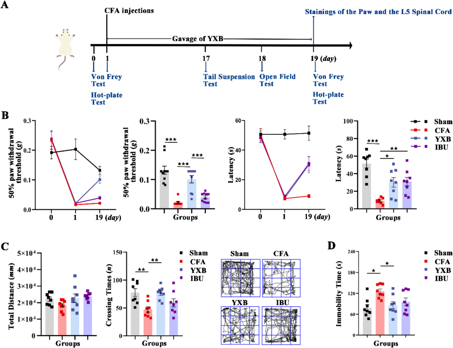
Effects of YXB on mechanical allodynia, heat hypersensitivity and depressive and anxious behavior in the CFA mice. A Schematic diagram of experimental design in this section. B The mechanical allodynia and heat hypersensitivity. C The Open Field Test (OFT) to evaluate anxious behavior. D The Tail Suspension Test (TST) to evaluate depressive behavior. All data are presented as (Mean ± S.E.M), *P < 0.05, **P < 0.01, ***P < 0.001
Experimental animals8-week-old male SPF grade mice from Institute of Cancer Research (ICR), weighing 30 ± 2 g, were purchased from Spearfish (Beijing) Laboratory Animal Technology Co. [certificate no. SCXK (Jing) 2019-0010]. The experimental environment was the Animal Experiment Center of Institute of Basic Theory for Chinese Medicine, China Academy of Chinese Medical Sciences [license No. SYXK (Jing) 2021-0017]. The breeding environment was maintained at a temperature of 20 °C–24 °C, humidity of 40–60%. The mice were fed in separated cages, 5 mice per cage with free access to water and food. The operations involved in this experiment were in accordance with the requirements and standards of the Ethics Committee of the Institute of Chinese Materia Medica, China Academy of Chinese Medical Sciences.
CFA animal modelAs described [25], complete Freund’s adjuvant (CFA, 20 μL, Sigma-Aldrich, F5881) was injected subcutaneously into the left hind paws of mice. Sham group were injected with equal volume of saline.
Experimental design and groupingAccording to the objectives, the following design and grouping were carried out in our study: (1) To verify whether the analgesic effect of YXB in CFA mice is associated with the modulation of oxylipin-mediated inflammation resolution, the mice were randomly split into 4 groups: Sham, CFA, YXB-H (optimal dosage) [23, 24] and IBU.
(2) To verify the effect of YXB on the expression of LXA4, FPR2, TRPA1, Leukotriene A4 hydroxylase (LTA4H) in the paws of CFA mice, they were randomly divided into six groups: Sham, CFA, IBU, YXB-L, YXB-M and YXB-H.
(3) To further demonstrate whether the analgesic effect of YXB on CFA mice is related to the “LXA4-FPR2-TRPA1” pathway, two batches of animal experiments were performed. On the one hand, to assess whether the analgesic effect of YXB can be impaired after injection of FPR2 antagonist, the mice were randomly divided into four groups: Sham, CFA, YXB (YXB-H) and YXB-WRW4 (FPR2 antagonist, Selleck, S9818). In YXB-WRW4 group: for the CFA model, YXB was administered orally for 19 days, and on days 17–19, 45 min after the administration of YXB, WRW4(0.5 μg/μL) [26] was injected into the left hind paws of the mice. On the other hand, to evaluate whether TRPA1 is downstream of the YXB regulatory, the mice were randomly divided into three groups: Sham, AITC (TRPA1 agonist, Sigma-Aldrich, W203408) and YXB-AITC. Based on literature [27] and experimental experience, AITC (50 μM, 10 μL) was injected into the left hind paws of mice to construct a model. While for YXB-AITC group, AITC was injected into the left hind paws of mice 19 days after the pre-administration of YXB.
Von Frey testMechanical allodynia thresholds were assessed by the Von Frey test. Briefly, the mice were acclimatized in the test frame for 30 min. When in the resting state, the mice were vertically stimulated with 0.008–0.4 g of Von Frey fiber wire at the plantar area of the left hind paws for 6–7 s. The presence of paw contraction, shaking or licking was recorded as positive. The operation was performed six times cumulatively, with an interval of 5 min. The 50% paw withdrawal threshold (PWT) was analyzed by up-down method [28].
Hot plate testHeat hypersensitivity was evaluated by the hot plate method. The test was performed before and after modeling and 1 h after drug administration on day 19. The mice were placed on the hot plate that was preheated to 53 °C, and the latency time was recorded when the mice showed reactions of foot lifting or licking [29].
Tail suspension testThe depressive behavior of mice was assessed by Tail-suspension test (TST). As previously described [30], the mice were wrapped with tape around the tail tip at 1.5 cm and hung upside down on a tail suspension box for 6 min. The first 2 min served as the adaptation stage, and the accumulated time for the mouse to stop struggling was recorded in the last 4 min.
Open Field testThe anxiety behavior of mice was evaluated by Open Field Test (OFT). Mice were placed in black cubic open-top box for free movement, the trajectories of mice were tracked and data were collected within 5 min by video analysis software (Shanghai Jiliang Software Technology Co., Ltd.). At the end of each batch of experiments, the field was wiped with ethanol to prevent the remaining odor from affecting the rest of the mice [31].
Quantitative analysis of oxylipinsLeft paws from five or six mice per group were chosen for oxylipins. According to the previous literature [32], supernatant was extracted from the samples after homogenization, centrifugation, re-extraction, elution, concentration, and re-dissolution. Liquid chromatography tandem-mass spectrometry (LC–MS / MS) platform (SCIEX Corporation, USA) and multiple reaction monitoring (MRM) mode were employed to perform qualitative and quantitative analyses of oxylipins in extracted samples.
In terms of chromatographic separation, the Waters ACQUITY UPLC HSS T3 C18 column (2.6 μm, 2.1 mm × 100 mm i.d.) was employed. The separation utilized a mobile phase consisting of two components: Phase A was a mixture of acetonitrile and water (60/40, v/v) with 0.04% acetic acid (Sigma-Aldrich, Germany), and Phase B comprised equal volumes of acetonitrile (Merck) and isopropanol (Merck). The column temperature was kept constant at 40 °C, with a flow rate of 0.4 mL/min and an injection volume of 10 μL. The gradient elution commenced with a 99.9:0.1 (v/v) ratio of A to B, adjusting to 70:30 (v/v) at 2 min, 50:50 (v/v) at 4 min, and moving to a 1:99 (v/v) ratio between 5.5 and 7 min, before returning to the initial ratio at 7.1 min. For mass spectrometry analysis, the electrospray ionization source temperature was adjusted to 550 °C, with the mass spectrometric voltage set to 5500 V in positive ion mode and − 4500 V in negative ion mode. The curtain gas pressure was maintained at 35 psi. Within the Q-Trap 6500 + system, ion pairs were scrutinized using optimized declustering potential (DP) and collision energy (CE) settings, ensuring precise identification and quantification of analytes.
Analyst® 1.6.3 was used for processing mass spectrometric data. Mass spectrometric qualitative analysis of oxylipins relied on information from the oxylipin database. Differential metabolites were screened based on variable importance in projection (VIP > 1) using the orthogonal partial least squares discriminant analysis (OPLS-DA) model and independent samples t-test (P < 0.05). Subsequently, differential genes underwent heatmap analyses.
Enzyme linked immunosorbent assay (ELISA)Protein was extracted from the left paws of mice from each group, and the levels of LXA4 were measured according to the instructions of the ELISA kit (ELK Biotechology, China).
ImmunofluorescencePerfused with 4% paraformaldehyde, the left paws, L4 ~ L5 segments of the DRG and spinal cords were taken and placed in fixative (4% paraformaldehyde + 8% sucrose solution) for 24 h. After 48 h of gradient dehydration in 15% and 30% sucrose solutions, respectively, the tissues were blotted dry with filter paper and collected. Coronal frozen sections with the thickness of 18 μm were prepared after embedding the tissue with OCT. Subsequently, immunofluorescence staining was performed. Sections were eluted three times with phosphate buffered saline (PBS). After blocking with 5% BSAT (5% Bovine albumin + 0.5% Triton X-100) for 30 min, the primary antibodies, namely, rabbit anti-LAT4H (Immunoway YT6624 1:400), rabbit anti-TRPA1 (Immunoway YN3017 1:400), rabbit anti-TRPV1 (abcam ab305299 1:500) mouse anti-IBA1(Invitrogen GT10312 1:400), mouse anti-CD68 (Immunoway YM3050 1:200) and mouse anti-CGRP (1:400) were incubated, and rested overnight at 4 °C. Sections were eluted three times with PBST and incubated fluorescent secondary antibodies: dylight goat anti-rabbit 488 (Earthox E032220 1:400) and dylight goat anti-mouse 594 (Earthox E032210 1:400) away from light for 1 h at ambient temperature. Subsequently, the nuclei were stained by DAPI (Solarbio C0065 1:1000). Finally, sections were dripped with antifade mounting medium and sealed. Fluorescence images were captured using laser confocal microscope (Olympus FV1000, × 600) and analyzed by ImageJ software.
Flow cytometryIn line with literature [33] and our research group’s experience, fresh left paws of mice were processed to prepare a single-cell suspension through enzymatic digestion. Cells were aliquoted into flow cytometry tubes at a concentration of 1 × 10⁶ cells per tube, each supplemented with PBS up to 1 mL. The tubes were then centrifuged at 350 g for 5 min, the supernatant discarded, and the cells resuspended in 100 μL of stain buffer FBS 2 μL of Fc block was added, followed by a 5-min incubation at 4 °C. Antibodies CD45, CD11b, and F4/80 (BD Bioscience 463891, 557397, 566787) were then added, and the mixture was incubated for another 30 min at 4 °C. After adding 1 mL of PBS and another round of centrifugation at 350 g for 5 min, the supernatant was discarded, and the cells were resuspended in 500 μL of stain buffer FBS 2.5 μL of 7-AAD (BD Bioscience 559925) was added, and the mixture was incubated in the dark for 10 min, then filtered through a 70 μm mesh, and analyzed using a flow cytometer (BD Celesta). Statistical analysis that proportion of F4/80+CD11b+ cells among live cells in the paws was performed using FlowJo software.
Western blottingProtein was extracted from the left paws of mice, and its concentration was determined using the BCA method. After adding the loading buffer, the samples were boiled for 10 min to denature the proteins. Subsequently, the samples underwent 90-min electrophoresis with 80 V and 2-h wet transfer with 250 mA, followed by a 2-h blocking phase using 5% non-fat milk. Primary antibodies TRPA1 (1:1000) and β-actin (1:5000) were introduced and the mixture incubated overnight at 4 °C. The membranes were washed three times with 1% TBST, each wash lasting 15 min, and then incubated with secondary antibodies (1:5000) at room temperature for 2 h. Following three additional 15-min washes, the membranes were developed using an ECL detection reagent. β-actin served as the internal control. Protein bands were analyzed with Image J software to determine the relative expression levels of the TRPA1 protein.
Statistical analysisGraphPad Prism 8.0.2 was used for data analysis. Values are presented as mean ± standard error of the mean (x̄ ± S.E.M). Multigroup comparisons were made using analysis of variance. P < 0.05 indicated statistical significance.
留言 (0)