The current study aims to comprehensively analyze the proteomic profile of human CL lesions due to L. donovani infections in Sri Lanka and validate the biological pathways represented.
Sample collection and confirmation of diagnosis
This study received ethical approval from the Ethics Review Committee of the Faculty of Medicine, University of Kelaniya, Sri Lanka (P/99/06/2013) and was conducted adhering to the approved protocol and in agreement with the Helsinki Declaration. Patients and controls were recruited voluntarily and informed written consent was obtained before sample collection.
Patients with skin lesions suspected of CL were recruited from Base Hospital Padaviya and the Sri Lanka Army. Lesion biopsies with a diameter of 3–4 mm were obtained from the active edge of the CL lesion before starting treatment. The diagnosis was established with light microscopy of Giemsa-stained tissue impression smears and species diagnosis was confirmed using previously established molecular methods [29]. Control skin specimens were obtained from incision sites of patients with no signs or symptoms of leishmaniasis, who underwent minor surgical procedures due to unrelated surgical causes. Skin biopsy specimens were immediately submerged in RNAlater and stored at – 20 ℃ until further analysis. Eight patient and eight control skin specimens were processed for proteomic profiling by mass spectrometry.
Samples for immunohistochemical (IHC) validation of the unfolded protein response (UPR) pathway were selected from previously archived formalin fixed paraffin embedded lesion and control specimens. Thirty lesion specimens from leishmaniasis-confirmed patients and six control specimens from patients undergoing minor surgical procedures for unrelated surgical causes were used for IHC staining. This part of the study received ethical approval from the Ethics Review Committee of the Faculty of Medicine, University of Kelaniya, Sri Lanka (Ref. No. P/21/03/2021).
Specimen transport
Specimens were transported to the Department of Pathology, Wexner Medical Center, Ohio State
University, USA, submerged in RNAlater. Dry ice was used to maintain the temperature below − 20 ℃. The amount of dry ice required for the shipment was calculated according to the time taken for air transportation, using guidelines available at http://dryiceinfo.com/shipping.htm. Triple packaged samples were shipped as Category B biological substances.
Sample processing for proteomics
Sample preparation was carried out at the Campus Chemical Instrument Center (CCIC) Mass Spectrometry and Proteomics Facility, Ohio State University, Columbus, Ohio, USA. Sample preparation was done in a Class II type A2 biosafety cabinet (NuAir, Minnesota, USA). Tissue samples stored in RNAlater were removed from the reagent, blotted on a filter paper to remove traces of RNAlater, and placed in new 1.5 ml microcentrifuge tubes (Fisher Scientific, New Hampshire, USA). Protein digestion was done using RapiGest SF Protein Digestion Surfactant (Waters, Milford, MA, USA), and samples were regarded as proteolytic resistant or hydrophobic proteins as for the manufacturer instructions and processed accordingly. To each of the tubes, 100 µL of 0.2% RapiGest SF Protein Digestion Surfactant (Waters, Milford, MA, USA) in 50 mM NH4HCO3 was added. Samples were sonicated in a Sonic Dismembrator (Fisher Scientific, New Hampshire, USA) at speed 6 for 4 times and speed 5 twice, each sonication lasting 3 s. Sonicated samples were then heated at 105 ℃ in a heat block for 30 min, following which they were cooled on ice for 5 min. Samples were then vortexed for 5 min, following which they were heated at 70 ℃ for 2 h. Dithiothreitol (DDT) (ThermoFisher Scientific, USA) was added at a final concentration of 5 mM and heated at 60 ℃ for 30 min. Following this, iodoacetamide (IAA) (Acros Organics, NJ, USA) was added at 15 mM final concentration and incubated at room temperature, in the dark for 15 min to inhibit proteases. DTT reduces disulfide bonds between cysteine residues, converting them into free sulfhydryl (-SH) groups, which is essential for fully denaturing the protein and making it more amenable to enzymatic digestion, while IAA alkylates the free sulfhydryl groups to prevent them from reforming disulfide bonds, thus stabilizing the protein's denatured state and ensuring consistent and reliable analysis. For digestion of proteins, 1 µg of sequencing grade trypsin (Promega, Wisconsin, USA) was added and samples were incubated at 37 ℃ overnight.
Rapigest was precipitated by adding trifluoroacetic acid (Fisher Scientific, New Hampshire, USA) to a final concentration of 0.5% and incubating at 37 ℃ for 30 min. Samples were then centrifuged at 13,000 g for 15 min. The supernatant was then transferred to a microcentrifuge tube (Eppendorf®) and dried in a speed vac (Eppendorf Vacufuge Plus, Hamburg, Germany). Samples were stored at − 80 ℃ until analysis. They were then re-suspended in 50 mM acetic acid (Ultrex II Ultrapure Reagent, J.T. Baker™) and peptide concentrations were determined from their absorbance at 280 nm using a Nanodrop 1000 spectrophotometer (Thermo Fisher Scientific, USA).
Instrument protocol- tandem mass spectrometry
Tandem mass spectrometry (MS2) was carried out using Thermo Orbitrap fusion HPLC MS–MS system (Thermo Fisher Scientific, USA). Prior to tandem mass spectrometry, samples were subjected to two –dimensional liquid chromatography (2-D LC) separation using a Thermo Scientific 2D rapid separation liquid chromatography (RSLC) high-pressure liquid chromatography (HPLC) system. A sample volume consisting of 12 ug of peptides was first separated on a 5 mm × 300 μm Ethylene Bridged Hybrid (EBH) C18 column with 5 μm particle size and 130 Å pore size. Solvent A was composed of 20 mM ammonium formate (Fisher Scientific New Hampshire, USA) at pH 10, and solvent B was 100% HPLC grade acetonitrile (Sigma Aldrich, Missouri, USA). Peptides were eluted from the column in eight successive fractions using 9.5, 12.4, 14.3, 16.0, 17.8, 19.7, 22.6 and 50% solvent B. Each eluted fraction was then trapped, diluted, neutralized, and desalted on a µ-Precolumn Cartridge (Thermo Fisher Scientific) for the second-dimension separations performed with a 15 cm × 75 cm PepMap C18 column (ThermoFisher Scientific, Waltham, MA) with 3 μm particle size and 100 Å pore size. For the Thermo Scientific 2D RSLC HPLC system, the flow rate for the analytical column was 500 μL/min. The gradient was 0 to 5 min, 2% solvent B; 5 to 38 min, 35% solvent B; 38 to 46 min, 35–55% solvent B; 46 to 47 min, 55–90% solvent B. Mobile Phase B was kept at 90% for 1 min before quickly brought back to 2%. The system was equilibrated for 11 min for the next separation.
Tandem mass spectrometry data was acquired with a spray voltage of 1.7 kV and the capillary temperature used was 275 ℃. The scan sequence of the mass spectrometer was based on the preview mode data dependent TopSpeed™ method: the analysis was programmed for a full scan recorded between m/z 400–1600 and an MS2 scan to generate product ion spectra to determine the amino acid sequence in consecutive scans starting from the most abundant peaks in the spectrum in the next 3 s. To achieve high mass accuracy mass spectrometry determination, the full scan was performed in Fourier Transformation (FT) mode and the resolution was set at 120,000. The automatic gain control (AGC) target ion number for the FT full scan was set at 2 × 10⁵ ions, the maximum ion injection time was set at 50 ms, and the micro scan number was set at 1. Tandem mass spectrometry was performed using ion trap mode to ensure the highest signal intensity of MS2 spectra using both collision-induced dissociation (CID) for 2 +and 3 +charges and electron-transfer dissociation (ETD) for 4 + to 6 + charges. The AGC target ion number for the ion trap MS2 scan was set at 1000 ions, the maximum ion injection time was set at 100 ms, and the micro scan number was set at 1. The CID fragmentation energy was set to 35%. Dynamic exclusion is enabled with an exclusion duration of 15 with a repeat count of 2 within 30 s and a low mass width and high mass width of 10 ppm.
Sequence information from the MS2 data was processed by converting.raw files into a mgf file using MS convert (ProteoWizard) and then mgf files from each of the fractions was merged into a merged file (mgf) using Merge mgf (ProteinMetrics). Isotope distributions for the precursor ions of the MS2 spectra were de-convoluted to obtain the charge states and mono-isotopic m/z values of the precursor ions during the data conversion. The resulting mgf files were searched using Mascot Daemon by Matrix Science version 2.5.1 (Boston, MA, USA) and the database was searched against the human database. The mass accuracy of the precursor ions was set to 10 ppm, the accidental pick of one 13C peak was also included in the search. The fragment mass tolerance was set to 0.5 Da. Considered variable modifications were oxidation (Methionine), deamidation (Asparagine and Glutamine), acetylation (Lysine), and carbamidomethylation (Cysteine) was considered as a fixed modification. Four missed cleavages for the enzyme were permitted. A decoy database was also searched to determine the false discovery rate (FDR) and peptides were filtered according to the FDR. Only proteins identified with < 1% FDR as well as a minimum of 2 peptides were reported.
Bio-informatics analysis of proteomics data
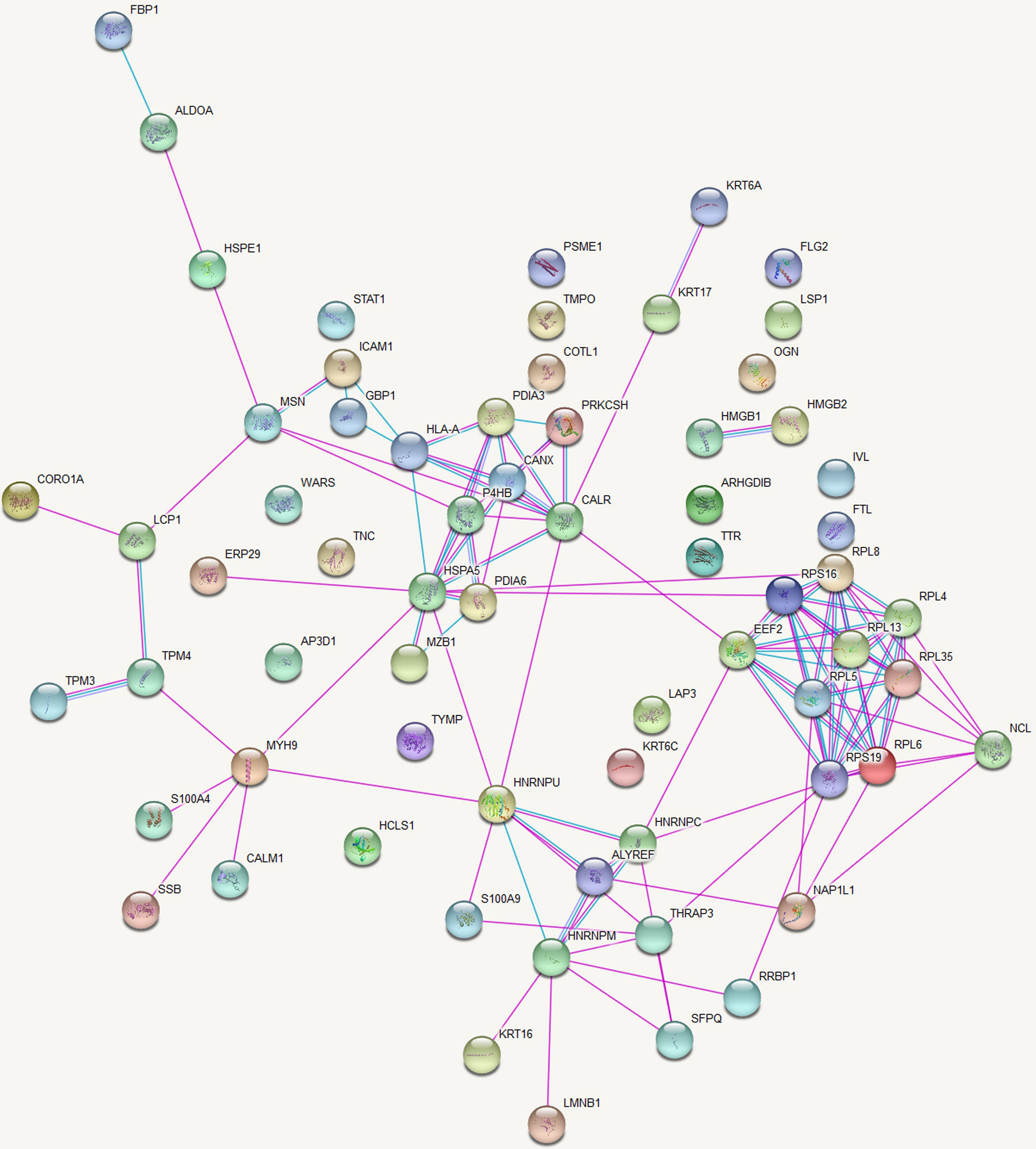
For this analysis raw data on MS/MS spectral counts were used. If a protein had a spectral count of < 6 in ≥ 90% of samples that protein was filtered out from the data analysis [30]. After filtering, 388 protein identities were left for further analysis. The Voom normalization was applied to normalize the data across all samples to reduce the bias in signal intensities from run to run. Comparison between groups was done by the ‘analysis of variance’ (ANOVA) method. The p-value obtained was adjusted for multiple corrections using the Benjamini–Hochberg procedure. All the proteins with an adjusted p-value < 0.01 were considered as significantly expressed between the groups compared. Significantly expressed proteins thus identified were entered into the UniProt human database [31] and converted to their corresponding gene names. Protein–protein interactions were assessed using the database ‘STRING: functional protein association networks’, Version 10.5 (https://string-db.org/) [32]. Pathway analysis was done using the Reactome pathway portal, version 3.2 (http://www.reactome.org) [33].
Immunohistochemical validation of IRE1, ATF6 and PERK
Immunohistochemical staining for IRE1, ATF6, and PERK was performed at Lanka Hospital Diagnostics (Pvt) Limited (PV90884), Colombo 05, Sri Lanka on paraffin-embedded tissue samples in all selected cases and controls. Tissue sections of 4 µm were cut using a microtome and the sections were mounted on positively charged slides and dried overnight in an oven at 60 ℃. The slides were dewaxed in xylene and rehydrated with 100% ethanol and 90% ethanol for 10 min each. The slides were then washed with deionized water two times for 5 min. The slides were subjected to a 25-min microwave-boiling process for antigen retrieval, using an appropriate buffer and pH as specified in Table 1.
Table 1 Immunohistochemical protocols and scoring for IRE1, PERK, and ATF6 markersEndogenous peroxidase was blocked by incubation in 3% hydrogen peroxide for 10 min followed by washing in deionized water and wash buffer (1 × TBST). Non-specific binding was blocked by adding the blocking solution for 1 h at room temperature in a humidified chamber. The tissue sections were then incubated overnight at 4 ℃ with primary antibodies.
The antigen–antibody complex was detected by the Labeled Streptavidin–Biotin (LSAB) staining method using a biotinylated goat anti-rabbit antibody (DakoCytomation), subsequently conjugated with streptavidin horseradish peroxidase (HRP) and visualized by reacting with 3,3′-diaminobenzidine for color detection. The tissue sections were counterstained with hematoxylin. Dehydration was done by submerging the slides in 95% ethanol, 100% ethanol, and xylene twice for 10 min each respectively. Finally, the sections were mounted using a mounting medium and observed under the microscope.
The IHC-stained tissue samples were evaluated by light microscopic examination for the expression of IRE-1, PERK and ATF-6. The intensity of staining for each of the markers and the proportion of cells that expressed attaining were evaluated in 5 random fields (400 × magnification) for each section. IRE-1 was assessed for cytoplasmic staining. PERK and ATF-6 were evaluated for both cytoplasmic and nuclear staining. The proportion score was calculated as: 0, 0–5% cells were stained; 1, 6–30% cells were stained; 2, 31–70% cells were stained, and 3, 71–100% cells were stained. The staining intensity was scored as follows: 0, no staining; 1 weak staining; 2, moderate staining; 3, strong staining [34].
The overall score for IRE-1 was calculated as:
The overall score for ATF-6 and PERK was calculated as [34, 35]:
$$(\text\times \text) + (\text\times \text)$$
The cases that showed staining for each of the markers, IRE1, PERK, and ATF-6 were further categorized as low positivity and high positivity based on the score obtained for each of the markers. IRE -1—low positive < = 4 and high positive > 5, PERK—low positivity < = 8 and high positivity > 9, ATF-6—low positivity < = 2 and high positivity > 3. The selection of these arbitrary cut-off values is made specifically for this study, as there is no uniform threshold established in the relevant literature.
Statistical analysis
Statistical analyses were carried out using SPSS (version 25.0, SPSS Inc, Chicago, IL. USA) software. The association between the degree of staining for each marker and the clinical and pathological features was assessed using the Chi-square test. A p value < 0.05 was considered statistically significant.


留言 (0)