
記住我

The genes of the glypican family are highly conserved across animal species and play important roles in fundamental biological processes such cell survival, motility, and differentiation(Dwivedi et al. 2013; Kamimura and Maeda 2021). Therefore, it is not surprising that change in the expression of glypican genes has been reported in multiple human cancers(.McGough et al. 2020; Melo et al. 2015; Zhou et al. 2018). Recently, much attention had been paid to the role of GPC2 in tumors. GPC2 was shown to be highly expressed in colon adenocarcinoma (COAD) and was associated with advanced tumor stage and poor prognosis. GPC2 deficiency induced cell cycle arrest and apoptosis in COAD cell lines, as well as inhibiting cell proliferation, migration, and invasion(.Lin, He, Ni, Zhang, Liu, Mao, Huang and Lin 2022). In early-stage pancreatic ductal adenocarcinoma (PDAC), GPC2 expression was higher in tumor tissues when compared to adjacent normal tissues. Moreover, survival analysis based on TCGA database revealed that higher expression of GPC2 was correlated with unfavorable survival in PDAC(.Liu et al. 2020). In neuroblastoma (NB), GPC2 was reported to be upregulated in tumor tissues and was selectively expressed on the cell surface. More importantly, GPC2 was minimally expressed in normal tissues, making it an attractive target for immunotherapy of neuroblastoma. Chimeric antigen receptor (CAR) T cells targeting GPC2 showed excellent tumor inhibitory role in neuroblastoma in vivo(.Li et al. 2021). GPC2 antibody-drug conjugate could change the tumor microenvironment (TME) to a proinflammatory state in neuroblastoma, and might promote immunogenic cell death and enhance antitumor immune response(.Pascual-Pasto et al. 2022). In another report, Raman and his colleague constructed a GPC2-directed antibody-drug conjugate (ADC). Further experiments confirmed that this ADC could inhibit neuroblastoma and small-cell lung cancers tumor regression, which was associated with DNA damage, cell apoptosis, and bystander cell killing(.Raman, Buongervino, Lane, Zhelev, Zhu, Cui, Martinez, Martinez, Wang, Upton, Patel, Rathi, Navia, Harmon, Li, Pawel, Dimitrov, Maris, Julien and Bosse 2021). There studies indicated that GPC2 might be an ideal therapeutic target in cancer, while GPC2 role in prostate cancer remains unclear.
In this work, our findings revealed that GPC2, which was higher in prostate cancer than that in adjacent normal tissue, was positively correlated with clinical stage and lymphatic metastasis in prostate cancer. Kaplan-Meier survival analysis indicated that higher expression of GPC2 predicted worse clinical outcome in patients with prostate cancer, suggesting a potential role of GPC2 in prognostic prediction in prostate cancer. A real-world cohort of prostate cancer patients needs to be collected to further test the predictive ability of GPC2 in prostate cancer. To explore the functional role of GPC2 on malignancy of PC cells, we performed in vitro experiments to examine the effect of GPC2 knockdown or overexpression on cell proliferation, migration, and invasion. Our results indicated that GPC2 served as an oncoprotein in prostate cancer, and the knockdown of GPC2 impaired the abilities of cell proliferation, migration, and invasion, while overexpression of GPC2 had the opposite effect. Taken together, these in vitro experiment results that we have presented so far indicated that GPC2 might be a potential therapeutic target in prostate cancer, further exploration should be conducted in vivo to confirm the anti-tumor efficiency of GPC2-based target therapy.
To investigate the potential molecular mechanism underlying the effect of GPC2 on cell proliferation, migration, and invasion, we employed bioinformatic analysis on the gene expression matrix of the prostate cancer tissues in TCGA database. We identified GPC2-related DEGs by separating the gene expression matrix into GPC2High and GPC2Low groups and further KEGG functional enrichment analysis revealed that these GPC2-related DEGs were particularly enriched in PI3K/AKT pathway, which attracted our attention due to its vital role in prostate carcinogenesis and progression(.Chen et al. 2016). The phosphatidylinositol 3-kinase (PI3K), comprised by three subunits, the regulatory subunits p85 and p55, and the catalytic subunit p110, functions as a plasma membrane-associated protein kinase(.Toren and Zoubeidi 2014). Once activation by the upstream receptor tyrosine kinases (RTKs) or non-RTKs, PI3K phosphorylates the PIP2 to produce PIP3, which further activates intracellular signaling by recruiting pleckstrin homology (PH) domain-containing proteins, including AKT, to the cell membrane. Then, the activated AKT proteins were translocated into cytoplasm and nucleus, and leading to the activation of downstream targets that participated in regulating cancer survival, angiogenesis, and metastasis(.Alzahrani 2019; Chen, Zhou, Wu, Li, Wen, Sha and Wen 2016, He et al. 2021; Karar and Maity 2011; Yang et al. 2019). To detect whether there were association between GPC2 and PI3K/AKT signaling, we examined the expression level of PI3K, p-PI3K, AKT, and p-AKT in prostate cancer cells by western blot assay. As expected, our results showed that knockdown of GPC2 resulted in decreased expression of p-PI3K and p-AKT, while GPC2 overexpression had the opposite effect, suggesting that GPC2 positively regulated the activation of PI3K/AKT signaling pathway.
Then, we asked by which means that GPC2 regulated PI3K/AKT signaling pathway. We first analyzed the interacting proteins of GPC2 through STRING database and found that MDK might be a direct target of GPC2. Midkine (MDK), encoded by MDK gene, was a multifunctional heparin-binding protein that could be secreted into the blood, urinary, and cerebrospinal fluid(.Cerezo-Wallis et al. 2020; Filippou et al. 2020; Muramatsu and Kadomatsu 2014). Recently, researches on MDK had escalated, especially in the field of cancer. MDK was reported to be upregulated in multiple cancers than that in healthy individuals, and this aberrant expression was associated with tumor progression and patients’ prognosis(.Lu et al. 2018; Xia et al. 2022). Functionally, MDK could be served as critical regulator of malignant behaviors of cancers, including cell proliferation, survival, metastasis, angiogenesis, stemness, and chemoresistance by various pathways(.Donia and Jönsson 2021; Kishida and Kadomatsu 2014; Kishida et al. 2013; Tang et al. 2022). Of note, the PI3K/AKT signaling was one of the most important downstream pathways of MDK that exerted roles in cancer. For example, Hu et al.(.Hu, Qin, Li, Wei, Mo, Fan, Lei, Wei and Zou 2021) reported that MDK promoted glioblastoma cell proliferation, migration, and invasion via activating the PI3K/AKT signaling. In breast cancer, MDK was identified as a direct downstream protein of miR-1275. Silence of miR-1275 leads to upregulation of MDK and further activating PI3K/AKT signaling to enhance the properties of cancer stem cells and promote chemoresistance(.Han, Li, Xu, Fu, Wang, Wang, Xia, Wang and Ma 2023). To explore whether GPC2 promotes the activation of PI3K/AKT signaling via MDK in prostate cancer, we first confirmed the physical interaction between GPC2 and MDK through immunoprecipitation assay. Then, we manipulated the expression of MDK after knocking down of GPC2 and detected the protein levels of p-PI3K and p-AKT. Our results revealed that overexpression of MDK could attenuate GPC2 knockdown induced downregulation of p-PI3K and p-AKT, therefore suggesting that GPC2 positively regulated PI3K/AKT signaling pathway at least partly through MDK. Moreover, rescue experiments showed that overexpression of MDK could attenuate the inhibitory effect of GPC2 knockdown on prostate cancer proliferation, migration, and invasion. To simplify, our research reveals that GPC2 promotes prostate cancer progression via MDK-mediated activation of PI3K/AKT signaling pathway, and GPC2 might be a potential therapeutic target in prostate cancer.
Our study had some limitations. First, GPC2 expression in prostate cancer tissues should also be examined by experiments to make our result more convincing. Second, in vivo experiments needed to be performed to investigate the inhibitory effect of GPC2-based target therapy in prostate cancer.
In summary, our study identified GPC2 as an oncoprotein in prostate cancer. GPC2 was upregulated in tumor tissues and correlated with clinical stage, tumor metastasis, and patients’ prognosis. GPC2 promotes prostate cancer cell proliferation, migration, and invasion via MDK-mediated activation of PI3K/AKT signaling pathway. GPC2 might be a promising prognosis predictor and potential therapeutic target.
Fig. 1
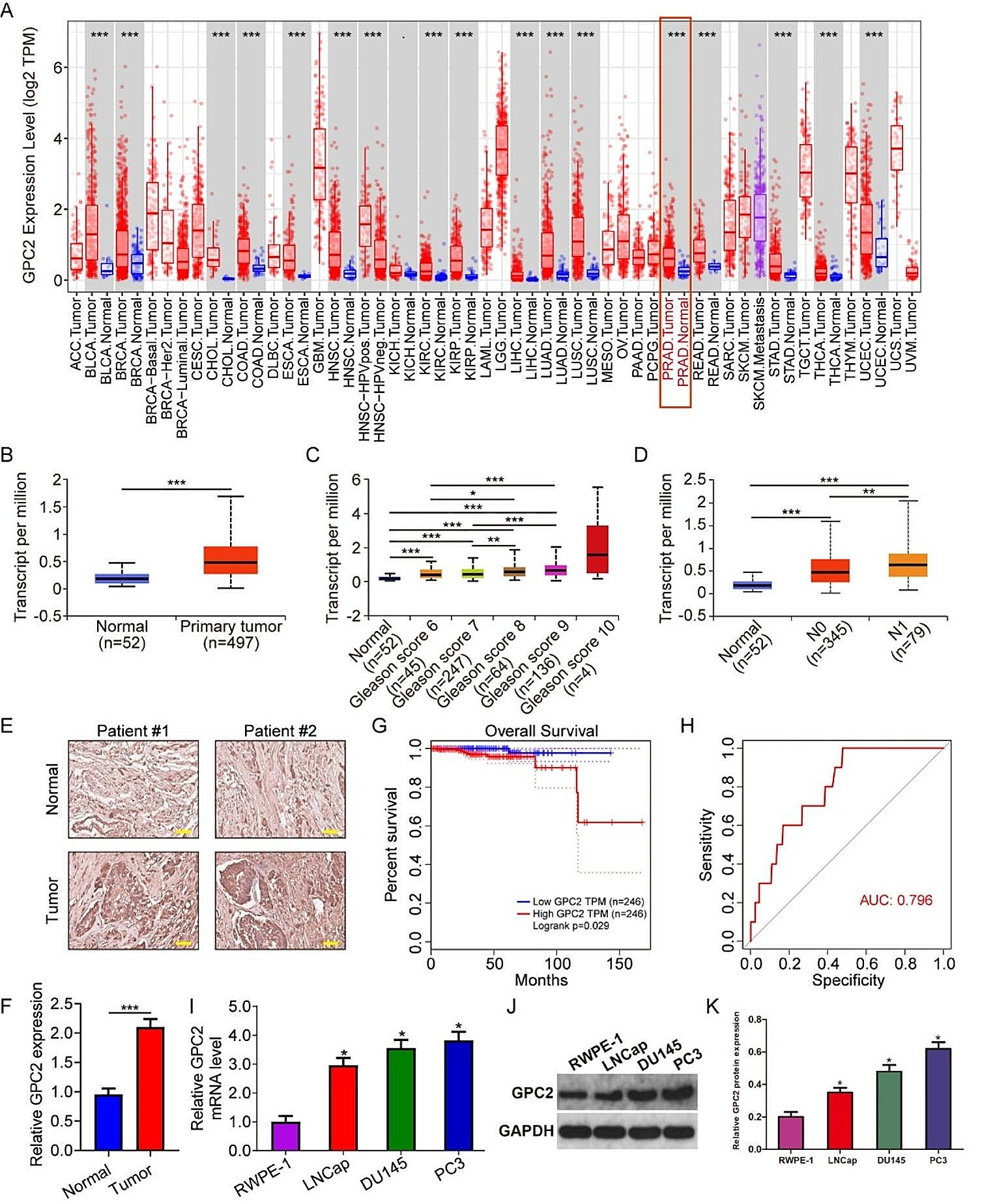
The expression of GPC2 was upregulated in prostate cancer and was negatively correlated with patients’ prognosis. (A) The expression level of GPC2 across various tumor types. ACC, adrenocortical carcinoma; BLCA, bladder urothelial carcinoma; BRCA, breast invasive carcinoma; CESC, cervical squamous cell carcinoma; CHOL, cholangiocarcinoma; COAD, colon adenocarcinoma; DLBC, diffuse large B-cell lymphoma; ESCA, esophageal carcinoma; GBM, glioblastoma multiforme; HNSC, head and neck squamous cell carcinoma; KICH, kidney chromophobe; KIRC, kidney renal clear cell carcinoma; KIRP, kidney renal papillary cell carcinoma; LAML, acute myeloid leukemia; LGG, brain lower grade glioma; LIHC, liver hepatocellular carcinoma; LUAD, lung adenocarcinoma; LUSC, lung squamous cell carcinoma; MESO, mesothelioma; OV, ovarian serous cystadenocarcinoma; PAAD, pancreatic adenocarcinoma; PCPG, pheochromocytoma and paraganglioma; PRAD, prostate adenocarcinoma; READ, rectum adenocarcinoma; SARC, sarcoma; SKCM, skin cutaneous melanoma; STAD, stomach adenocarcinoma; TGCT, testicular germ cell tumors; THCA, thyroid carcinoma; THYM, thymoma; UCEC, uterine corpus endometrial carcinoma; UCS, uterine carcinosarcoma; UVM, uveal melanoma. (B) Comparison of GPC2 expression level in prostate cancer tissues and adjacent normal tissues. (C) Higher expression of GPC2 was correlated with increased Gleason score. (D) Higher expression of GPC2 was correlated with lymphatic metastasis. (E-F) IHC analysis of GPC2 expression in normal and tumor tissues, and quantitative analysis. Scale bar, 200 µM. (G) Kaplan-Meier survival analysis and Log-rank test revealed that the higher GPC2 expression predicted worse overall survival. (H) ROC curve analysis of the predictive performance of GPC2 expression level in prostate cancer. (I-K) The mRNA and protein expression level of GPC2 in normal (RWPE-1) and cancerous prostate epithelial cell lines including DU145, PC-3, and LNCap. *P < 0.05, ***P < 0.001
Fig. 2
Silence of GPC2 inhibited cell proliferation, migration, and invasion in prostate cancer cells. (A) qRT-PCR assay confirmed the knockdown efficiency of shRNA targeting GPC2. (B) Western blot assay examined the protein level of GPC2 in shNC and shGPC2 groups and quantitative analyses. (C-D) CCK-8 and colony formation assays showed decreased proliferation ability after silencing GPC2. (E) Wound healing assay revealed that silence of GPC2 inhibited cell migration. Scale bar, 100 μm. (F) Transwell invasion assay revealed that silence of GPC2 inhibited cell invasion. Scale bar, 200 μm. Differences between groups were compared using Student’s t-test, *P < 0.05
Fig. 3
Overexpression of GPC2 promoted cell proliferation, migration, and invasion in prostate cancer cells. (A) qRT-PCR assay showed the efficiency of overexpressing GPC2 in DU145 and PC-3 cells. (B) Western blot assay examined the protein level of GPC2 in LV-Ctrl and LV-GPC2 groups and quantitative analyses. (C-D) CCK-8 and colony formation assays showed increased proliferation ability after overexpressing GPC2. (E) Wound healing assay revealed that overexpression of GPC2 promoted cell migration. Scale bar, 100 μm. (F) Transwell invasion assays revealed that overexpression of GPC2 promoted cell invasion. Scale bar, 200 μm. Differences between groups were compared using Student’s t-test, *P < 0.05. *P < 0.05
Fig. 4
Bioinformatic analysis identified GPC2-related DEGs and functional enrichment analysis. (A) Volcano plot showing the differentially expressed genes between GPC2High and GPC2Low group. The criteria of DEG were set as |log2(fold change) |>0.5 and FDR < 0.05. (B) Heatmap showing the expression profiles of GPC2-related DEGs in prostate cancer tissues of TCGA database. (C) GO enrichment analysis of GPC2-related DEGs. (D) KEGG enrichment analysis of GPC2-related DEGs showing PI3K/AKT signaling pathway was markedly enriched. (E) Construction of a PPI network using the GPC2-related DEGs. (F) The top thirty ranking genes in the PPI network
Fig. 5
GPC2 positively regulated PI3K/AKT signaling pathway through MDK. (A-B) Western blot assay was performed to detect the phosphorylation of PI3K and AKT in LV-Ctrl and LV-GPC2 groups of prostate cancer cells, and quantitative analyses. (C-D) The effect of MDK overexpression on the protein levels of PI3K, p-PI3K, AKT, and p-AKT in prostate cancer cells after knocking down of GPC2, and quantitative analyses. *Indicates a significant difference between the shGPC2 and shNC groups. #Indicates a significant difference between the shGPC2 + MDK and shGPC2 groups. *P < 0.05. #P < 0.05
Fig. 6
Overexpression of MDK reversed the inhibitory effect of GPC2 knockdown on cell proliferation, migration, and invasion in prostate cancer. (A) CCK-8 assay was conducted to assess cell proliferation ability in prostate cancer cells transfected with shNC, shGPC2, and shGPC2 + MDK. (B-C) Would healing and transwell invasion assays were performed to detect cell migration and invasion abilities in prostate cancer cells transfected with shNC, shGPC2, and shGPC2 + MDK. *Indicates a significant difference between the shGPC2 and shNC groups. #Indicates a significant difference between the shGPC2 + MDK and shGPC2 groups. *P < 0.05. #P < 0.05
留言 (0)