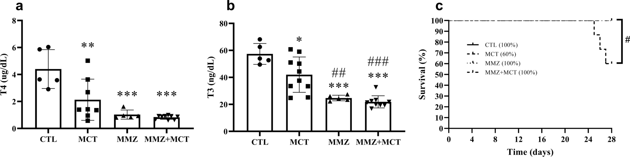
In this study, we initially tried to evaluate the effect of long-term LDTN stimulation on ventricular electrophysiological stability and further explored its possible mechanisms in a canine model of chronic MI. We found that the increased sympathetic components of HRV and LSG neuronal discharge activity were suppressed by LDTN stimulation. In addition, LDTN stimulation resulted in a considerable reduction in VAs susceptibility by prolonging ventricular ERP and APD90, decreasing the dispersion of ERP and APD90, and increasing VFT. Notably, the increased neural apoptosis of LSG was observed in the MI + LDTN group compared with that in the other groups. Taken together, our study provides direct evidence for a potential role of LDTN stimulation in improving ventricular electrical remodeling in a canine model of chronic MI through neural apoptosis of LSG.
It is widely recognized that the autonomic nervous system plays a key role in regulation of cardiac electrophysiology. Briefly, sympathetic activation results in the instability of cardiac electrophysiology; conversely, sympathetic inhibition or vagus nerve activation exhibits an opposite effect [13, 14]. The Poincaré plot of R–R intervals is a geometrical method for visually and quantitatively describing the heart rate variability [15]. The LF value indicates the sympathetic tone, and the HF value indicates the parasympathetic tone, while the LF/HF ratio indicates the relative balance between sympathetic and parasympathetic nerves [16]. In this present study, a significant increase in the LF (nu) value and LF/HF ratio, and a reduction in HF (nu) value were observed in the MI group compared with that in the sham group. However, LDTN stimulation led to a significant reduction in the LF (nu) value and LF/HF ratio and an increase in the HF (nu) value. These results indicated that LDTN stimulation can improve the imbalance of autonomic nervous system function.
It is well known that the function and activity of LSG play a key role in the regulation of ventricular electrophysiology; therefore, we evaluated the effect of LDTN stimulation on the function and activity of LSG. In this study, the LSG discharge activity was increased in the MI group compared with that in the sham group; however, the LDTN stimulation decreased the LSG discharge activity. The results indicated that LDTN stimulation could inhibit the LSG activity. Furthermore, cresyl violet staining results showed that LDTN stimulation decreased the number of neurons, while IF staining results showed that LDTN stimulation decreased the percentage of TH positive neurons. Taken together, our results indicated that LDTN stimulation could inhibit the LSG function and activity, which may be a novel method for heart sympathetic denervation; however, this still needs further investigation and verification.
Previous studies indicated that sympathetic stimulation induces a decrease in ventricular ERP and APD, along with an increase in dispersion of repolarization [17, 18]. Thus, we evaluated the effects of LDTN stimulation on ventricular electrophysiological properties in this study. We found that the ERP and APD90 were decreased in the MI group, and the dispersions were increased or showed an increasing trend; however, the LDTN stimulation prolonged the ERP and APD90 and decreased their dispersions. The results indicated that ventricular electrophysiological properties were improved by LDTN stimulation. The possible reason may be that the protective effect of LDTN stimulation on ventricular electrical remodeling can be partly resulted from the regulation of sympathetic remodeling. However, further investigation is required to elucidate the exact molecular mechanism underlying the improvement in ventricular electrical remodeling following LDTN stimulation.
As mentioned above, LDTN stimulation decreased the activity of LSG and the percentage of TH positive cells. Therefore, we aimed to identify whether the above results were caused by the apoptosis of LSG neurons. Previous studies have shown that vagus nerve stimulation can cause neuronal damage in the stellate ganglion. It is mainly due to the fact that vagus nerve fibers contain some sympathetic nerve fibers originated from the stellate ganglion. However, stimulating the vagus nerve increases the excitability of the stellate ganglion [8, 19]. The rapid and sustained stimulation can lead to the excitotoxicity of neurons and therefore result in damage and apoptosis to neurons [20]. The LDTN is connected to the LSG and contains the postganglionic sympathetic nerve components originating from the LSG. A recent study has shown that long-term stimulation of LDTN leads to damage of the LSG neurons and therefore attenuates its activity [7]. Consistent with previous findings, our results showed that LDTN stimulation also inhibited the LSG discharge activity; however, the underlying molecular mechanism has not been elucidated. Therefore, we subsequently identify whether LDTN stimulation inhibiting LSG activity was related to neuronal apoptosis and explored the exact molecular mechanisms.
The Bcl-2 protein family is well recognized to regulate mitochondrial integrity and apoptosis at each physiological or functional level, including pro-apoptotic and anti-apoptotic members [21]. A recent study showed that Shuxuening injection can reduce the apoptosis of hippocampal neurons induced by cerebral ischemia–reperfusion injury in rats by inhibiting the activation of Bax/Bcl-2 (22). Another study has shown that leptin protects against hyperglycemia-induced neural damage by inhibiting Bax/Bcl-2 ratio [23]. In our study, the IHC result showed that LDTN stimulation increased the expression of pro-apoptotic Bax and reduced the expression of anti-apoptotic Bcl-2. Furthermore, the WB assay results were consistent with the IHC results. These results collectively indicated that LDTN stimulation increased the pro-apoptotic Bax, and decreased the anti-apoptotic Bcl-2, thereby resulting in the LSG apoptosis. Additionally, the β-catenin was increased in the MI group compared with that in the sham group. Conversely, the MI + LDTN group exhibited a greater reduction in β-catenin levels compared with the MI group. These results indicated that LDTN stimulation could inhibit the activated Wnt/β-catenin signaling pathway, thereby resulting in the neuronal apoptosis of LSG through up-regulation of pro-apoptotic Bax, and down-regulation of anti-apoptotic Bcl-2.
Together, we initially aimed to investigate the effect of LDTN stimulation on ventricular arrhythmias after MI, and finally found that the LDTN stimulation could improve ventricular electrical remodeling and exhibit some cardioprotective effect on the heart by inhibiting the discharge of LSG. Additionally, the LDTN is a subcutaneous nerve, which is simple to isolate with less damage and has some advantages when compared with traditional SG block or resection if LDTN stimulation is further defined clinically effective. However, the present study has several potential limitations. First, the sample size of experimental animals is relatively small. Although beagle dogs with similar age and weight were selected for the experiment, there remain some individual differences, which need to be further studied by expanding the sample size. Second, some in vivo experiments are carried out under anesthesia in dogs, which may have certain effects on the heart rate and blood pressure of experimental animals. However, all experimental animals have similar anesthesia methods and degrees, so the effects of anesthesia can be almost offset between groups. Third, there are some differences between beagles and human structure and the tolerance, which may limit the interpretation of experimental results. In addition, the optimal parameters, duration, and timing of LDTN stimulation were not explored. Of course, these are also the direction of our future research. We will expand the sample size of the study, deepen the mechanism research, and conduct further research on different parameters, different stimulation duration and optimal stimulation timing.

留言 (0)