Reagents and chemicals
Unless otherwise stated, the chemicals were of analytical and molecular grade and purchased from Merck KGaA (Darmstadt, Germany). Kanamycin sulfate was obtained from Calbiochem (San Diego, CA, USA). Soluble starch from potatoes was purchased from Kanto Chemical Co., Inc. (Tokyo, Japan). Tapioca and sago starches were of food grade and procured from THC Sdn. Bhd. (Penang, Malaysia). Amylose from potato and β-limit dextrin from maize were purchased from Megazyme (County Wicklow, Ireland, UK). High-grade (≥99% purity) glucose (G1), maltose (G2), maltotriose (G3), maltotetraose (G4), maltopentaose (G5), maltohexaose (G6), and maltoheptaose (G7) were obtained from Elicityl (Crolles, France).
Bioinformatics analysis of AmyJM
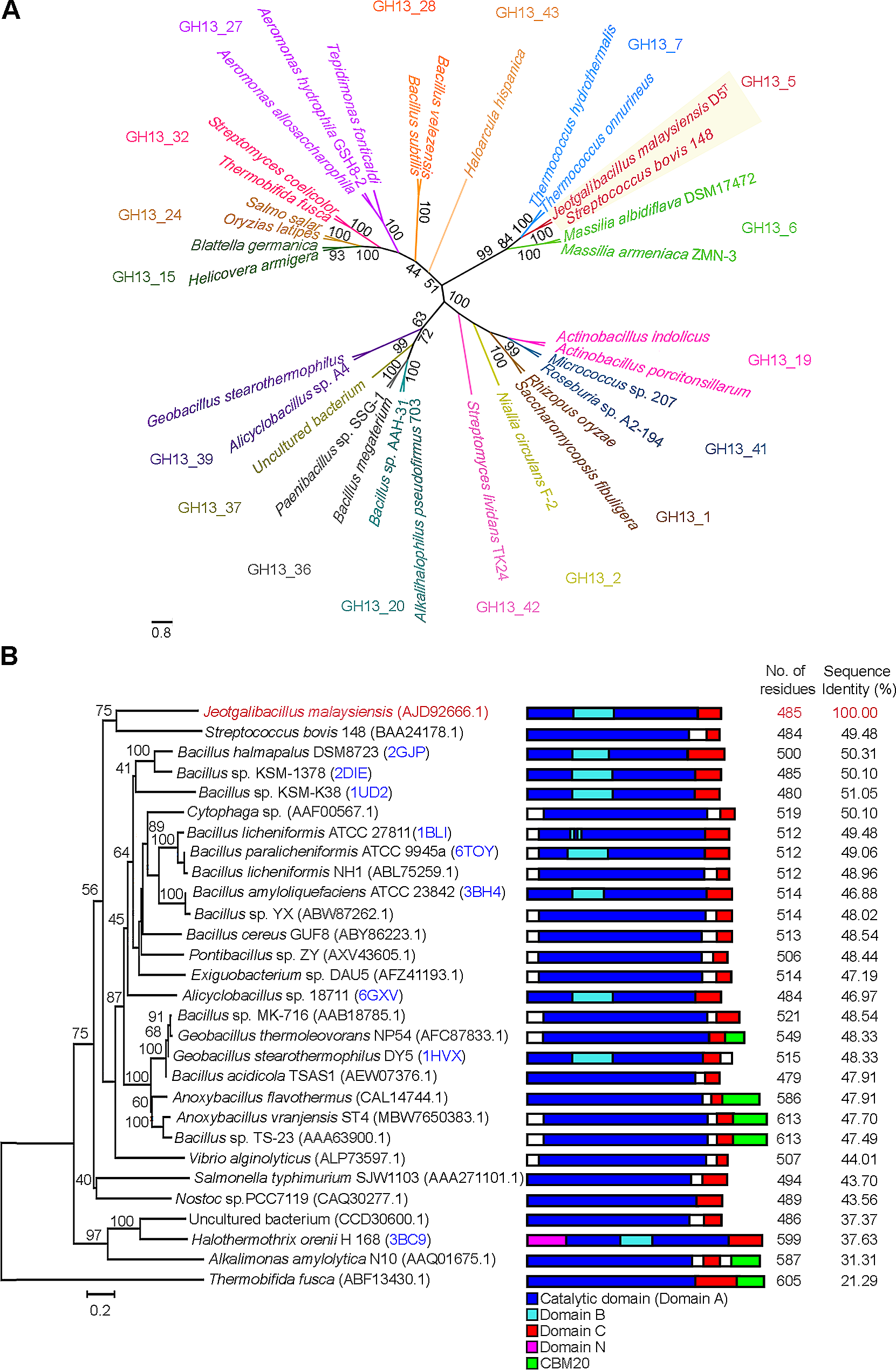
A putative α-amylase amino acid sequence was derived from the annotated complete genome of J. malaysiensis D5T. The α-amylase is designated as AmyJM, with accession number A0A0B5ARF3 in the UniProtKB database (Consortium 2023). Based on the dbCAN3 CAZy meta server (Zheng et al. 2023) family classification, AmyJM was classified in the subfamily GH13_5 (accessed on February 1, 2024). Sequences homologous to AmyJM were extracted from the CAZy database (Drula et al. 2022), focusing on biochemically characterized and crystallized α-amylases of subfamily GH13_5 (available as of February 1, 2024). Additional α-amylase sequences were obtained by NCBI BLASTp searches against the “non-redundant protein sequences (nr)” database. Multiple protein sequence alignments were performed using the Clustal Omega web server (Madeira et al. 2022). Phylogenetic trees were generated by the neighbor-joining method using Molecular Evolutionary Genetic Analysis (MEGA v.11.0.13) with a bootstrap value of 1,000 replicates (Tamura et al. 2021). Sequence logos of eight conserved sequence regions (CSRs) were created using the WebLogo3 online server (Crooks et al. 2004). Putative protein domains were predicted using the InterProScan v.5.56-89.0 online server (Paysan-Lafosse et al. 2023). The 3D homology model of AmyJM was retrieved from the AlphaFoldDB protein structure database (model number: AF-A0A0B5ARF3-F1) (Varadi et al. 2023); the quality of the AmyJM model was verified using the Structural Analysis and Verification Server (SAVES v.6.0). The AmyJM homology model was viewed and analyzed using PyMol v11 (Schrödinger, New York, USA). Default parameters were used for all software tools unless otherwise specified.
Expression and purification of recombinant AmyJM
The amyJM gene was synthesized by the GenScript Corporation (Piscataway, NJ, USA). The synthetic gene was cloned into pET-28a(+) (Novagen/Merck KGaA) using the BamHI and XhoI restriction sites. The pET-28a(+) construct was transformed into Escherichia coli BL21(DE3) (New England BioLabs, Ipswich, MA, USA). To express AmyJM, recombinant E. coli BL21(DE3) was grown on Luria–Bertani (LB) agar (pH 7.0) supplemented with 50 µg/mL kanamycin sulfate (kan) at 37 °C for 24 h. A single colony of recombinant E. coli BL21(DE3) was inoculated into 50 mL of LB/kan medium (pH 7.5) in a 250 mL flask and cultured under shaking at 200 rpm at 37 °C for 24 h. A 2 mL inoculum (equivalent to 1% v/v) was transferred into 200 mL of fresh LB/kan medium (pH 7.5) in a 1 L flask and incubated at 37 °C, 200 rpm. At periodic intervals, culture medium absorbance at 600 nm (A600) was recorded using an Ultrospec 2100 pro UV/Visible Spectrophotometer (Cytiva, Marlborough, MA, USA). When the A600 reached 0.5, enzyme expression was induced by adding a final concentration of 0.4 mM isopropyl-β-D-thiogalactopyranoside and further incubation at 37 °C, 200 rpm for 4 h. Then, the culture was centrifuged at 5,000 × g, 4 °C for 10 min, and the cell pellet was collected. To obtain crude AmyJM, the pellet was lysed using a B-PER™ Bacterial Protein Extraction Reagent Kit (Thermo Fisher Scientific, Rockford, IL, USA), according to the manufacturer’s instructions. The cell-free lysate was dialyzed against 100 mM sodium phosphate buffer (pH 7.5) at 4 °C for 18 h using SnakeSkin dialysis tubing with a 10-kDa molecular weight cut-off, MWCO (Thermo Fisher Scientific).
The crude AmyJM was purified using a pre-packed 1 mL HisPur™ nickel-nitrilotriacetic (Ni-NTA) chromatography cartridge (Thermo Fisher Scientific). The cartridge was equilibrated with 20 mM sodium phosphate buffer, 300 mM NaCl, 55 mM imidazole, pH 7.4. The bound enzyme was eluted with a linear gradient of 55–300 mM imidazole. The active fractions were pooled and dialyzed against 100 mM sodium phosphate buffer (pH 7.5) at 4 °C for 18 h using SnakeSkin dialysis tubing, 10-kDa MWCO (Thermo Fisher Scientific). The purified AmyJM was used for subsequent analyses.
Enzyme and protein assays
α-Amylase activity was determined using the 3,5-dinitrosalicylic acid (DNS) method (Miller 1959). A reaction mixture consisting of 0.1 mL of enzyme (5.0 U/mg; 1.0 mg/mL) and 0.9 mL of 1% (w/v) soluble starch in 100 mM sodium phosphate buffer (pH 7.5) was incubated at 40 °C for 15 min. DNS reagent (1 mL) was then added to the mixture, followed by boiling (100 °C) for 5 min. Subsequently, A540 was measured using the Ultrospec 2100 pro UV/Visible Spectrophotometer. As a control, an unreacted mixture was incubated and analyzed under the same conditions. Maltose was used as the assay standard. One unit (U) of α-amylase activity was defined as the amount of enzyme that generated 1 µmol of reducing sugar per minute per milliliter at 40 °C. The protein concentration was quantified using a PIERCE™ bicinchoninic acid (BCA) Protein Assay Kit (Thermo Fisher Scientific) with bovine serum albumin as the standard. The assays were performed at least in triplicate unless otherwise specified.
Characterization of AmyJMGel electrophoresis and zymography
The molecular mass and purity of AmyJM were determined using 12% (v/v) sodium dodecyl sulfate-polyacrylamide gel electrophoresis (SDS-PAGE) analysis. Imperial™ Protein Stain (Thermo Fisher Scientific) was used to stain the protein bands, which were compared with Benchmark™ Protein Ladder (Life Technologies, Carlsbad, CA, USA) to estimate the molecular mass. Zymogram staining for the detection of AmyJM starch-degrading activity was performed as previously described (Yang et al. 2004), except that 1% (w/v) soluble starch was dissolved in 100 mM sodium phosphate buffer (pH 7.5) and incubated at 40 °C for 15 min.
Effects of pH, buffer, and temperature
The optimum pH for AmyJM was determined at 40 °C using the following buffers (100 mM each): glycine-HCl (pH 2.0–3.0), sodium acetate (pH 4.0–5.5), sodium phosphate (pH 6.0–7.5), Tris-HCl (pH 8.0–9.0), and glycine-NaOH (pH 10.0–11.0). To measure the pH stability of AmyJM, the enzyme was incubated in each buffer without substrate at 25 °C for 20 min, and residual activity was measured under standard assay conditions.
The effects of different buffers on AmyJM activity were determined by reacting the enzyme with soluble starch dissolved in five different buffers (100 mM each, pH 7.5): sodium phosphate, potassium phosphate, Tris-HCl, HEPES-NaOH, MOPS, at 40 °C, and measuring the residual activity.
The optimum temperature for AmyJM was evaluated at 10–90 °C in the optimum enzyme buffer (100 mM sodium phosphate buffer, pH 7.5). To investigate its thermostability, the enzyme was pre-incubated at different temperatures for 20 min without substrate, and residual activity was measured. The thermostability of AmyJM was further evaluated by pre-incubating the enzyme with or without 5 mM CaCl2 at 40–50 °C for 150 min, taking samples at periodic intervals, and measuring residual activity under standard assay conditions.
Kinetic parameters
The kinetic parameters were assessed by measuring maltose formation by AmyJM for different concentrations of soluble starch (2–40 mg/mL) in 100 mM sodium phosphate buffer (pH 7.5) at 40 °C. The values of the Michaelis constant (Km), maximum velocity (Vmax), and turnover number (kcat) of AmyJM were determined using the GraphPad Prism v.9.0.0 software (GraphPad Software Inc., La Jolla, CA, USA).
Effects of metal ions and chemical reagents
The influence of various additives on AmyJM activity was investigated using varying concentrations of chloride salts (5 and 10 mM each): calcium chloride, magnesium chloride, sodium chloride, potassium chloride, ammonium chloride, zinc chloride, copper (II) chloride, nickel (II) chloride, cobalt (II) chloride, manganese (II) chloride, and iron (III) chloride. Besides, effect of various chemical reagents on AmyJM activity was evaluated using (5 and 10 mM each): ethylenediaminetetraacetic acid (EDTA), urea, and β-mercaptoethanol, and (5% v/v and 10% v/v each): Triton X-100, Tween-20, Tween-80, dimethyl sulfoxide (DMSO), and sodium dodecyl sulfate (SDS). All the additives were added to the standard enzymatic assay and incubated at 40 °C in 100 mM sodium phosphate buffer (pH 7.5). Residual activity was measured, and enzyme activity without the additives was used as a reference (100%).
Analysis of reaction products from gelatinized substrate
Purified AmyJM was concentrated using an Amicon® Ultra-15 (10-kDa MWCO) Centrifugal Filter Unit (Merck KGaA). The concentrated AmyJM was used in subsequent reaction product analysis. All substrates used in the analysis were gelatinized by boiling (100 °C) in 100 mM sodium phosphate buffer (pH 7.5) with continuous stirring for 10 min, followed by cooling in a water bath at 40 °C for 10 min. The hydrolytic ability of AmyJM was determined by separately incubating the concentrated AmyJM (40 U) with various gelatinized substrates (1% w/v each), including soluble starch, wheat starch, tapioca starch, sago starch, potato starch, rice starch, corn starch, pullulan, amylose, amylopectin, β-limit dextrin, glycogen, and β-cyclodextrin (β-CD). All reactions were conducted for 24 h in a water bath at 40 °C, shaking at 100 strokes per min. The reactions were stopped by boiling (100 °C) for 10 min. Insoluble particles were filtered through a 0.45-µm nylon membrane syringe filter (Millex-GN/Merck KGaA) and subjected to high-performance liquid chromatography with evaporative light-scattering detection (HPLC-ELSD) analysis.
The reaction products were analyzed using an Agilent 1260 Infinity HPLC system with an Agilent 1260 Infinity ELSD (Agilent Technologies, Santa Clara, CA, USA). The column employed was a 0.5-µm Zorbax carbohydrate analysis (NH2) column, 4.6 × 150 mm (Agilent Technologies). The column temperature was maintained at 30 °C. The ELSD nebulizer and evaporator temperatures were maintained at 30 °C, and the nitrogen gas flow was maintained at 1.6 L/min. Acetonitrile-water (75:25, v/v) was used as the mobile phase at a 1 mL/min flow rate. High-grade (≥ 99% purity) glucose (G1), maltose (G2), maltotriose (G3), maltotetraose (G4), maltopentaose (G5), maltohexaose (G6), and maltoheptaose (G7) were used as standards. Unreacted substrates were also injected under the same chromatographic conditions as controls.
Raw starch degradation by AmyJMDetermination of starch amylose/amylopectin composition
The amylose/amylopectin ratios in wheat, tapioca, sago, potato, rice, and corn starches were determined using an Amylose/Amylopectin Assay Kit (Megazyme), according to the manufacturers’ instructions.
Adsorption, hydrolysis, and morphology of raw starch
The adsorption and hydrolytic abilities of AmyJM toward raw starches were assessed by incubating AmyJM (40 U) with 1% (w/v) of various raw starch granules (wheat, tapioca, sago, potato, rice, and corn) in 100 mM sodium phosphate buffer (pH 7.5) in a final volume of 5 mL. All reactions were conducted in a shaking water bath (40 °C, 100 strokes per min) for 3 h. After centrifugation at 12,000×g, 4 °C for 3 min, residual activities in the supernatants were measured under the standard assay conditions. As a control, an unreacted mixture was incubated and analyzed under the same conditions. The percentage adsorption was calculated using the following formula (Nisha and Satyanarayana 2015):
$$Percentage\, absorption\, (\%) = 100-[(C/C_)]\times100]$$
where C is the enzyme activity in the supernatant after binding, and C0 is the initial enzyme activity.
The degree of raw starch hydrolysis (Rh) was defined using the following formula, (Shofiyah et al. 2020):
$$R_\,(\%)=(A_/A_)\times100.$$
where A1 is the total amount of sugars in the supernatant after the reaction, and A0 is the amount of raw starch before the reaction.
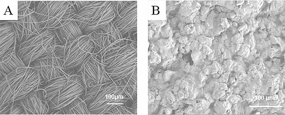
Field emission scanning electron microscopy (FESEM) was used to observe the raw starch granules before and after hydrolysis by AmyJM. The pellets recovered from the aforementioned reactions were treated as previously described (Fang et al. 2019). The samples were then mounted on a specimen holder using a silver plate, sputtered with gold, and viewed under a high-resolution FEI Quanta 650 FEG field emission scanning electron microscope (Thermo Fisher Scientific, Hillsboro, OR, USA) operated at 10 kV.
Analysis of the hydrolysis products
To analyze the ability of AmyJM to hydrolyze 13 different raw substrates, AmyJM (40 U) was mixed with 1% (w/v each) of soluble starch, wheat starch, tapioca starch, sago starch, potato starch, rice starch, corn starch, pullulan, amylose, amylopectin, β-limit dextrin, glycogen, or β-CD in 100 mM sodium phosphate buffer (pH 7.5). Subsequently, the reaction mixtures were incubated in a shaking water bath (40 °C, 100 strokes per min) for 24 h. The enzymatic reactions were stopped by boiling (100 °C) for 10 min, filtered through a 0.45-µm nylon-membrane syringe filter, and subjected to HPLC-ELSD under the aforementioned conditions. Non-reacted substrates served as controls.
Statistical analysis
The enzymatic assays and HPLC-ELSD analyses were analyzed using SYSTAT v.12.02.00 software (Systat Software, San Jose, CA, USA). Student’s t-test yielded a probability value (p value) of < 0.05, confirming that the data were adequate to test all hypotheses.

留言 (0)