RT–PCR and plasmids
The open reading frames of mouse (m) PRDX6, PRDX1, SEPHS2, GPX4 (cytosolic form), SARS1, PSTK, SEPSECS, SCLY and FSP1, and human (h) PRDX6, SEPHS2, SCLY, eEFSec, SBP2 and SEPHS2-3′UTR(SECIS) were amplified by RT–PCR. Single-guide RNA (sgRNA) resistant constructs of mPRDX6 and mSEPHS2, and mutants of mPRDX6 (H26A, D31A, S32A, C47S and D140A), hPRDX6 C47S, mGPX4 U46C, mSEPHS2 U/C and hSEPHS2 U/C were generated by two-step PCR. GFP-3′SECIS (WT, C70U and S175U) was generated from the amplified open reading frames of GFP and the 3′-SECIS of GPX4. DNAs were ligated into the appropriate epitope-tag sequences and then cloned into pMXs-IP, pMXs-IRES-Bsr, pMXs-neo and pT7-7 vectors. Luciferase C258U-3′SECIS was generated from the luciferase open reading frame and the 3′-SECIS of GPX4 and then ligated into the pEU vector. CRISPR sgRNA sequences were selected from CHOPCHOP or Benchling. The oligonucleotide sequences preceding the protospacer motif were as follows:
mFBXL5, GAGGAAGATGGGTTATACCTG; mACSL4 #1, GGTTCTACGGGCCGCCCCAA; mACSL4 #2, GACCGATCACAATCTCACCTC; mLPCAT3 #1, GCCGGTGACTACGATATCAAGTGG; mLPCAT3 #2, GTAATAACGGCAGTGACGGTGCGG; mPDSS2 #1, GATGCCGGCTGTCGTGCACGA; mPDSS2 #2, GCGGCATAACCTACAACTGCG; mFSP1 #1, GCCAGCGCTCACAATTCATCG; mFSP1 #2, GCGCTCACAATTCATCGTGG; mPRDX6 #1, GATAGAACTATACCTTGCTCC; mPRDX6 #2, GCTCACCACACGGGCCGTCAC; mPSTK, GAAAGTCGACTTTCCGGCCGC; mSEPHS2, GAACCCGTGGATTATCATCGG; mMETAP1, GTGTACCGATAGCCTGCCCA; mSNX27, GTTGACAATGCGCACGACCCG; mUBE2N, GTGGGGACCACTTATCTATGA; mCINP, GGCCCCCTCTATTTCACACG; mCENPE, GCGCTATTTATCAGAGCGATG, mSTX5A, GTATGCGACTCGATGATCCCG; mDDX10, GAGCGATTAAAGCTCCGCACC; mKLF5, GACCTGAGGACTCATACGGGT; mSCLY, GTAATGAGACCGGCGTCATCA; mGPX4, GACGATGCACACGAAACCCC; control gRNA, GCGAGGTATTCGGCTCCGCG; hPRDX6, GATGCGGCCGACGGTGGTAT. Guide sequences were inserted into pX459 (ref. 58) or lentiCRISPRv2 (ref. 59) (puro, bsr or hygro).
Antibodies and reagents
The following antibodies were used: anti-PRDX6 (Proteintech, 13585-1-AP; western blotting (WB), 1:2,000; PLA, 1:100), anti-SEPHS2 (Proteintech, 14109-1-AP; WB, 1:2,000), anti-GPX4 (Proteintech, 67763-1-AP; WB, 1:2,000), anti-FSP1 (Proteintech, 20886; WB, 1:2,000), anti-SCLY (Proteintech, 67606-1-Ig; WB, 1:2,000), anti-GPX4 (Santa Cruz, sc-166570; WB, 1:2,000), anti-SELN (Santa Cruz, sc-365824; WB, 1:2,000), anti-GPX1/2 (Santa Cruz, sc-133160; WB, 1:2,000), anti-PSTK (Santa Cruz, sc-373991; WB, 1:2,000), anti-FBXL5 (Santa Cruz, sc-390102; WB, 1:2,000), anti-FTH1 (Santa Cruz, sc-376594; WB, 1:300), anti-ACSL4 (Santa Cruz, sc-365230; WB, 1:2,000), anti-PDSS2 (Santa Cruz, sc-515137; WB, 1:2,000), anti-ferritin (Sigma-Aldrich, F6136; WB, 1:2,000), anti-IRP2 (in-house; WB, 1:1,000), anti-LRP8 (Abcam, ab108208; WB, 1:2,000), anti-β-actin (Sigma-Aldrich, A5316; WB, 1:15,000), anti-tubulin (CEDARLANE, CLT9002; WB, 1:5,000), anti-Myc (Merck, 05-724; WB, 1:2,000; PLA, 1:200), anti-HA (MBL, M180-3; WB, 1:2,000), anti-ubiquitin K48-specific (Merck, ZRB2150; WB, 1:2,000), anti-p62 (Wako, 018-22141; WB, 1:2,000), HRP-linked anti-mouse IgG (Cell Signaling, 7076; WB, 1:10,000) and HRP-linked anti-rabbit IgG (GE Healthcare, NA934; WB, 1:10,000).
The following reagents were also used: ferric ammonium citrate (Sigma-Aldrich, F5879), liproxstatin-1 (Selleck, S7699), bafilomycin A1 (Selleck, S1413), IKE (Selleck, S8877), RSL3 (Selleck, S8155), E64d (Peptide Institute, 4321-v), pepstatin A (Peptide Institute, 4397-v), sodium selenite (Sigma-Aldrich, 214485), selenocystine (Tokyo Chemical Industry, E1368), cycloheximide (ALBIOCHEM, 239764), hydrogen peroxide (Santoku Chemical Industry, 18412), tert-butyl hydroperoxide (Sigma-Aldrich, 458139), Tris(2-carboxyethyl)phosphine (TCEP) (Nacalai, 07277), pyridoxal-5-phosphate (PLP) (Sigma-Aldrich, P9255) and biotin (WAKO, 021-08712).
Cell lines and cell culture
MEFs were generated in-house. HepG2 was gifted by K. Nakajima (Osaka City University), originally purchased from ATCC (HepG2: HB-8065). HEK293T was gifted by E. Nakamura (Kyoto University), originally purchased from RIKRN RBC (293T: RCB2202). PLATE was gifted by T. Kitamura (Tokyo University). A549, H226, H460 and H1975 were gifted by A. Sato (Kyoto University), originally purchased from ATCC (A549: CCL-185, H226: CRL-5826, H460: HTB-177, H1975: CRL-5908). SK-N-DZ and HeLa cells were purchased from ATCC (SK-N-DZ: CRL-2149, HeLa: CCL-2). PANC-1 and MIA Paca-2 cells were purchased from RIKEN RBC (PANC-1: RCB2095, MIA Paca-2: RCB2094). NB-1 cells were purchased from JCRB (NB-1: JCRB0621). All cells were cultured in a humidified incubator at 37 °C and 7.5% CO2. MEFs, HEK293T, PLATE, A549, HeLa, PANC-1, MIA Paca-2 and HepG2 cells were grown in DMEM supplemented with 10% FBS, 100 IU ml−1 penicillin and 100 μg ml−1 streptomycin. SK-N-DZ cells were grown in DMEM supplemented with 10% FBS, 1× non-essential amino acids (Gibco, 11140050), 100 IU ml−1 penicillin and 100 μg ml−1 streptomycin. GPX4, SEPHS2 and PSTK KO MEF cells were maintained in medium containing 2 μM of liproxstatin-1. H226, H460 and H1975 cells were grown in Roswell Park Memorial Institute (RPMI) 1640 medium supplemented with 10% FBS, 100 IU ml−1 penicillin and 100 μg ml−1 streptomycin. NB-1 cells were grown in 45% RPMI and 45% MEM medium supplemented with 10% FBS, 100 IU ml−1 penicillin and 100 μg ml−1 streptomycin.
Generation of FBXL5 KO MEFs
To generate FBXL5 KO MEFs, a NEPA21 electroporator (NEPAGENE) was used to electroporate MEFs with the pX459 plasmid containing an sgRNA sequence specific for mFBXL5. Cells were then treated with puromycin for 2 days. Following selection, the cells were seeded at a low density and isolated colonies were picked. To identify FBXL5 KO cells, expression of FBXL5 was analyzed by immunoblotting.
Lentivirus production and generation of CRISPR–Cas9-mediated KO cell lines
Lentivirus was produced by co-transfecting HEK293T cells with the LentiCRISPRv2 (puro, bsr or hygro)-containing guide sequences, the psPAX2 packaging plasmid and the VSV-G envelope plasmid using PEI MAX (Polysciences) transfection reagent. On the following day, the medium was replaced with fresh medium. After 2 days of culture, lentivirus-containing supernatant was collected and used to infect target cells overnight in the presence of 10 μg ml−1 polybrene (Merck). Infected cells were selected with puromycin, blasticidin or hygromycin.
Retroviral expression
pMXs-IP, pMXs-neo or pMXs-IRES-Bsr containing appropriate inserts were transfected into PLATE packaging cells or GP2-293 cells along with the pVSV-G plasmid. The resultant viruses were used to infect target cells in the presence of 10 μg ml−1 polybrene. Stably transduced cells were selected using puromycin, G-418 or blasticidin.
Cell lysis and western blotting
Cells were washed with PBS and lysed with lysis buffer (50 mM Tris-HCl (pH 7.5), 150 mM NaCl, 1% Triton X-100, 1 mM PMSF, and protease inhibitor cocktail (Sigma-Aldrich)). The lysates were clarified by centrifugation at 20,400g for 20 min at 4 °C. Protein concentrations were determined using the Bradford assay (Nacalai Tesque), and equal amounts of protein were mixed with SDS sample buffer. Samples were heated for 5 min at 95 °C, separated by SDS–PAGE and transferred onto PVDF membranes (Millipore). After blocking in Tris-buffered saline (TBS) containing 0.1% Tween-20 and 5% (w/v) nonfat dry milk, the membrane was incubated with the appropriate primary antibodies followed by appropriate secondary antibodies. The membranes were visualized using enhanced chemiluminescence and analyzed on a LAS4000mini or LAS3000 instrument (GE Healthcare).
Cell viability assay
Cells were plated in 96-well plates (in triplicate and at a density of 2,500–10,000 cells per well) in the presence or absence of FAC, RSL3 or IKE. After culture for 24–48 h, 10 μl of Cell Counting Kit-8 (DOJINDO) was added for 1–2 h. Absorbance at 450 nm was measured using a SpectraMax M5 microplate reader (Molecular Devices). Cell viability, monitored continuously using the iCELLigence or xCELLigence system (ACEA Bioscience), was expressed as an impedance-based cell index. GPX4 KO cells (4,500 cells per well) were plated onto an E-Plate L8 PET (ACEA Bioscience), and pancreatic ductal adenocarcinoma cells (3,000 cells per well) and SK-N-DZ cells (6,000 cells per well) were plated on an E-Plate 16 PET (ACEA Bioscience). SK-N-DZ cells were cultured in DMEM supplemented with 10% FBS, 1× non-essential amino acids and 0.25 mM l-glutamine (Fujifilm). The cell index was monitored continuously. Recording data were analyzed by RTCA software lite v.2.2.1.
Cell staining using BODIPY 581/591 C11, and iron staining
For BODIPY 581/591 C11 (Invitrogen) staining, cells were plated in a six-well plate and treated with FAC. After 1.5–24 h, the medium was removed and the cells were labeled with DMEM containing 10 μM BODIPY 581/591 C11 at 37 °C for 30 min. Cells were then washed three times with PBS and detached from the plate using trypsin. Initial cell population gating (FSC-Area versus FSC-height) was used to ensure doublet exclusion, and green fluorescence was measured by using FACS Canto II (BD Biosciences) and FACS Diva software v.6.1.2 (Becton Dickinson). Data were analyzed using FlowJo software (v.9.9.6). For iron staining, cells were plated in a 35-mm glass bottom dish and treated with 25 μg ml−1 FAC for 4 h. Cells were then washed three times with HBSS and labeled at 37 °C for 30 min with 1 μM FerroOrange (DOJINDO) followed by visualization under a Fv1000 confocal microscope (Olympus).
Genome-wide CRISPR screening
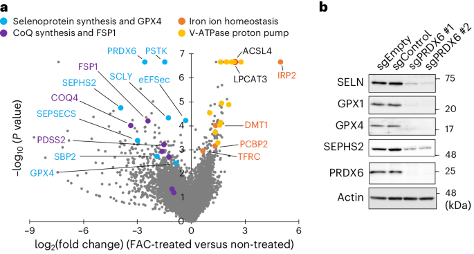
FBXL5 KO MEFs were infected with the mouse GeCKO v.2 library59 at a multiplicity of infection of 0.3 and selected with puromycin for 1 week post infection. The cells were then treated with 100 μg ml−1 FAC for 48 h and cultured for a further 24 h in fresh medium. The cells were lysed in NTE buffer (15 mM Tris-HCl (pH 7.5), 150 mM NaCl and 1 mM EDTA), and genomic DNA from non-treated and FAC-treated cells was prepared by phenol-chloroform extraction and isopropanol precipitation. sgRNA sequences were amplified from genomic DNA by PCR using Herculase II Fusion DNA polymerase. The resultant amplicons were gel-extracted and subjected to DNA sequencing on a Novaseq 6000 (Illumina) sequencer. Sequence data were analyzed using the MAGeCK pipeline.
PLA
The PLA was conducted using the Proximity Ligation Kit (Sigma-Aldrich). A549 cells, either empty or expressing Myc-SEPHS2 or Myc-SCLY, were cultured on micro cover glass slips in a 6-well plate. Cells were washed with PBS and fixed for 20 min with 4% formaldehyde in PBS at room temperature (25–27 °C). Cells were then washed twice with PBS and permeabilized with 0.1% Triton X-100 in PBS at room temperature for 10 min. Cells were washed twice with PBS and then incubated at 37 °C for 1 h with Duolink blocking solution. Cells were then incubated at room temperature for 1 h with primary antibodies targeting PRDX6 (rabbit polyclonal; 1:100) and Myc (mouse monoclonal; 1:200). Cells were washed twice for 5 min with Duolink wash buffer A at room temperature and then incubated with a secondary antibody (anti-mouse minus and anti-rabbit plus) at 37 °C for 1 h. Cells were washed twice at room temperature for 5 min with Duolink wash buffer A and then incubated with ligase solution at 37 °C for 30 min. Next, the cells were washed twice with Duolink wash buffer A and incubated with polymerase in amplification buffer at 37 °C for 100 min. Cells were washed twice at room temperature for 10 min with Duolink wash buffer B, followed by 0.01% Duolink wash buffer B for 1 min. Finally, the cover glasses were mounted with Duolink PLA mounting medium with DAPI. Protein–protein interactions were visualized under an Fv1000 confocal microscope (Olympus). PLA foci were counted by ImageJ (v.2.3.0).
TurboID pulldown
Cells were treated (or not) for 30 min with 50 μM biotin, washed with PBS and lysed with lysis buffer (50 mM Tris-HCl (pH 8.0), 150 mM NaCl, 1% Triton X-100 and 1 mM PMSF). The lysates were clarified by centrifugation at 20,400g for 20 min at 4 °C. Protein concentrations were determined using the Bradford assay, and equal amounts of protein and Streptavidin Sepharose (Cytiva) were incubated overnight at 4 °C by rotation. The beads were washed three times with lysis buffer and one time with PBS. Proteins were eluted from beads using SDS sample buffer supplemented with 2 mM biotin and then analyzed by western blotting.
Protein purification
His-TEV-PRDX6 WT or C47S, His-TEV-hSCLY, hSEPHS2-His, mouse His-TEV-eEFSec and His-TEV-SBP2 were expressed in E. coli strain BL21-CodonPlus (DE3)-RIPL (Agilent Technologies). Expression of His-TEV-PRDX6 WT or C47S was induced by the addition of 0.2 mM IPTG, and culture was continued at 30 °C for 3 h. Cells were collected by centrifugation and frozen rapidly. Subsequently, cells were resuspended at 4 °C for 30 min in buffer containing 50 mM Tris-HCl (pH 8.0), 150 mM NaCl, 10 mM 2-mercaptoethanol (Nacalai Tesque), 200 μg ml−1 lysozyme chloride (Nacalai Tesque), 10 μg ml−1 DNase (Roche), 2 mM PMSF and a protease inhibitor cocktail (Roche), followed by lysis at 4 °C for 20 min in the presence of 0.2% Triton X-100. Insoluble material was removed by centrifugation at 23,700g for 20 min at 4 °C. His-TEV PRDX6 WT or C47S were purified from the supernatant using Ni-NTA beads (QIAGEN). The eluted samples were incubated for 30 min at 4 °C with 10 mM dithiothreitol (DTT) and then desalted in 20 mM Tris-HCl (pH 7.5) buffer on a PD10 column.
Expression of His-TEV-hSCLY and hSEPHS2-His was induced by the addition of 0.2 mM IPTG, and culture was continued overnight at 15 °C. Cells were collected by centrifugation and frozen rapidly. Subsequently, the cells were resuspended in buffer containing 50 mM Tris-HCl (pH 8.0), 150 mM NaCl, 10 mM 2-mercaptoethanol, 2 mM PMSF and a protease inhibitor cocktail and then lysed by sonication at 4 °C. Insoluble material was removed by centrifugation at 23,700g for 20 min at 4 °C. Proteins were purified using Ni-NTA beads. The eluted samples were desalted in 20 mM Tris-HCl (pH 7.5) and 1 mM DTT buffer on a PD10 column.
Expression of His-TEV-eEFSec and His-TEV-SBP2 was induced by the addition of 0.3 mM IPTG, and culture was continued at 30 °C for 4 h. Cells were collected by centrifugation and frozen rapidly. Subsequently, the cells were resuspended in buffer containing 20 mM Tris-HCl (pH 8.0), 500 mM NaCl, 10 mM imidazole, 10% glycerol, 10 mM 2-mercaptoethanol, 1 mM PMSF and a protease inhibitor cocktail and then lysed by sonication at 4 °C. Insoluble material was removed by centrifugation at 23,700g for 20 min at 4 °C. Proteins were purified using Ni-NTA beads. Eluted samples were desalted on a PD10 column. eEFsec was stored in buffer containing 20 mM Tris-HCl (pH 7.5), 150 mM NaCl and 1 mM DTT, and SBP2 was stored in buffer containing 10 mM Tris-HCl (pH 7.5), 150 mM NaCl, 5% glycerol and 1 mM DTT.
GPX assay
GPX activity was measured using a GPX4 Inhibitor Screening Assay Kit (Cayman Chemical). First, 0.5 μg of GPX4 protein (included in the kit) or 5 μg of PRDX6 protein was dissolved in 50 μl of GPX4 assay buffer (included in the kit). Then, 20 μl of the GSH/GSH reductase mix (included in the kit) was added and mixed. Next, 20 μl of the NADPH solution (included in the kit) was added and mixed. Finally, 10 μl of cumene hydroperoxide (included in the kit), hydrogen peroxide (final concentration, 500 μM) or tert-butyl hydroperoxide (final concentration, 500 μM) was added and mixed, and absorbance at 340 nm was measured using SpectraMax M5 microplate reader (Molecular Devices).
Selenophosphate synthetase assay
A selenophosphate synthetase assay using SCLY and selenocysteine as a selenium source was performed in degassed buffer containing 50 mM Tris-HCl (pH 7.0), 100 μM ATP, 10 mM KCl, 10 mM MgCl2, 1 mM DTT, 0.5 μM SEPHS2-His, 20 μM PRDX6 WT or C47S, 1 μM SCLY, 1 μM PLP (Sigma-Aldrich) and 50 μM (Sec)2 at 30 °C for 45 min. The resultant AMP product was measured in an AMP-Glo assay (Promega) using a Nivo plate reader (PerkinElmer). A selenophosphate synthetase assay using selenide-bound PRDX6 as a selenium source was performed by incubating 200 μg of PRDX6 WT or C47S with 2 mM sodium selenite and 20 mM DTT in degassed 20 mM Tris-HCl (pH 7.5) buffer at room temperature for 30 min. Then, the solution was diluted 20 times in buffer containing 20 mM Tris-HCl (pH 7.5), 150 mM NaCl and 0.5% Triton X-100, and 30 μl of Ni-NTA magnetic beads was added. After rotation at 4 °C for 1 h, beads were washed three times and eluted with 300 mM imidazole in 20 mM Tris-HCl (pH 7.5) buffer. The eluted samples were incubated with a buffer mixture containing 50 mM Tris-HCl (pH 7.0), 100 μM ATP, 10 mM KCl, 10 mM MgCl2, 1 mM DTT and 0.5 μM SEPHS2-His under a layer of mineral oil at 30 °C for 30 min. The resultant AMP product was measured in an AMP-Glo assay.
RT–qPCR
RNA was isolated using the RNeasy Mini Kit (QIAGEN) and reverse transcribed using a high-capacity RNA-to-cDNA Kit (Applied Biosystems). RT–qPCR data were obtained by ABI ViiA7 Real-Time PCR system (Applied Biosystems) and analyzed by ViiA7 RUO Software v.1.2.3. RT–PCR of GPX4 was performed using the following primers:
mGPX4_Fwd, 5′-CCTCTGCTGCAAGAGCCTCCC-3′ and
mGPX4_Rev, 5′-CTTATCCAGGCAGACCATGTGC-3′. Control primers were
β-mActin_Fwd, 5′-ATGGATGACGATATCGCTC-3′ and
β-mActin_Rev, 5′-GATTCCATACCCAGGAAGG-3′.
Isolation of aa-tRNAs
aa-tRNAs were isolated from WT, PRDX6 KO and SEPHS2 KO MEF cells. In brief, cells were treated (or not) with 50 nM (Sec)2 for 105 min, followed by addition of 20 μg ml−1 cycloheximide for 15 min. Cells were then washed three times in PBS and detached using trypsin. After centrifugation at 200g, 3.2 ml of DPEC water, 0.8 ml of 5× T buffer (50 mM NaOAc, 3.25 M NaCl, 50 mM MgCl2 and 5 mM EDTA) and 2 ml of acid-phenol pH 4.2 (Nippon gene) were added to the cell pellets in that order. The solution was mixed briefly and centrifuged at 12,000g for 5 min at 4 °C. The aqueous phase was transferred to another tube and re-extracted with 2 ml of acid-phenol (pH 4.2). RNA was precipitated with 2.5 volumes of 100% ethanol, washed twice with 75% ethanol and air-dried for 5 min. The aa-tRNA pellet was resuspended in 5 mM NaOAc buffer.
The luciferase reporter Sec UGA assay for Sec-tRNA[Ser]Sec evaluation
Each reaction (20 μl) contained wheat germ lysate (10 μl; Promega), 20 μM amino acid mix, 8 U of RNasin Plus ribonuclease inhibitor (Promega), 52.5 mM potassium acetate, 0.2 μl of luciferase reporter mRNA and 2.25 μg of aa-tRNAs in the presence or absence of 1.2 μg of eEFSec and 0.6 μg of SBP2. Reactions were incubated at 30 °C for 90 min. Luciferase activity was measured in a Lumat Luminometer (Berthold).
ICP–MS
The reaction (100 μl) contained 40 μM sodium selenite, 160 μM GSH and 40 μM (or not) recombinant PRDX6 WT or C47S in degassed buffer containing 1 mM EDTA and 50 mM Tris-HCl (pH 7.0). Reactions were incubated at 25 °C for 5 min. Any selenium that did not bind to PRDX6 protein was removed using a NAP-5 column. The protein solution was then concentrated with an Amicon 3K concentrator. A small aliquot (50 μl) of the protein solution was put into a glass test tube, and the proteins were decomposed by addition of 0.1 ml of 60% HNO3 followed by heating at 170°C for 2 h. The digested samples were diluted with Milli-Q water, and the concentration of selenium was measured by ICP–MS (Agilent 8800 ICP–MS/MS; Agilent Technologies). The O2 mass-shift mode was used to monitor the signal intensity of 80Se16O, with a dwell time of 100 ms. Selenium concentrations were measured from a standard calibration curve.
Mass spectrometry analysis
The reaction contained recombinant PRDX6 WT (75 μM), sodium selenite (75 μM) and GSH (300 μM) in degassed buffer containing 50 mM Tris-HCl (pH 7.0), or PRDX6 WT (75 μM), Sec (75 μM) ((Sec)2 was pre-reduced with an equimolar amount of TCEP), SCLY (1.5 μM) and PLP (1.5 μM) in degassed buffer containing 50 mM Tris-HCl (pH 7.0). Reactions were incubated at 25 °C for 5 min. Then, PRDX6 was alkylated with 10 mM iodoacetamide in 0.02% ProteaseMAX surfactant at 37 °C for 10 min, followed by digestion with 27 mg ml−1 Trypsin Gold at 37 °C for 3 h. Products were analyzed using liquid chromatography–electrospray ionization–quadrupole time-of-flight tandem mass spectrometry (LC–ESI–Q-TOF MS/MS). LC–ESI–Q-TOF analysis was performed using a 6545XT AdvanceBio LC–Q-TOF apparatus (Agilent Technologies) connected to the Agilent HPLC system. Analysis of the modifications to the active center cysteine (Cys47) was performed using Agilent MassHunter BioConfirm software v.10.0. The modification levels in DFTPVCTTELGR peptide, which includes the active center cysteine residue, were detected by monitoring at m/z 698.3341 (for CysS-AM (AM, iodoacetamide adduct)), m/z 738.2942 (for CysSSe-AM) and m/z 862.3172 (for CysSSe-SG). The efficiency of trypsin-mediated protein digestion was normalized using the FHDFLGDSWGILFSHPR peptide (m/z 678.0041). The level of selenium modification in the fragment containing the active center cysteine was assessed by determining the relative ratio between the intensity of peptide-CysSSe-AM and that of peptide-CysS-AM. Samples of peptide-CysS-AM subjected to trypsin digestion followed by reduction with TCEP and subsequent alkylation with iodoacetamide were used to measure peptides corresponding to peptide-CysSSe-AM.
Statical analysis and reproducibility
Data are presented as the mean ± s.d. or mean ± s.e.m. GraphPad Prism9 v.9.4.0 was used to calculate P values using Student’s t-test, one-way ANOVA or two-way ANOVA, followed by Tukey’s multiple comparison test. The P values are presented in the figure legends. All experiments were reproduced at least twice, each with similar results (the expression checks shown in Extended Data Figs. 1g and 3a were single experiments).
Reporting summary
Further information on research design is available in the Nature Portfolio Reporting Summary linked to this article.


留言 (0)