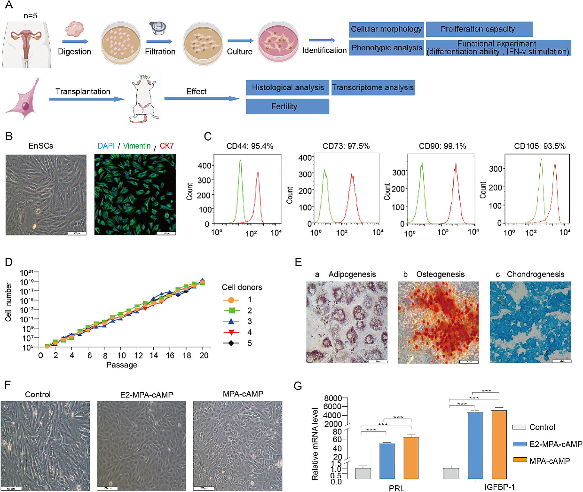
Isolation of human ASCs and cell culture
ASCs were isolated from the subcutaneous fat tissue of four non-diabetic, non-smoking female donors with an average age of 45 y (32–57 y) and an average body mass index of 24.6 (21.0–26.6) [20]. The study protocol has received approval from the Research Ethics Committee of National Taiwan University Hospital, and the informed consent was acquired from each adipose tissue donor participating in this study. This study was conducted in compliance with the institutional biosafety regulations. Small pieces of subcutaneous fat tissue were finely minced and subsequently rinsed with phosphate-buffered saline (PBS; Omics Biotechnology, Taipei, Taiwan), followed by enzymatic digestion using collagenase type I (Gibco, Carlsbad, CA, USA) for 1 h. After centrifuging the cell suspension, pellets were suspended and plated with Dulbecco’s modified Eagle’s medium (DMEM)/F-12 (Hyclone) which is supplemented with 10% fetal bovine serum (FBS; Hyclone, Logan, UT, USA), 1% penicillin–streptomycin (Biological Industries, Kibbutz Beit Haemek, Israel), and 1 ng/mL FGF2 (R&D systems, Minneapolis, MN, USA). The cells were cultured in a humidified atmosphere with 5% CO2 at 37 °C, and the medium was refreshed every 2–3 days. The cells were detached using 0.05% trypsin-EDTA (Biological Industries) upon reaching 90% confluence and re-plated until the third or fourth passage for various experiments. These cells have been previously tested to exhibit differentiation capabilities toward multi-lineages [21, 22]. Isolated human ASCs were pooled into a single population due to their similar surface marker expression and comparable differentiation potential, as exhibited by each ASC clone.
Fabrication of ASC sheets
ASC sheets were prepared and characterized as described previously [23]. Briefly, ASCs were seeded at a density of 1 × 10⁴ cells/cm2 in a 0.1% gelatin coated culture dish or plate. The culture medium consisted of DMEM-high glucose (DMEM-HG; Hyclone), 1% penicillin/ streptomycin (Biological Industries), 0.02% heparin (Sigma-Aldrich, St. Louis, MO, USA), 250 µM L-ascorbate 2-phosphate (A2-P; Sigma-Aldrich), and supplemented with 10% FBS or 5% HPL (UltraGRO™; HELIOS BioScience, Atlanta, GA). The culture medium was refreshed every 2 ~ 3 days. As for the harvest of conditioned medium, the cultured medium of cell sheet was switched to DMEM-HG for additional 2 days.
In vivo burn wound model
Male Wistar rat, aged 10–12 weeks, were purchased from BioLASCO Taiwan (Taipei, Taiwan). All experiments were performed in compliance with Guidance for Care and Use of Laboratory Animals and approved by the Taipei Medical University Administrative Committee on Animal Research (LAC-2018-0130) and ARRIVE (Animal Research: Reporting of In Vivo Experiments) 2.0 guidelines. All experiment procedures were conducted under general anesthesia (isoflurane, 3–5%, 0.5 L/min). After the hair at the rat dorsum was shaved, a stainless-steel cylinder (1 cm in diameter) was heated to 100 °C in a boiled water bath for 8 min, and then placed onto the back of rats for 15 s to induce partial-thickness burn wounds [24]. Four 1 cm-diameter round burn wounds were created on the back of each rat, and the wounds were immediately photographed using a digital camera (Ricoh, Ohta-ku, Tokyo, Japan). In each rat, one wound was selected for PBS injection as controls, and the other three wounds were treated with HPL-cultured or FBS-cultured ASC sheets respectively. Subsequently, the dorsal skin was covered with a transparent, semi-occlusive adhesive dressing (Tegaderm; 3 M, St. Paul, MN, USA) for wound protection. The wounds were observed and photographed on post-injury day 0, 7, 14, 21 and 28. Each wound was evaluated by four blinded investigators on a rating from 0 to 4 based on (1) brown discoloration and (2) scabbing/hardness (Additional file1: Table S1) [24]. The wound images were analyzed using ImageJ software to measure wound areas, with the wound size on day 0 defined as 100%. All animals were euthanized by carbon dioxide inhalation and the entire wound tissues were harvested on post-injury day 5 and day 28 for further analysis (n = 3 for each time point in each group).
Histological analysis and immunohistochemistry
Wound tissue samples were snap frozen in liquid nitrogen for frozen sections or fixed in 4% paraformaldehyde overnight and dehydrated before paraffin embedding. Sections were made perpendicular to both the wound surface and the anterior-posterior axis, cutting into 4 μm for histological analysis. In order to visualize the histological changes and collagen deposition, Hematoxylin and Eosin (H&E) and Masson’s trichrome staining were employed to stain the sections according to the manufacturer’s protocol (Sigma-Aldrich). Three sections per group were randomly chosen, and five high-power fields (hpfs) in each section were randomly selected and measured for skin thickness and collagen density.
For immunohistochemical staining, rat wound tissue sections were deparaffinized in xylene and rehydrated in a series concentration of ethanol. The antigen was unmasked using pH 9.0 Tris-EDTA buffer (Abcam, Cambridge, UK) at 95 °C for 30 min. Tissue sections were blocked with 3% bovine serum albumin (BSA; BioShop, Burlington, Ontario, Canada) before the incubation with primary antibodies. Then the sections were labeled with the primary antibodies against CD31 (Abcam), CD68 (Abcam), α-smooth muscle actin (α-SMA; Abcam), and transforming growth factor-β1 (TGF-β1; Santa Cruz Biotechnology, Dallas, Texas, USA) at 4 °C overnight. The secondary antibody linking to horseradish peroxidase (R&D systems) was incubated for 1 h at room temperature. All specimens were visualized by a 3,3’-Diaminobenzidine chromogen kit (BIOTnA Biotech, Kaohsiung, Taiwan), and the nuclei were counterstained with hematoxylin solution (Abcam). The stained sections were scanned by TissueFAXS scanning system (TissueGnostics, Vienna, Austria) and analyzed using StrataQuest software. The signals were quantified in 5 randomly selected high-power fields per section from three different sections.
Immunofluorescence staining
The paraffin-embedded tissues were cut into 4-µm sections, mounted on slides, deparaffinized, dehydrated, and the antigen was retrieved by pH 9.0 Tris-EDTA buffer at 70°C for 20 min. The sections were incubated in 3% BSA with 0.1% triton (Sigma-Aldrich) for blocking non-specific protein binding. The staining of transplanted human ASCs was conducted using anti-human nuclear antigen (HNA; Merck Millipore, Darmstadt, Germany) overnight at 4°C, followed by incubation with a fluorescence-conjugated secondary antibody (Alexa Fluor 594-conjugated goat anti-mouse IgG; BioLegend, San Diego, CA, USA) at room temperature for 1 h. Nuclei were counterstained with 4’, 6-diamidino-2-phenylindole (DAPI; Santa Cruz Biotechnology). To rule out non-specific labeling, negative controls lacking primary antibodies were also employed. The sections were analyzed using a fluorescent microscope (EVOS™ M7000 Imaging System; Invitrogen, Waltham, MA, USA), and the immunolabeled cells were quantified in 3 randomly selected high-power fields per section from three different sections.
Microarray processing and gene expression analysis
Total RNA from FBS-cultured or HPL-cultured ASC sheets was prepared using TRIzol™ Reagent (Invitrogen). Residual genomic DNA was removed by DNase I digestion. RNA quantification was performed using a NanoDrop-1000 spectrophotometer (Thermo Fisher Scientific, Wilmington, DE, USA), and its quality was assessed through agarose electrophoresis and Bioanalyzer 2100 with an RNA 6000 Nano Kit (Agilent Technologies, Santa Clara, CA, USA). RNA expression in HPL and FBS-cultured ASC sheets was analyzed using SurePrint G3 Human Gene Expression 8 × 60 K Microarray (Agilent Technologies). RNA was amplified and labeled using Low Input Quick-Amp Labeling kit (Agilent Technologies) following the manufacturer’s instructions. Then, the correspondingly fragmented labeled RNA was pooled and hybridized to Agilent SurePrint Microarray. The chip was scanned with an Agilent microarray scanner and analyzed by Feature extraction 10.5.1.1 software (Agilent Technologies). The raw microarray data were submitted to NCBI Gene Expression Omnibus (GEO) repository with accession number GSE252798. For gene expression analysis, the microarray raw signals underwent normalization using the quantile method. Differential expression analysis was conducted using the NOISeq R package [25]. Genes with a probability of differential expression equal to or greater than 0.8 were considered as differentially expressed genes. Pre-rank gene-set enrichment analysis (GSEA) was performed utilizing the functions of the R package clusterProfiler [26]. Gene-sets from MSigDB (v7.4) were employed for this analysis [27]. Genes were ranked based on the product of their rank and probability values, obtained from the NOISeq results. This approach enhances the sensitivity of GSEA by incorporating both the degree of differential expression and the statistical confidence in the ranking.
Angiogenesis array
Analysis of secretion profile in the conditioned medium of FBS or HPL-cultured ASC sheets was performed using a Human Angiogenesis Array C1 (RayBiotech, Norcross, GA, USA) according to the manufacturer’s instructions, allowing semi-quantitative determination of protein levels of a variety of pro-angiogenic factors.
RNA isolation and quantitative polymerase chain reaction
Total RNA was extracted from FBS or HPL-cultured ASC sheets utilizing a RNeasy Mini Kit (Qiagen, Valencia, CA, USA) in accordance with the manufacturer’s protocols. Total RNA concentration was determined by optical density at 260 nm (OD260) using a spectrophotometer (Tecan, Männedorf, Switzerland) and reverse-transcribed into complementary DNA (cDNA) using High-Capacity cDNA Reverse Transcription Kits (Applied Biosystems, Foster City, CA, USA). Quantitative PCR was performed using a FastStart Universal SYBR Green Master (Roche, Indianapolis, IN, USA) and CFX Connect Real-Time PCR Detection System (Bio-Rad, Hercules, CA, USA). The gene expression level was normalized to glyceraldehyde 3-phosphate dehydrogenase (GAPDH) for each cDNA sample. A comparative threshold cycle method (ΔΔCt) was adopted to analyze the relative quantity (RQ) of mRNA expression between treated and corresponding control samples. The sequences of the used primers are shown in Additional file1: Table S2.
Enzyme-linked immunosorbent assay (ELISA)
The quantitative analysis of C–C motif chemokine ligand 5 (CCL5) and angiogenin in the conditioned medium from FBS or HPL-cultured ASC sheets was performed using ELISA (Quantikine; R&D Systems). Optical densities were determined using a spectrophotometer (Tecan, Männedorf, Switzerland) at 450 nm, with wavelength correction set to 570 nm. Values were presented as the concentration of secreted CCL5 or angiogenin per 105 cells at the time of harvest.
In vitro proliferation assay
Human umbilical vein endothelial cells (HUVECs; PromoCell, Heidelberg, Germany) were seeded in a 0.1% gelatin-coated 96 well plate with serum supplemented endothelial cell growth medium 2 (EGM2; PromoCell). After 24 h-cell attachment, the cells were treated by CCL5 (R&D systems; 1 ng/mL) or angiogenin (R&D systems; 1, 10 and 50 ng/mL) in a serum-free medium composed of endothelial basal medium (EBM; PromoCell) and DMEM-HG (EBM/DMEM, 1:1). The conditioned medium from the HPL sheet was mixed with equal volume of EBM. The HUVECs were incubated for additional 24 h. The aforementioned basal medium (EBM/DMEM) served as a negative control. The proliferation was assessed by Alamar Blue assay (AbD Serotec, Kidlington, United Kingdom). The fluorescence signals were measured at an excitation wavelength at 560 nm and an emission wavelength at 590 nm by a spectrometer (Tecan). The activity index of ASCs was defined as the proliferative rate of each group relative to the control.
In vitro tube formation assay
The capability of HUVECs to form capillary-like structures was further evaluated in the tube formation assay as described previously [23]. In brief, HUVECs were seeded on pre-chilled µ-slides (Ibidi, Grafelfing, Germany) coated with growth factor reduced Matrigel basement membrane matrix (Corning, Lowell, MA, USA) at a density of 7,500 cells/well. HUVECs were treated by CCL5 (1, 10, 50 ng/mL) or angiogenin (1, 10, 50 ng/mL) in the aforementioned EBM/DMEM medium. The conditioned medium from FBS sheet or HPL sheet was mixed with equal volume of EBM before treatment. The neutralizing CCL5 antibody (R&D systems; 75 ng/mL) was applied in the HPL sheet derived conditioned medium to examine the contribution of CCL5. The aforementioned basal medium (EBM/DMEM) served as a negative control, while serum supplemented EGM2 was used as a positive control. Formation of tube-like structures was visualized by a phase-contrast microscope after 6 h incubation. The images were analyzed using ImageJ software as described previously [28].
Statistical analysis
All measurements are reported as means ± standard deviation. Statistical significance was evaluated using one-way analysis of variance (ANOVA) followed by Tukey’s multiple comparison tests or Student’s t test to compare the results of control and the experimental groups. All experimental data were analyzed using GraphPad Prism 8 (GraphPad Software, Boston, MA, USA). Statistical significance was defined as p < 0.05.


留言 (0)