
記住我


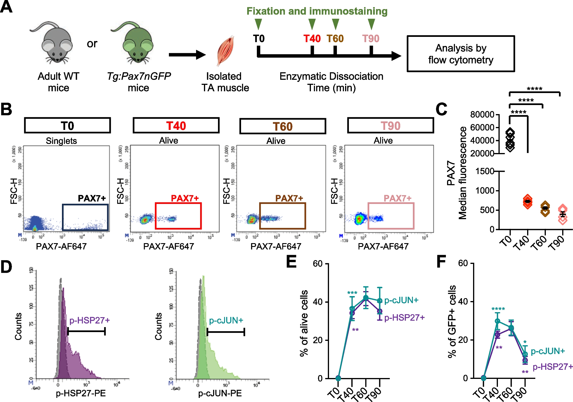
Recent studies have highlighted profound transcriptomic changes in MuSC during muscle tissue dissociation, notably a reduction in Pax7 transcripts [19]. We therefore sought to investigate the dynamics of PAX7 regulation at the protein level during this process, by flow cytometry (Fig. 1). The level of PAX7 protein in quiescent MuSC was determined by analyzing mononucleated cells from single Tibialis Anterior (TA) muscle fixed directly upon harvest (T0). For other timepoints, mononucleated cells were recovered after 40, 60 and 90 min of dissociation (T40, T60 and T90), and immediately fixed and immunostained for PAX7 as previously described [40] (Fig. 1A). Between T0 and T40, we noted a strong reduction in PAX7 protein levels in the entire population of MuSC, evidenced by the significant reduction in the median of fluorescence of PAX7 staining (Figs. 1B, C and S1A, B). Subsequently, from T40 to T90, the medians of fluorescence continue to decrease, albeit in a much less pronounced manner. In addition, at T40, PAX7 + population was well defined and grouped, showing a homogenous level of PAX7 protein. Conversely, from T60 onwards, the PAX7 + population appeared more dispersed, demonstrating a progressive loss of PAX7 protein but at different rates among the MuSC population (Fig. 1B). Overall, our observations revealed that PAX7 proteostasis is rapidly affected upon dissociation leading to a two-step decrease. A rapid and strong decrease occurring in the entire population of PAX7 + MuSC, followed by a more heterogeneous and progressive decline.
Fig. 1
PAX7 protein declines drastically in MuSC concomitant with activation of stress signaling pathways upon dissociation. A Isolated TA muscles were either fixed directly after harvest (T0) or dissociated for different times (T40, T60 and T90) before fixation and immunostaining. B Representative density scatter plots showing PAX7 expression after the different times of dissociation. Debris, doublets and dead cells were excluded from the analysis, except for T0 when muscles were directly fixed. C Median fluorescence of PAX7 immunostaining analyzed by flow cytometry (n = 6 mice for each time point). D Representative histograms showing phospho-HSP27 (p-HSP27, purple) and phospho-c-JUN (p-cJUN, green) cells among alive cells at T60. Positivity was determined based on FMO (grey). E, F Proportions of alive cells (E) and GFP + cells (F), with detectable levels of p-HSP27 or p-cJUN in TA muscles from Tg:Pax7nGFP mice, determined by flow cytometry (n = 8 mice for T40, T60 and T90, n = 3 mice for T0). Values are the mean ± SEM of independent experiments. One- and Two-Way ANOVA with Tukey’s multiple comparisons test, *p < 0.05, **p < 0.01, ***p < 0.001 and ****p < 0.0001 (versus preceding time point for E and F)
Since P38 MAPK and JNK pathways are prominently induced pathways during the dissociation process [17, 30], we next evaluated whether they could be involved in the downregulation of PAX7 protein. We first investigated by flow cytometry the activation kinetics of both pathways by monitoring the phosphorylation status of HSP27 and c-JUN (referred to as p-HSP27 and p-cJUN), known downstream targets of P38 MAPK and JNK, respectively. For these analyses, we used Tg:Pax7nGFP mice, which enabled us to follow the myogenic lineage even if PAX7 expression was lost through perdurance of the GFP protein [33]. We assessed the proportion of p-HSP27 + and p-cJUN + cells, among the whole population of mononucleated cells (alive cells), and the GFP + myogenic cells (Figs. 1D–F and S1C–F). At T0, almost all the cells were negative for p-HSP27 and p-cJUN consistent with the homeostatic state of cells in adult muscle. The proportions of mononucleated cells p-HSP27 + and p-cJUN + significantly increased at T40 compared to T0, continued to increase until T60, and finally began to decrease at T90. Activation of P38 MAPK and JNK pathways is therefore coordinated and peaks at 60 min of dissociation in muscle mononucleated cells (Fig. 1E). A concurrent activation of these two pathways was also observed in GFP + myogenic cells between T0 and T40 (Fig. 1F). However, in contrast to mononucleated cells, the levels of GFP + p-HSP27 + or p-cJUN + cells, were significantly diminished at T90 compared to T60. Overall, these experiments demonstrated that P38 and JNK pathway activation is a more transitory event in MuSC than in other mononucleated cells of the muscle (Fig. 1E, F).
Since P38 MAPK and JNK activation occurred concomitantly to the drastic decrease of PAX7 protein in MuSC, we next sought to determine whether these two phenomena were directly linked. We therefore added pharmacological inhibitors (PI) of these two pathways from the harvest of the muscle to the fixation step (Fig. 2). SB202190 (SB) and SP600125 (SP) were used alone or in combination as inhibitors of P38 MAPK and JNK, respectively (Fig. 2A). We first verified the efficacy of our treatments by assessing the proportion of GFP + p-HSP27 + and p-cJUN + cells in TA muscles from Tg:Pax7nGFP mice dissociated with vehicle, SB, SP or SB + SP for 40 min, by flow cytometry (Fig. 2B, C). We observed that in conditions with SB alone or with SB + SP, the proportion of GFP + p-HSP27 + cells was significantly reduced compared to vehicle. We also noticed a reduction in the proportion of GFP + p-HSP27 + with SP treatment that likely results from a direct effect of JNK on HSP27 phosphorylation state, as previously reported [41, 42]. In agreement, the combination of SB + SP had a synergistic inhibitory effect on the proportion of GFP + p-HSP27 + cells compared to SB treatment (Fig. 2C). Upon exposure with SP alone or in combination with SB, the proportion of GFP + p-cJUN + cells was reduced compared to control or SB conditions (Fig. 2C).
Fig. 2
Inhibition of P38 MAPK and JNK pathways does not prevent initial drop of PAX7 protein. A Inhibitors of P38 MAPK (SB202190, SB) and JNK (SP600125, SP) or vehicle were added from the muscle harvest to the fixation. Tg:Pax7nGFP mice were used to monitor GFP + myogenic cells and WT mice to monitor PAX7 levels. B Representative histograms showing phospho-HSP27 (p-HSP27) and phospho-cJUN (p-cJUN) positive cells among the GFP + population from TA digested with vehicle (grey) or SB + SP (blue). C Proportion of GFP + p-HSP27 + and GFP + p-cJUN + cells after 40 min of dissociation with vehicle or the inhibitors (n = 5 mice/group). D Median fluorescence of PAX7 immunostaining, determined by flow cytometry (n = 6 mice/group). E Representative density scatter plots showing the PAX7 + cells among GFP + population. F Proportion of PAX7 + cells among GFP + population (n = 4 to 5 mice/group). Debris, doublets and dead cells were excluded from the analysis. Values are the mean ± SEM of independent experiments. One Way and Two Way ANOVA and Tukey’s multiple comparisons test, *p < 0.05, **p < 0.01, ***p < 0.001 and ****p < 0.0001
Having confirmed the inhibitory effect of our treatments, we next monitored the level of PAX7 protein in MuSC from TA muscles dissociated with vehicle, SB, SP or SB + SP (Fig. 2D). We observed that inhibition of P38 and/or JNK pathways did not prevent the drastic loss of PAX7 protein during the initial phase of muscle dissociation and that the mean level of PAX7 protein was similar between vehicle and treated conditions. Nevertheless, using the Tg:Pax7nGFP reporter line, we showed that treatment with inhibitors (alone or in combination) during the dissociation phase increased the proportion of GFP + cells exhibited detectable amounts of PAX7 (Fig. 2E, F). Our findings demonstrate that the sharp decline of PAX7 protein occurring in the initial phase of dissociation is independent of P38 MAPK and JNK increased activities. However, these pathways may rather contribute to the progressive and heterogenous loss of PAX7 observed later on.
Inhibition of P38 MAPK and JNK throughout dissociation and cell sorting mitigates stemness markers down-regulation and early activation of MuSCSince MuSC purification requires a cell sorting step after muscle dissociation, we next extended our inhibitory strategy to the entire process, including during immunostaining and FACS sorting (Fig. 3A). To uncouple the detrimental impact of prolonged enzymatic dissociation from the role of these pathways in early MuSC activation, we limited the dissociation time to 40 min. For these experiments, MuSC were recovered from hindlimb muscles of wildtype adult mice and purified as shown in Fig S2. We performed a transcriptomic profiling of MuSC purified from muscles digested and sorted in the presence of SB, SP, SB + SP or vehicle, by RT-qPCR. We first assessed the level of transcripts of immediate and early response genes (Egr1, 2, 3) and members of the AP-1 family genes (cJun, Junb, Fos, Fosb and Fosl1), previously shown to be strongly induced during MuSC isolation [17, 19, 20] (Fig. 3B). Among Egr genes, only Egr3 transcripts were significantly downregulated in SP and SB + SP conditions compared to vehicle indicating that activated JNK was primarily involved in their increase during MuSC isolation (Fig. 3B). In agreement with previous observations, Fos transcript levels were stable regardless of the treatment [16]. Fosb and Fosl1 transcripts exhibited a more pronounced decrease with SB + SP treatment, demonstrating the involvement of both JNK and P38 MAPK in their induction. Junb transcripts were similarly downregulated with SB and the combination SB + SP, proving that P38 MAPK was the main player. Overall, our data demonstrated that during dissociation, P38 MAPK and JNK contribute to the up-regulation of only a subset of immediate and early response genes.
Fig. 3
Lowering P38 and JNK activities preserves stemness marker expression and limits early activation of MuSC. A Hindlimb muscles from adult WT mice were digested for 40 min. Mononucleated cells were immunostained and MuSC purified by FACS (see Fig. S2). Vehicle or inhibitors were added during the whole process. Transcript levels of B Egr and AP-1 genes, C genes up-regulated upon activation, D genes coding for stemness markers, analyzed by RT-qPCR. Values are the mean ± SEM of 4 to 5 independent experiments. E Representative density scatterplots showing the expression of CD34 and SCA1 among the Lin- fraction, and of ITGA7 among CD34 + SCA1 + population. Graphs showing F the median level of fluorescence for CD34 and ITGA7 stainings, G the purification yield of murine MuSC (mMuSC) per g of muscle, H the percentage of viable mMuSC. Values are the mean ± SEM of 6 independent experiments (n = 6 mice/group). One-way ANOVA analysis and Dunnett’s multiple comparisons test, with *p < 0.05, **p < 0.01, ***p < 0.001 and ****p < 0.0001
We next analyzed the expression of markers described to be upregulated in early activated MuSC relative to quiescent MuSC, including Myod1, Cdkn1a (p21) and Hspb1 (HSP27) (Fig. 3C). SB + SP treatment had the more marked effect on the transcript levels of these 3 genes, indicating that both P38 MAPK and JNK participate in their induction. Interestingly, the inhibition profiles obtained for Myod1 transcripts suggested that JNK was predominantly involved. Overall, these results demonstrated that simultaneous inhibition of P38 MAPK and JNK during MuSC isolation restricts their premature activation. Of note, by flow cytometry analysis, we detected rare MYOD + cells in equivalent proportions under all digestion conditions, consistent with the timescale of purification (Fig. S2A, B). Moreover, SB + SP treatment synergistically increased the transcript levels of the stemness markers, Pax7 and Calcr, while the levels of Spry1 and Cd34 transcripts remained unchanged (Fig. 3D). Interestingly, upon FACS sorting, we noticed an increased level of CD34 surface protein in SP and SB + SP conditions whereas of α7-integrin (ITGA7) level was similar (Figs. 3E, F and S2C–E). JNK induction during dissociation seems to affect CD34 protein level, which is essential for murine MuSC functionality and quiescent state [43]. This effect could rely on a post-translational regulation since a putative phosphorylation site by JNK was identified in the intracellular domain of CD34 [44].
Lastly, the purification yield of MuSC was significantly improved in all conditions with inhibitors, but more markedly with SP and SB + SP treatments (Fig. 3G). We hypothesize that this improved purification yields could be attributed to a better survival of the cells (Fig. 3H), associated with a better detection of CD34 + cells during the sorting step.
Constant inhibition of P38 MAPK and JNK signaling pathways ameliorates survival and amplification rate of MuSC in vitroWe next sought to assess the functionality of MuSC isolated with inhibitors by in vitro assays (Fig. 4A). After 4 days of culture in growth conditions, MuSC isolated in the presence of the inhibitors showed an increased amplification rate compared to those isolated with vehicle, this effect being more pronounced with SB + SP combination (Fig. 4B). However, only the cells purified with SB + SP formed larger colonies relative to control (Fig. 4C). Surprisingly, 48 h after seeding, we noticed that twice as many cells had proceeded to their first division when cells were isolated with inhibitors compared to vehicle (Fig. 4D). We hypothesized that restricting stress pathway activities during isolation preserved MuSC in a better state making them more inclined to adhere and divide. In line with this hypothesis, we observed that exposure to inhibitors during isolation and especially to SB (SB alone or SB + SP), led to an increased number of colonies (Fig. 4E), associated with a decreased proportion of dead cells, 48 h after plating (Fig. 4F). Lastly, after 3 days in differentiation conditions, the fusion index was similar in all conditions, indicating that exposure to the inhibitors did not affect the fusion ability of the cells (Fig. 4G, H). Interestingly, we noticed that further addition of SB + SP during the culture could potentialize the positive effect of digestion with SB + SP on MuSC amplification rate (Fig. S3A–D). Altogether, our data demonstrated that limiting P38 MAPK or JNK activation during the isolation process has a beneficial effect on MuSC function in vitro. Inhibition of P38 MAPK appeared critical for MuSC survival upon seeding, whereas combined inhibition of P38 MAPK and JNK allowed a better survival and increased amplification rate in vitro.
Fig. 4
P38 and JNK inhibition during isolation elicits increased survival and amplification of MuSC in vitro. A MuSC were purified with vehicle or inhibitors and plated at low density in Growth Medium (GM) for 4 days or at high density for fusion (DM: Differentiation Medium). Graphs showing, B the total number of cells after 4 days of amplification, C the size of the colonies, D the proportion of cells having proceeded to their first division at 48 h, E the total number of colonies per cm2, F the proportion of dead cells relative to seeded cells 48 h after plating. G Representative images of the myotubes obtained after 3 days in DM, immunostained for Myosin Heavy Chain (MHC). Nuclei were stained with DAPI. Scale bar: 100 µm. H Fusion Index (%). Values are the mean ± SEM of minimum 3 independent experiments (n = 3–6 mice/group). One-way ANOVA analysis, with *p < 0.05, **p < 0.01, ***p < 0.001 and ****p < 0.0001
MuSC exposed to P38 MAPK and JNK inhibitors during isolation exhibit increased engraftment potential in vivoSince SB + SP treatment had the most beneficial effect on MuSC function in vitro, we next assessed its effect on their engraftment potential in vivo. MuSC were purified by FACS from two models of reporter mice, Tg:Pax7nGFP and RosanT−nG, in the presence of vehicle or SB + SP. Freshly isolated MuSC were transplanted to TA muscles of recipient mice previously injured by CTX injection and partially depleted of endogenous MuSC to optimize the efficacy of injected MuSC engraftment (Fig. 5A) [38, 39]. Indeed, in this recipient mouse model, Cre-driven recombination induces Diphtheria Toxin Receptor (DTR) expression specifically in PAX7 + MuSC, following tamoxifen administration. Next, intramuscular injection of Diphtheria Toxin (DT), an inhibitor of protein synthesis, induces a reproducible depletion of 75% of the endogenous MuSC (Fig. S4A–C). Two weeks post-transplantation, the number of engrafted mononucleated GFP + cells was determined by flow cytometry (Fig. 5B and Fig. S4D–G). We observed that the total number of GFP + cells/TA as well as the number of GFP + cells normalized per mg of muscle, was significantly increased when MuSC had been processed with SB + SP during the whole isolation process (Fig. 5C). The mean proportion of PAX7 + cells among the GFP + population was equivalent between vehicle and SB + SP (Fig. 5D). Moreover, regardless of the treatment more than 90% of the GFP + PAX7 + cells were KI67- and thus presumably in a quiescent state (Fig. 5D). Based on these observations and in vitro data, we concluded that the increased number of GFP + cells in SB + SP condition, was mainly due to an improved survival and/or amplification of the transplanted MuSC rather than an increased self-renewal capacity. We next analyzed the engraftment efficacy 1-month post-transplantation of RosanT−nG MuSC isolated with SB + SP or its vehicle, by immunofluorescence (Figs. 5E and S4H, I). We noticed that the mean number of Tomato + nuclei (nTom +) per myofiber and the size of these myofibers were significantly increased in SB + SP compared to vehicle condition (Fig. 5F, G). Consistent with our data at 2 weeks, we observed a higher number of sub-laminal nTom + MCadherin + cells in SB + SP condition than in vehicle (Figs. 5H, I and S4J). Overall, these experiments demonstrate that combined inhibition of P38 MAPK and JNK during isolation preserves the regenerative potential of MuSC.
Fig. 5
MuSC isolated with SB + SP inhibitors exhibit an increased engraftment potential. A Recipient mice received 4 consecutive tamoxifen (TMX) injections, followed by intramuscular injection of cardiotoxin (CTX). Diphtheria toxin (DT) was injected 3 days later (see also Methods and Fig. S4A–C). Muscles from donor mice (Tg:Pax7nGFP or RosanT−nG) were dissociated with vehicle or SB + SP, MuSC were purified by FACS and transplanted, 8 h after DT injection. Transplanted TA muscles were harvested after 2 weeks for flow cytometric analysis or 1-month for immuno-histological analysis. B Representative scatter plots showing the expression of PAX7 and KI67 among GFP + cells (see also Fig. S4). Quantification of C the total number of viable GFP + cells/TA and the number of GFP + cells/mg of TA, D the proportion of PAX7 + cells among the GFP + population and the proportion of KI67 + cells among the GFP + PAX7 + cells. Values are the mean ± SEM of 4 independent experiments (n = 4 mice/condition). E Representative images of transversal sections of TA grafted with RosanT−nG. MuSC purified with vehicle or SB + SP, 1-month post-transplantation, immunostained for Tomato and Laminin. Scale bars: 100 µm and 50 µm. Quantification of F the mean number of Tom + nuclei per fiber, G the cross-sectional area and the diameter of myofibers with Tom + nuclei. H Representative images of TA cross-sections immunostained for Tomato, M-Cadherin and Laminin. Arrowheads point to nTom + MCad + cells. Scale bars: 20 µm. I Number of nTomato + Mcadherin + cells normalized by 100 fibers. Values are the mean ± SEM of 3 independent experiments (n = 3 mice/condition). Unpaired T test and One-way ANOVA, with *p < 0.05 and **p < 0.01
An optimized version of muscle tissue dissociation protocol enabling high purification yield of human MuSC while preserving their stemnessWe next sought to determine whether our findings were relevant to human MuSC (hMuSC). To alleviate the detrimental impact of long-term enzymatic dissociation, we optimized the standard protocol used in the field. We restricted the dissociation time to 40 min, performed an intermediate recovery of mononucleated cells at 20 min, and added SB + SP during the whole isolation process. hMuSC were sorted by FACS using CD29, CD56, and ITGA7 as selection markers (Fig. 6A and Fig. S5A–D). Given the small size and the rarity of human biopsies, to assess the effect of this optimized purification protocol, we compared the transcriptome of hMuSC purified following this protocol (referred to as hMuSC-40/PI) to that of hMuSC isolated after 120 min of dissociation without inhibitors previously published (hMuSC-120/no PI) [25], by RNA sequencing. Principal component analysis (PCA) plot of gene expression revealed distinct transcriptomic profiles between the 2 groups (Fig. 6B), with more than 2000 differentially expressed genes (DEG) (Fig. 6C). Interestingly, hMuSC-40/PI samples coming from 2 distinct donors clustered together, whereas the 2 hMuSC-120/no PI samples corresponding to a biological replicate of the same donor were more distant, suggesting that our isolation strategy could reduce the variability between samples. By comparing the expression profiles of the stress core genes [17], we identified two gene sets (Fig. 6D, E). Genes which expression was reduced in hMuSC-40/PI, including known target genes of P38 MAPK and JNK (HSPB1, HSP90A, GADD45, ATF2), as well as FOSL1 and CDKN1a that were also down-regulated in mMuSC isolated with SB + SP (Fig. 6E). In contrast, EGR and AP-1 gene expression was globally higher in hMuSC-40/PI, likely due to the different dissociation times (Fig. 6E). Indeed, the expression of FOS and JUN genes is peaking at 40 min of dissociation, whereas it is declining at 120 min [17, 32]. Expression of genes involved in MuSC stemness and quiescent state was systematically higher in hMuSC-40/PI including, PAX7, CDH15, SPRY1, COL5a, TENM4, Notch receptors and downstream target genes (HES1, HEY1, HEYL) [45]. hMuSC-40/PI exhibited increased expression of anti-apoptotic genes (BCL2, BCL2L1, BCL2L2 and MCL1). Transcripts of MYF5, DEK and inflammation-related genes were up-regulated in hMuSC-120/no PI, as previously described in an early activated population of hMuSC [46] (Figs. 6F and S5E, F). Lastly, pathways associated with protein translation and ribosomes were enriched in hMuSC-120/no PI, whereas pathways related to gene transcription were enriched in hMuSC-40/PI (Fig. 6G). Overall, this comparative study indicates that our protocol favors the purification of hMuSC with preserved expression of stemness markers, better survival and in a less activated state than standard protocols, in agreement with our data on mMuSC.
Fig. 6
Optimized muscle dissociation protocol enables marked improvement of purification yield of hMuSC with preserved stemness. A Human muscle biopsies were processed with SB + SP from the harvest to the FACS sorting. hMuSC (Lin-CD34-CD29 + CD56 + ITGA7 + , see Fig . S5A–D) purified with this protocol are referred to as hMuSC-40/PI (40 min dissociation, with Pharmacological Inhibitors). B–G Comparative analysis of the transcriptome of freshly isolated hMuSC-40/PI to that of hMuSC isolated without PI previously published (hMuSC-120/no PI) [25], analyzed by RNAseq. B PCA plot of gene expression data. C MA plot of differentially expressed genes (DEG). D Volcano plot of 100 stress core genes differentially expressed. Heatmap clustering profiles of E stress core target genes, F quiescence and anti-apoptotic genes, based on log2 transformed counts. G Top biological pathways enriched hMuSC-40/PI versus hMuSC-120/no PI, based on differentially expressed genes and ranked by p value. H Comparison of hMuSC purification yield, when muscles were digested without inhibitors (no PI) or with SB + SP (PI). Grey and blue filled symbols represent our data (Biopsies 1–5, B#1 to B#5), open black circles correspond to data extracted from the literature. GAS: Gastrocnemius. See also Table S1. Values are the mean ± SEM of 3–5 independent biopsies, One-way ANOVA and Tukey’s multiple comparisons test, ***p < 0.001 and ****p < 0.0001
To assess whether these beneficial effects could translate into a better purification yield of hMuSC, we took advantage of previously published data [13, 14, 47] (Table S1). We noticed a marked improvement of hMuSC purification yield when human biopsies were processed following our protocol, compared to data extracted from the literature or to the data we previously obtained with standard dissociation protocol (Fig. 6H). Therefore, our purification strategy could greatly ameliorate hMuSC purification yield (up to 14-fold).
留言 (0)