
記住我


Commercial anti-ZBP1 antibodies are only available for humans, while, the homology is only 69.9% of nucleotide sequences between humans and swine ZBP1 (Fig. 1A), and is 55.1% of amino acid (Fig. 1B). To ensure protein detection in following experiments, anti-porcine ZBP1 IgG was prepared. ELISA showed that rabbit anti-porcine ZBP1 serum has excellent specificity until dilutions reach 1:16,000 (Fig. 1C). The prepared anti-porcine ZBP1 IgG was used as the primary antibody to detect the recombinant His-ZBP1 by western blotting. The results show that the prepared IgG has favorable specificity (Fig. 1D).
Fig. 1
SVA infection up-regulates ZBP1 expression in 3D4/21 cells. A, B The homology of ZBP1 nucleotide and protein between swine, human, and mouse were aligned. C The titers of serum were detected by ELISA, positive: immune serum, negative: unimmunized serum. D The recombinant His-ZBP1 protein were detected by western blotting using prepared rabbit polyclonal anti-porcine ZBP1 IgG. E 3D4/21 cells were infected with SVA, protein levels of ZBP1 and VP2 were detected by western blotting. F 3D4/21 cells were infected with SVA and heat-SVA at an MOI of 1, mock-infected served as a control. Cells were harvested at 6, 12, 24 and 36 h.p.i, the mRNA expression levels of ZBP1 were measured by qRT-PCR. G The mRNA expression levels of ZBP1 were measured by qRT-PCR in 3D4/21 cells infected with SVA at MOIs of 0.5, 1, 2, and 3 for 24 h. All experiments were repeated three times independently. *P < 0.05, **P < 0.01
3D4/21 cells were infected by SVA (MOI of 1), western blot showed that SVA infection promoting the expression of ZBP1 from 12 to 36 h.p.i (Fig. 1E). 3D4/21 cells were infected with SVA (MOI of 1) or heat-SVA, and mock-infected served as a control. The expression levels of ZBP1 mRNA were measured by qRT-PCR at 6, 12, 24, and 36 h.p.i. The results showed that the mRNA of ZBP1 was significantly up-regulated from 6 to 36 h.p.i (P < 0.01) (Fig. 1F). While, the heat-SVA could not induce changes expression levels of ZBP1, indicating that the expression of ZBP1 depends on SVA replication. 3D4/21 cells were infected with SVA at different dose (MOIs of 0.5, 1, 2, and 3) for 24 h and the expression of ZBP1 was induced by SVA infection (P < 0.01) (Fig. 1G). These results indicate that SVA significantly up-regulates the expression of ZBP1 in 3D4/21 cells.
ZBP1 inhibits the replication of SVATo investigate the role of ZBP1 on SVA proliferation, 3D4/21 cells with ZBP1-overexpression and -interference expression were used for SVA infection. In ZBP1-overexpression cells, pEGFP-N1 or pEGFP-N1-ZBP1 was transfected into 3D4/21 cells, respectively (Fig. 2A). The results of qRT-PCR showed that ZBP1 inhibited the copy number of SVA VP2 gene from 12 to 48 h.p.i compared with the control (P < 0.01) (Fig. 2B). Western blots showed that ZBP1 overexpression significantly inhibited the expression of SVA VP2 protein (P < 0.05) (Fig. 2C, D). Transfection of pEGFP-N1-ZBP1 resulted in a significant decrease in viral titers of approximately three- to tenfold at 6 (P < 0.05), 12 (P < 0.05), 24 (P < 0.01), 36 (P < 0.01), and 48 h.p.i (P < 0.01) compared to the control (Fig. 2E). In ZBP1-interference expression cells, shZBP1 and shNC was transfected into 3D4/21 cells, respectively, and the expression of ZBP1 was only 0.5 in cells with ZBP1-interference expression (Fig. 2F, G). The mRNA levels of SVA VP2 gene was significantly higher in ZBP1-interference group from 12 to 48 h.p.i compared to the control (P < 0.01) (Fig. 2H). The results of western blot revealed that VP2 protein expression were also raised in cells with impeded ZBP1 expression (P < 0.01) (Fig. 2I, J). Virus titers in ZBP1-interference cells were significantly higher at 6 (P < 0.05), 12 (P < 0.01), 24 (P < 0.01), 36 (P < 0.01), and 48 h.p.i (P < 0.01) compared to the control, and approximately tenfold increase at 24 h.p.i (Fig. 2K). These results show that ZBP1 inhibits the replication of SVA.
Fig. 2
ZBP1 inhibits the replication of SVA. A The expression levels of ZBP1 were detected by western blotting in 3D4/21 cells transfected with pEGFP-N1 or pEGFP-N1-ZBP1 for 24 h. B The mRNA expression of SVA VP2 gene was analyzed by qRT-PCR at 6, 12, 24, 36, and 48 h.p.i in SVA (MOI of 1) infected 3D4/21 cells after transfected with pEGFP-N1 or pEGFP-N1-ZBP1 for 24 h. C, D The expression levels of VP2 protein were detected by western blotting at 24 h.p.i and quantitatively analyzed by Image J. E Virus titers were determined in 3D4/21 cells using the Reed-Muench method. F, G The expression of ZBP1 was detected by western blotting in 3D4/21 cells transfected with shNC or shZBP1 for 24 h and analyzed by Image J. H The expression of SVA VP2 gene were analyzed by qRT-PCR in SVA (MOI of 1) infected 3D4/21 cells after transfected with shNC or shZBP1 for 24 h. I, J The expression levels of VP2 protein were detected by western blotting at 24 h.p.i and quantitatively analyzed by Image J. K Virus titers were determined in 3D4/21 cells. All experiments were repeated three times independently. *P < 0.05, **P < 0.01, ns: not significant
ZBP1 promotes the expression of IFNs, ISGs, and pro-inflammatory cytokines in SVA infection3D4/21cells with ZBP1-overexpression and -interference expression were used to determine whether ZBP1 had an effect on the expression of type I IFNs and pro-inflammatory cytokines in SVA infection. In ZBP1-overexpression cells, the mRNA expression of IFN-α, IFN-β, PKR, ISG15, IFIT1, IL-6, and IL-8 were significantly higher at 6, 12, and 24 h.p.i (P < 0.01), TNF-α was significantly higher at 6 (P < 0.01), 12 (P < 0.05), and 24 h.p.i (P < 0.01) compared to the control (Fig. 3A–H). While, the opposite results were shown in ZBP1-interference expression cells. Impeded ZBP1 expression decreased the expression of IFN-α, IFN-β, PKR, IL-6, IL-8 and TNF-α from 6 to 24 h.p.i (P < 0.01), ISG15 at 6 (P < 0.01), 12 (P < 0.05), and 24 (P < 0.01) h.p.i, IFIT1 at 6 (P < 0.01), 12 (P < 0.01), and 24 (P < 0.05) h.p.i, compared to the control (Fig. 3I–P). These results indicate that ZBP1 promotes the expression of type I IFNs, ISGs, and pro-inflammatory cytokines in SVA infection.
Fig. 3
ZBP1 promotes the expression of IFNs, ISGs, and pro-inflammatory cytokines in SVA infection. A–H SVA (MOI of 1) infected 3D4/21 cells after transfected with pEGFP-N1 or pEGFP-N1-ZBP1 for 24 h, the mRNA expression of IFN-α, IFN-β, PKR, ISG15, IFIT1, IL-6, IL-8, and TNF-α were detected by qRT-PCR at 0, 6, 12, and 24 h.p.i. I–P SVA (MOI of 1) infected 3D4/21 cells after transfected with shNC or shZBP1 for 24 h, the mRNA expression of IFN-α, IFN-β, PKR, ISG15, IFIT1, IL-6, IL-8, and TNF-α were detected by qRT-PCR at 0, 6, 12, and 24 h.p.i. All experiments were repeated three times independently. *P < 0.05, **P < 0.01, ns: not significant
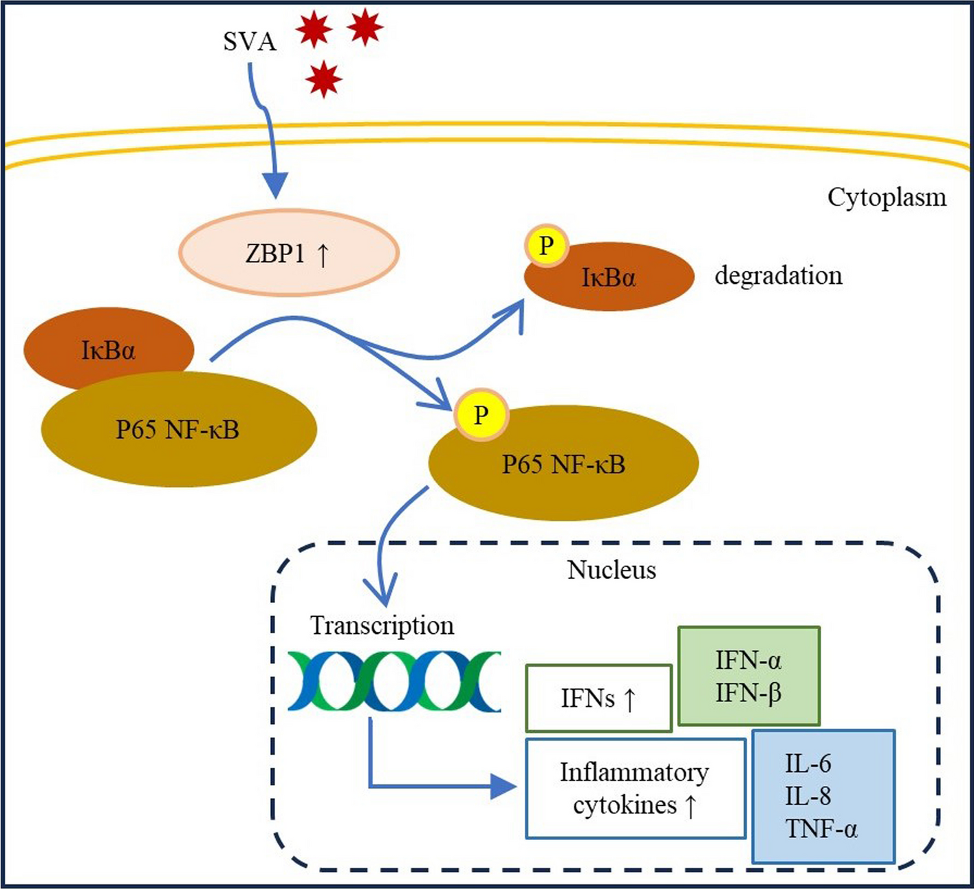
ZBP1 activates NF-κB signaling pathway in SVA-infected cellsNF-κB is a key regulator of IFN activation and inflammatory cytokine production [23]. To verify whether inhibitory effect of ZBP1 on SVA replication is approached via NF-κB signaling pathway, infection experiments was carried out in 3D4/21 cells with ZBP1-over and -interference expression. In overexpression group, SVA infection increased the expression of p-NF-κB p65 (P < 0.05) and p-IκBα (P < 0.05), and decreased the expression of IκBα (P < 0.01), compared to pEGFP-N1 group; ZBP1-overexpression increased the expression of p-NF-κB p65 (P < 0.05) and p-IκBα (P < 0.01), and decreased the expression of IκBα (P < 0.01), compared to pEGFP-N1 + SVA, no significant difference in the protein levels of NF-κB at 24 h.p.i (Fig. 4A, B). In interference expression group, SVA infection increased the expression of p-NF-κB p65 (P < 0.01) and p-IκBα (P < 0.05), compared to shNC; impeded ZBP1 expression decreased the expression of p-NF-κB p65 (P < 0.05) and p-IκBα (P < 0.05), and increased the expression of IκBα (P < 0.05), compared to shNC + SVA, no significant difference in the protein levels of NF-κB at 24 h.p.i (Fig. 4C, D). These results indicate that ZBP1 activates NF-κB signaling pathway in SVA-infected cells.
Fig. 4
ZBP1 activates NF-κB signaling pathway in SVA-infected cells. A, B SVA (MOI of 1) infected or unifected 3D4/21 cells after transfected with pEGFP-N1 or pEGFP-N1-ZBP1 for 24 h, the expression levels of NF-κB p65, p-NF-κB p65, IκBα, and p-IκBα protein were detected by western blotting and quantitatively analyzed by Image J. C, D SVA (MOI of 1) infected or unifected 3D4/21 cells after transfected with shNC or shZBP1 for 24 h, the expression levels of NF-κB p65, p-NF-κB p65, IκBα, and p-IκBα protein were detected by western blot and quantitatively analyzed by Image J. All experiments were repeated three times independently. *P < 0.05, **P < 0.01, ns: not significant
ZBP1 activating NF-κB signaling pathway was verified by the specific inhibitorAn NF-κB-specific inhibitor, BAY 11–7082, was used to confirm the activation of ZBP1 in NF-κB signaling pathway in SVA infection. In cytotoxicity assays, 3D4/21 cells were treated with BAY 11–7082 at concentrations of 0.5, 1, 2, and 5 μM for 24 h. The results showed that BAY 11–7082 below 5 μM had no obvious effect of cell viability (Fig. 5A). Then, 3D4/21 cells were transfected with pEGFP-N1-ZBP1 and incubated by inhibitor (5 μM) for 16 h. The qRT-PCR showed that ZBP1 inhibited the mRNA expression of the SVA VP2 gene (P < 0.01), while, when BAY 11–7082 was present, the inhibition effect of ZBP1 on SVA was reduced (Fig. 5B). The results of western blot showed that the VP2 protein levels were increased in BAY 11–7082 group compared with the ZBP1-SVA-infected group, which was consistent with the mRNA expression levels of SVA VP2 gene (Fig. 5C). In ZBP1-SVA-infected group, ZBP1 significantly increased IFN-α, IFN-β, IL-6, IL-8 and TNF-α (P < 0.01) compared to SVA-infected cells, while this facilitation was attenuated when the inhibitor was present, the expression of IFN-α (P < 0.01), IFN-β (P < 0.01), IL-6 (P < 0.05), IL-8 (P < 0.01) and TNF-α (P < 0.01) were decreased compared to the ZBP1-SVA-infected group (Fig. 5D–H). These results show that the inhibitory effect of ZBP1 on SVA replication is dependent on NF-κB.
Fig. 5
ZBP1 activating NF-κB signaling pathway was verified by the specific inhibitor. A The cytotoxic effect of BAY 11–7082 in 3D4/21 cells was determined by CCK-8 assay. B 3D4/21 cells were transfected with pEGFP-N1 or pEGFP-N1-ZBP1, and pre- treated with DMSO or BAY 11–7082 (5 μM) for 16 h, then, infected with SVA at an MOI of 1 for 24 h. The expression of SVA VP2 gene was analyzed by qRT-PCR. C The expression levels of VP2 protein were detected by western blot and quantitatively analyzed by Image J. D–H The expression of IFN-α, IFN-β, IL-6, IL-8, and TNF-α were detected by qRT-PCR. All experiments were repeated three times independently. *P < 0.05, **P < 0.01
留言 (0)