
記住我


Three to four weeks old female BALB/c mice were acquired from the Laboratory Animal Services Centre of The Chinese University of Hong Kong. All mice were maintained in fully accredited facilities in the Animal Unit at the university and fed with shrimp-free diet. Ethical approval for animal experimentation was obtained from the Animal Experimentation Ethics Committee of The Chinese University of Hong Kong (Ref. No. 21–243-NIH). All animal experiments were conducted under licenses granted by the Department of Health, HKSAR Government, China.
Preparation of Shrimp ProteinsRecombinant shrimp TM (rTM) expressed in pET30a (carrying N-terminal His-Tag/thrombin/S-Tag and C-terminal His-Tag) was prepared as previously described [13]. Metapenaeus ensis (greasyback shrimp) acquired from local market was used to prepare raw and boiled extracts. To prepare raw extract, the abdomen muscle isolated from five shrimp were blended in (1:1 wt/vol) phosphate-buffered saline (PBS) and sonicated for 5 min using an ultrasonic probe followed by centrifugation for 10 min at 12,000 rpm at 4 °C. Supernatant was obtained as raw shrimp extract. The boiled extracts were prepared by boiling abdomen muscle of five shrimp for 10 min in boiling water (100 oC). The samples were then sonicated and centrifuged as described above to collect the supernatant as boiled shrimp extract. Protein concentrations of raw and boiled extract, as well as rTM were determined by spectrophotometry on NanoDrop OneC (Thermo Fisher Scientific) at A280.
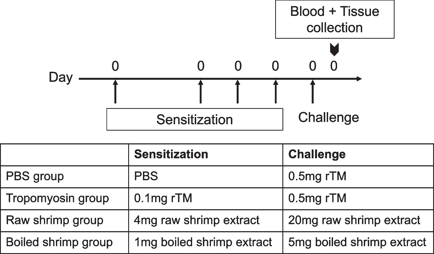
Sensitization and Challenge of MiceBALB/c mice were randomly divided into four groups: negative control, rTM group, raw shrimp group and boiled shrimp group. The experiment protocol for sensitization and challenge of the animals is shown in Fig. 1 as previously described [5]. Mice were intragastrically sensitized with 0.1 mg rTM, 4 mg (total protein content) of raw shrimp extract or 1 mg of boiled shrimp extract, respectively, on days 0, 12, 19 and 26 with 10 µg cholera toxin (CT) per sensitization. The sensitization doses were determined based on the same quantity of tropomyosin in the sensitizing agent as estimated by their relative quantity on the resolved protein gel using ImageLab (Bio-Rad). On day 33, mice sensitized with rTM, raw shrimp extract and boiled shrimp extract were challenged intragastrically with 0.5 mg rTM, 20 mg raw shrimp extract or 5 mg boiled shrimp extract, respectively. Mice in the negative control group were given PBS throughout the experiment. All mice were sacrificed on day 34 post-challenge for blood collection and harvesting of spleen and intestine. The experiment was repeated for five times, with a total of 19–23 animals per group.
Fig. 1
Experimental design of shrimp allergy mouse model. 3–4 weeks old BALB/c mice (n = 16–23 in each group) were sensitized intragastrically with recombinant tropomyosin (rTM), raw shrimp extract and boiled shrimp extract respectively using cholera toxin as adjuvant on days 0, 12, 19 and 26, followed by a 5-fold challenge on day 33. Mice fed with PBS served as negative control. Mice were sacrificed on day 34 that blood sample, small intestine and spleen were harvested for analysis
Assessment of Systemic Anaphylaxis and DiarrheaSystemic allergic responses and condition of feces (i.e. sign of diarrhea) were evaluated 30 to 45 min after oral challenge, according to a scoring system for determining IgE-mediated responses as previously described [5, 14].
Levels of Serum IgE AntibodiesAllergen-specific IgE levels in blood were measured by ELISA. Briefly, 96-well plates (Nunc, Thermo Scientific) were coated with 100 µl rTM, raw or boiled shrimp extract (5 µg/ml) in 0.05 M carbonate buffer overnight at 4 °C. Diluted serum samples (1:10) were incubated overnight at 4 °C after blocking the plates in 5% FBS/PBS. Bound IgE antibodies were detected using Biotin Anti-Mouse IgE (1:1000 dilution, BD Pharmingen), followed by HRP avidin D (1:1000 dilution, Vector Labs). Upon signal development with TMB substrate (BD Biosciences) and reaction termination with 0.1 M sulfuric acid, the optical density (OD) at 450 nm was measured using a microplate reader (BioTek). All assays were performed in duplicate. Positive reaction was defined as OD > 0.34 (maximum value + SD of IgErTM in the negative control group).
ImmunoblottingQuantity of tropomyosin in extracts, IgE-binding protein profiles of the sensitized animals and IgE reactivity of tropomyosin were determined by immunoblot. Briefly, rTM, raw shrimp extract or boiled shrimp extracted were resolved on 10% SDS-PAGE and transferred to PVDF membrane with Trans-Blot Turbo (Bio-Rad). Non-specific binding was blocked with 5% non-fat dry milk, followed by incubation with serum pool comprising sera of eight mice per experimental group or sera of shrimp allergic subjects (Supplementary Table 1) at 1:10 dilution. Samples from mice with rTM-specific IgE > 0.34 OD, or OD of IgEextract > OD of IgErTM on ELISA were selected. Membranes were then incubated with HRP-conjugated anti-mice IgE antibody, followed by SuperSignal West Pico PLUS Chemiluminescent Substrate (Thermo Fisher Scientific). Band signal was acquired with ChemiDoc MP Imaging system (Bio-Rad).
Histological AnalysisIntestinal sample processing and histological staining were performed as per our previously published protocols [6, 15]. The entire intestine was collected, flushed by ice-cold PBS to remove luminal contents, filled with ice-cold 4% paraformaldehyde (PFA) and pre-fixed in PFA for 4 h. Intestine was then divided into three parts equally as duodenum, jejunum and ileum. Intestine segments were cut longitudinally and prepared according to the Swiss-roll method. The segments were fixed in 4% PFA overnight and embedded in paraffin. 4-µm tissue sections were stained with Naphthol AS-D chloroacetate esterase staining kit (Sigma Aldrich) to identify mucosal mast cells. Five randomly selected areas of the three intestinal segments were counted for each tissue sample. Mast cells were quantified per square mm using the software cellSens. Goblet cells were identified by periodic acid-Schiff (PAS) staining (Sigma Aldrich & Leica Microsystems) for detection of mucus-containing cells [16]. The number of goblet cells and epithelial cells were counted in ten randomly selected villi per sample. The percentage of goblet cells were expressed as the number of goblet cells divided by the total number of epithelial cells counted.
Intestinal Cytokine ExpressionIleum sections were collected during tissue collection and stored in ice-cold RNAlater RNA Stabilization Reagent (Qiagen) immediately. Total RNA was extracted using TRIZOL (Invitrogen). The purity and concentration of purified total RNA were determined by measuring the absorbance at 260/280 nm ratio and 260 nm respectively. Total RNA was reversely transcribed using QuantiNova Reverse Transcription Kit (Qiagen) to synthesize cDNA. Quantitative real-time PCR was then performed to determine the expression of Th2-associated genes with specific primers, using the ABI7500 Fast Real-Time PCR system (Applied Biosystem) with QuantiNova SYBR Green PCR Kit (Qiagen). Relative quantification of mRNA expression was calculated by △△ cycle threshold method. The Ct (cycle threshold) value of each gene was normalized to Ct of house-keeping gene HRPT-1.
Shrimp Allergic SubjectsShrimp allergic subjects were recruited at the Prince of Wales Hospital, Hong Kong, with inclusion criteria of documented history of immediate allergic reactions within 2 h of shrimp consumption on at least two occasions over the past 5 years. Serum samples were collected at the time of recruitment during regular clinic visits for immunoblotting. Patients also underwent a double-blind placebo-controlled food challenge (DBPCFC) against black tiger prawn as described [17]. Skin prick test (SPT) was performed on day 1 before DBPCFC over the patients’ volar forearm with raw and heated shrimp extracts prepared in house (as described above), together with histamine (ALK-Abelló, 10 mg/ml) and normal saline as positive and negative controls respectively. Allergen-induced average wheal diameter (mm) was calculated as mean value of the longest and the midpoint orthogonal diameter of the wheal. SPT reaction was considered positive in case of a mean wheal diameter of 3 mm. Demographics of the subjects are shown in Supplementary Table 1. Participants in the study gave written-informed consent. Ethics approval was obtained from Joint Chinese University of Hong Kong—New Territories East Cluster Clinical Research Ethics Committee (no. 2018.484).
Data AnalysisIntensity of IgE binding against tropomyosin on immunoblot was compared by densitometry analysis on Image Lab (Bio-Rad). The data were presented as mean ± SEM. Statistical significance of data was determined by one-way analysis of variance (ANOVA) followed by Kruskal–Wallis test, using Prism (GraphPad). Statistical difference of SPT wheal diameter was determined by paired t-test. The difference was considered as significant at a p value of < 0.05.
留言 (0)