
記住我


Patients with acute respiratory distress syndrome (ARDS) exhibit inflammatory pulmonary edema resulting from changes in endothelial and epithelial permeability, leading to organ damage. The severity of ARDS determines the application of different types of mechanical support. Superimposed hemodynamic impairment may complicate patient management, worsening outcomes. Therefore, a comprehensive evaluation of ARDS patients involves careful respiratory and hemodynamic monitoring, encompassing both invasive and noninvasive technologies, along with clinical and laboratory data. This approach is crucial for tailoring therapeutic strategies to individual patients and minimizing lung injury.
This manuscript reviews strategies for respiratory and hemodynamic monitoring in ARDS patients, highlighting the most recent data and clinical utility in daily management, as synthesized in Fig. 1.
Fig. 1
Respiratory and hemodynamic monitoring in patients affected by ARDS. Recent evidence about respiratory and hemodynamic monitoring in mechanically ventilated patients is available. VeCORR corrected minute ventilation, EtCO2 end-tidal CO2, SpO2 peripheral oxygen saturation, FiO2 fraction of inspired oxygen, PaO2 arterial oxygen partial pressure, mL milliliters, kg kilograms, IBW ideal body weight, TVc tidal volume challenge, PEEP positive end expiratory pressure, ΔScvO2 central venous oxygen saturation increase
Respiratory monitoringCareful respiratory monitoring is essential in patients affected by ARDS. This approach allows the application of an adequate intensity of treatment and reduces injuries caused by mechanical ventilation (MV).
Gas exchange efficiencyGas exchange is directly affected by pulmonary alterations induced by ARDS. In this section, we review the renewed role of pulse oximetry and useful surrogate indices of dead space.
Pulse oximetryPulse-oximetry exploits the principle of spectrophotometry to quantify the amount of oxygenated hemoglobin in blood, allowing continuous noninvasive monitoring of arterial saturation [1]. The difference between arterial oxygen saturation (SaO2) measured via blood gas analysis and oxygen saturation measured via pulse-oximetry (SpO2) is normally less than 3% [2]. However, the accuracy of SpO2 may be lower among patients with darker skin pigmentation, thus overestimating arterial oxygen saturation. This phenomenon, as recently demonstrated by Henry et al., possibly increases the incidence of occult hypoxemia, i.e., patients in which SaO2 is lower than 88% with an SpO2 higher than 92% [3]. The clinical consequences of occult hypoxemia have also been investigated during the recent pandemic. In COVID-19 patients, occult hypoxemia is more frequent in Asian, Black and non-Black Hispanic patients than in White patients, with lower treatment eligibility for these three ethnicities [4].
The ratio of pulse-oximetric oxygen saturation to the fraction of inspired oxygen (SpO2/FiO2) is an acceptable surrogate of the ratio of the partial pressure of arterial oxygen to FiO2 (PaO2/FiO2). Its use has been described both in invasively and noninvasively ventilated patients [5,6,7,8,9]. The SpO2/FiO2 ratio is a good outcome predictor both in patients with coronavirus disease (COVID-19) and non-COVID-19 ARDS patients [10, 11]. In patients with COVID-19-associated pneumonia requiring oxygen therapy, the SpO2/FiO2 ratio at admission showed an area under the curve (AUC) of 85% for the prediction of ARDS occurrence [12]. Kim et al. showed that the SpO2/FiO2 ratio can predict high-flow nasal cannula (HFNC) failure [13]. Moreover, SpO2/FiO2 shows a good correlation with PaO2/FiO2 in invasively ventilated COVID-19 ARDS patients, and when computed on day 2 and day 3, it is associated with outcome [11]. These data confirm the reliability of pulse oximetry for evaluating gas exchange in ARDS patients and for following this trend, as pulse oximetry is continuously measurable. It is easy to measure and is thus especially valid in contexts in which a blood gas analyzer is not promptly available.
The optimal SpO2 concentration for ARDS treatment is still a matter of debate, ranging from 88% to 96–100% to balance the risk of hyperoxia and hypoxia. In a recent large randomized controlled trial (RCT), Semler et al. showed that, in mechanically ventilated patients, the use of a lower (90%, range from 88 to 92%), intermediate (94%, 92–96%) or higher (98%, 96–100%) SpO2 target does not affect either ventilator-free days or hospital outcomes [14].
Dead spacePhysiological dead space is the inspired volume of air that does not participate in gas exchange. It includes anatomic and alveolar dead space [15]. In mechanically ventilated patients, the anatomic dead space remains relatively constant, while the alveolar dead space can significantly increase according to alterations in the ventilation/perfusion (V/Q) ratio [16, 17].
In a seminal study, Nuckton et al. demonstrated that physiological dead space is significantly higher in non-ARDS survivors than in survivors [18]. Like in ARDS patients, COVID-19 pneumonia is characterized by an increase in minute ventilation and an increase in the dead space fraction [19, 20]. Additionally, in COVID-19 ARDS patients, there is a significant association between the amount of dead space computed in the first 7 days and mortality [21]. According to a secondary analysis of the PRoVENT COVID-19 study, the dead space fraction is significantly greater in nonsurvivors and increases more during the first four days than in survivors, suggesting that dynamic changes during the initial week in the intensive care unit (ICU) are crucial for evaluating outcomes [22]. These recent data underline the strong prognostic role of dead space and strengthen the rationale for its use in ARDS patients.
Corrected minute ventilationThe corrected minute ventilation (VEcorr) is a simple and easy-to-calculate surrogate of the dead space fraction that does not require the expired carbon dioxide (CO2) measurement. VEcorr is calculated as the ventilation required to achieve a PaCO2 value of 40 mmHg. In mechanically ventilated COVID-19 ARDS patients, Fusina et al. found a strong correlation between VEcorr and the dead space fraction, with a higher VEcorr in nonsurvivors, which was independently associated with mortality [23].
Ventilatory ratioIn recent years, in addition to dead space fraction computation, the ventilatory ratio (VR) has been proposed as an additional, easy-to-calculate estimation of ventilation efficiency [24]. VR is computed as the product of minute ventilation and arterial carbon dioxide weighed on the patient’s predicted body weight [24]. It is a unitless ratio, being approximately one in healthy subjects. In ARDS patients, Sinha et al. reported a positive relationship between VR and alveolar dead space. Furthermore, VR is more common in nonsurvivors than in survivors [24] and is associated with increased odds of hospital mortality (OR 2.07, confidence interval [CI] 1.53–2.83). As recently shown by Siegel et al., the ventilatory ratio, in association with the APACHE III score at admission, has an area under the curve (AUC) of 0.81 (95% CI 0.68–0.92) in predicting hospital mortality and is significantly better than the APACHE III score alone [25]. Changes in VR within the first 4 h after prone positioning in ARDS patients predict weaning from mechanical ventilation, with an AUC of 0.64 (95% CI 0.53–0.75) [26].
VR reliability can be affected by venous admixture (Qva/Q) and the amount of patient CO2 produced (VCO2). Indeed, these two factors may increase the difference between alveolar and arterial PCO2, with the latter being used for VR calculations. Maj et al. showed that the predictive value of the VR decreases in most severe patients, who are affected by greater Qva/Q impairment [27]. To investigate the effect of VCO2, Monteiro et al. performed a post hoc analysis of the PETAL-ROSE trial [28]. The authors showed that the presence of neuromuscular blockade, a factor influencing skeletal muscle CO2 production, did not significantly affect the relationship between VR and mortality [29].
In mechanically ventilated patients, dead space should be continuously assessed as an additional measurement of gas exchange impairment, together with the PaO2/FiO2 or SpO2/FiO2 ratio. The use of surrogates, which are easier to calculate, seems to be reliable and should encourage the use of such measures to predict patient outcomes. Caution must be used in moderate and severe ARDS patients with major Qva/Q impairment when assessing VR. In these situations, dead space might be overestimated.
ETCO2 to arterial PCO2A further parameter to estimate gas exchange efficiency is the computation of the end-tidal-to-arterial PCO2 ratio (PETCO2/PaCO2), which measures the influence of venous admixture and alveolar dead space on lung performance. Ideally, this ratio should be equal to one. Bonifazi et al. showed that the PETCO2/PaCO2 ratio significantly decreases from mild to severe ARDS [30]. Additionally, PETCO2/PaCO2 is strongly correlated with the amount of nonaerated tissue measured via computed tomography (CT) and respiratory compliance [30]. A subsequent study revealed a relationship between the PETCO2/PaCO2 ratio, alveolar ventilation and hospital mortality [31]. For every 0.01 increase in the PETCO2/PaCO2 ratio, the risk for mortality decreases by 1%.
Currently, weaning from venous extracorporeal membrane oxygenation (VV-ECMO) lacks well-defined criteria and is often based on acceptable blood gas analysis and the absence of excessive inspiratory effort. In a recent multicenter study, Lazzari et al. showed that the PETCO2/PaCO2 ratio, with a cutoff of 0.83, is able to predict weaning [32].
Ventilation and patient self-induced lung injuryMechanical ventilation and spontaneous inspiratory effort may be harmful. The mechanical power, its normalization (i.e., the mechanical power ratio) and the measurement of the esophageal pressure are crucial to minimize these sources of lung injury in patients affected by ARDS. Indices of recruitment are helpful for adequately establishing mechanical ventilation.
The mechanical powerMechanical power refers to the energy dissipated in the respiratory system while moving a specific volume at a given PEEP. It is typically expressed in Joules per minute (J/min) [33]. This energy dissipation within the respiratory system plays a crucial role in modulating and potentially promoting ventilator-induced lung injury (VILI). The mechanical power is a unifying indicator computed considering the major ventilatory variables generated from the interaction between the patient and ventilator. It can be assessed under passive conditions and categorized based on ventilation modality (pressure or volume-controlled ventilation) using algebraic equations [34]. The newest intensive care mechanical ventilators now offer the possibility to directly measure mechanical power, with acceptable accuracy compared to traditional algebraic methods [35].
Recent studies have demonstrated that mechanical power at admission is associated with hospital mortality across a heterogeneous range of patients [36,37,38]. Urner et al. further explored the relationship between the intensity of mechanical power throughout the intensive care stay and mortality, revealing an increased risk of death with each additional day of exposure to mechanical power equal to or greater than 17 J/min [39]. Pozzi et al. analyzed the clinical course of ventilatory variables in ARDS patients during the initial three days of MV and identified the mechanical power ratio at admission as the only variable associated with intensive care mortality [40]. By day 3, the mechanical power ratio, alveolar dead space, and PaO2/FiO2 were associated with the outcome. Therefore, in ARDS patients, assessing ventilatory variables during the initial days of mechanical ventilation seems to be crucial for predicting outcomes.
Concerning the different components of mechanical power, Costa et al. showed a stronger association with mortality for the dynamic component (i.e., the respiratory rate and the driving pressure) than for the total mechanical power [41]. However, the impact of similar values of mechanical power on lung injury can vary significantly based on factors such as ventilated lung size, respiratory system compliance, or the amount of aerated tissue at a given PEEP. Coppola et al. demonstrated that normalizing the mechanical power at admission to the compliance of the respiratory system and the amount of ventilated tissue, as computed by lung CT, provides a better predictive measure for outcomes in ARDS patients [36].
Esophageal pressure and diaphragmatic ultrasoundPreserving spontaneous breathing over invasive ventilation offers advantages [42, 43]. However, elevated inspiratory efforts are associated with high negative esophageal pressure (Pes) swing and positive transpulmonary pressure, which may lead to patient self-inflicted lung injury (PSILI). PSILI is associated with organ dysfunction and increased mortality [44,45,46]. Additionally, excessive inspiratory effort cannot be detected simply by monitoring airway pressure [47].
Computing the changes in esophageal pressure during inspiration (ΔPes) as the difference between the esophageal pressure at the beginning of inspiration and its lowest value is the easiest way to measure inspiratory effort. In the presence of acute respiratory failure, several noninvasive respiratory support methods, such as HFNC therapy, continuous positive airway pressure (CPAP), and noninvasive ventilation (NIV), should improve gas exchange and decrease inspiratory effort. Menga et al., in a crossover study comparing noninvasive support, showed that only NIV delivered by a helmet is able to reduce delta pes [48]. In a large group of COVID-19 ARDS patients receiving helmet CPAP, total stress, defined as the sum of the transpulmonary pressure generated by the patient and the end expiratory airway pressure, is independently associated with a negative outcome [49].
Transpulmonary pressure measurements allow clinicians to evaluate lung recruitment efficacy. With this aim, it can be useful to evaluate the effects of awake prone positioning, as performed in COVID-19 ARDS patients. Prone positioning leads to a reduction in ventral alveolar hyperinflation and dorsal atelectasis, thus promoting homogenization of transpulmonary pressure and improvement in oxygenation. Additionally, as demonstrated in a cohort of COVID-19 ARDS patients assisted with helmet CPAP, prone positioning significantly reduces the amount of work involved in breathing [50]. The role of esophageal pressure manometry in evaluating inspiratory effort and preventing PSILI is increasingly recognized, and this technique is always recommended for ARDS patients.
Another possible way to evaluate inspiratory effort is the use of ultrasound. However, Steinberg et al. show poor correlation between diaphragmatic thickening fraction (DTI), diaphragmatic excursions and esophageal swing in a cohort of 46 mechanically ventilated patients affected by Covid-19 ARDS [51]. Similarly findings are available from Poulard et al. [52]. Delta Pes monitoring remains therefore essential to evaluate PSILI in patients undergoing assisted mechanical ventilation.
Nevertheless, diaphragmatic ultrasound remains a valid tool to predict weaning from MV, and recent studies strengthen this evidence. Mawla et al. find a possible cutoff of 13.5% for DTI as accurate to predict weaning from MV [53]. Another original investigation shows how the association of different diaphragmatic ultrasound indexes has an area under the curve of 0.77 in predicting extubation success [54].
Recruitment: the recruitment/inflation ratio and the EIT-based PEEP titrationChen et al. proposed the recruitment-to-inflation ratio (R/I ratio) as a noninvasive method to compute the potential for lung recruitment at different PEEP levels [55]. Subsequently, the R/I ratio has been clinically validated to be accurate in detecting lung recruitment in ARDS patients in the supine position [56, 57]. In a secondary analysis of a previous study [58], the R/I ratio at two levels of PEEP, both in the supine and prone positions, correlated with lung recruitment computed by CT scan [59]. In addition, the overall data confirm high variability in lung recruitability among ARDS patients, with different effects on gas exchange, respiratory mechanics and hemodynamics. Zerbib et al. reported that an R/I ratio > 0.62 predicts lung recruitability with an AUC of 0.80 in COVID-19 ARDS patients [60]. Patients with high recruitability show an improvement in both oxygenation and respiratory system compliance, while in patients with low recruitability, an increase in oxygenation is associated with a decrease in cardiac output. These data confirm that the R/I ratio is a valuable aid for physicians to select an adequate level of PEEP, to improve respiratory mechanics and oxygenation, and to monitor hemodynamics and cardiac output.
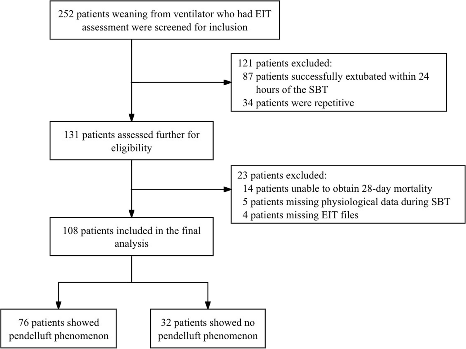
New interesting evidences are available about electrical impedance tomography (EIT) as an effective tool to titrate PEEP in patients affected by ARDS. In an original article on 108 Covid-19 ARDS patients, PEEP titration was performed during EIT monitoring, via decrementing PEEP trials [61]. The authors identify the best PEEP as the one corresponding to the crossing point of the collapse–overdistension curves. They also determine the PEEP with the best regional distribution of ventilation. Interestingly, EIT-based PEEP found at the collapse-overdistension crossing point well correlates to the PEEP with the highest compliance, while PEEP with the best EIT ventilation distribution is higher than the previous ones [61]. Jimenez et al. show that EIT-based PEEP setting allows to decrease mechanical power in ARDS patients, thus being potentially able to reduce VILI in this population [62]. Robust data on clinical outcomes of PEEP titration techniques are still lacking in the literature. A multicenter randomized controlled trial is actually going on to find out differences on clinical outcomes in ARDS patients whose PEEP is titrated using either EIT-based techniques or PEEP/FiO2 tables [63].
Cardiac monitoringIn ARDS patients, hemodynamic instability and low cardiac output may further decrease oxygen delivery and promote tissue hypoxia [64]. Strategies aimed at increasing cardiac output often involve fluid administration and vasoactive agents [65]. Therefore, hemodynamic monitoring is crucial in ARDS patients to optimize fluid administration and cardiac output [66].
Dynamic indexes of fluid responsivenessAs also recently highlighted by the Surviving Sepsis Campaign, the intravenous fluids of choice for critically ill patients are balanced crystalloids [67]. The risks of net fluid accumulation in critically ill patients have also recently been advocated [
留言 (0)