
記住我

A boy, 19 months old, was admitted to the hospital with the chief complaint of the right hip joint flexion for a year.
History of present illnessA year before admission, the boy developed right hip joint flexion which could not be straightened without obvious cause. The parents did not pay attention to it at the very beginning of the abnormality and assumed that it was caused by the right plantar hemangioma, and did not have systematic examination and treatment conducted, resulting in progressive exacerbation. Ten days prior to admission, it was found that the right hip joint flexion was further aggravated than before. The lower limb of the affected side was atrophied compared with the opposite, so he was sent to a hospital. The family visited our hospital for the sake of systematic diagnosis and treatment. The outpatient clinic admitted this case to our department on the diagnosis of abdominal mass.
History of past illnessThe boy was born at the gestational age of 28 weeks and was admitted to our hospital with the diagnose of a preterm with very low birth weight. Systematic treatment was for 46 days and was discharged after getting better.
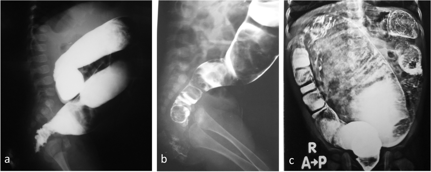
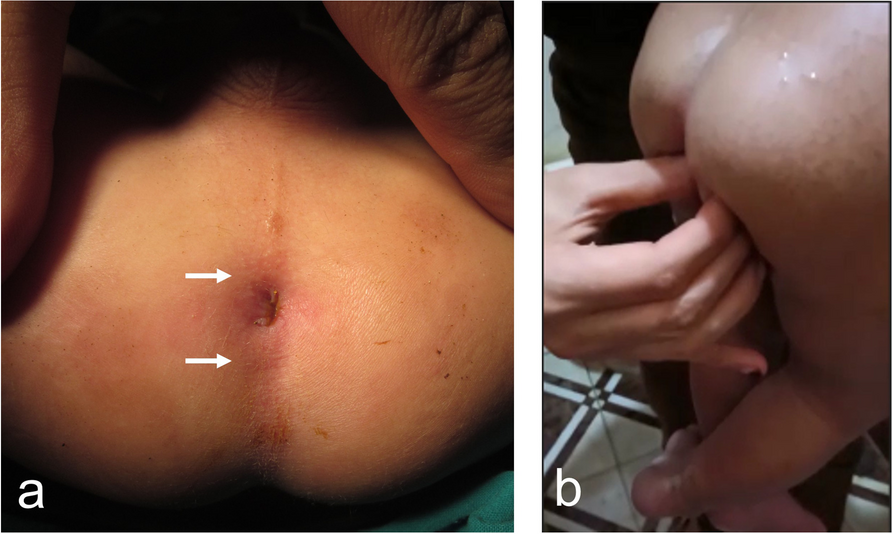
Physical examinationWhen the boy was admitted, he had lucid mind, clear speech, and no rash nor pigmentation; his pupils were equal and round, he had normal light reflex, no congestion in the pharynx, no abnormality in cardiopulmonary examination, and soft abdomen; his liver and spleen under the ribs untouched, no peristaltic wave or bowel pattern were seen, and he had normal bowel sounds. The right hip joint was flexed, could not be straightened, slight internal rotation, the leg circumference of the affected limb was smaller than the opposite side, and the muscle tone of both lower limbs was fair. About 2 × 2 cm of hemangioma can be seen on the right foot sole, the color faded on pressing, with no obvious pain and itching. There were no abnormalities in neurologic examination. Auxiliary examination: CT of the lower abdomen showed a right iliopsoas muscle mass, and the size was about 79 × 62 × 40 mm (A); full length bone X-ray indicated that the right femur, tibia, and fibula were thinner than the opposite, and the right lower limb was bent and rotated (B) (Fig. 1). Before the operation, blood routine examination, biochemical, and coagulation function were tested. Contraindications for operation, such as failure of systems and organs, hematological disorders, and coagulation disorders, were checked; no abnormalities were identified in the results. Preoperative assessment revealed that the abdominal neoplasm was large with speedy progress, the neoplasm boundary was not clear, and there was possibility that the neoplasm was a malignant one. It was also possible that the abundant blood vessels around the neoplasm were present and the risk of intraoperative bleeding might be high; therefore, it was necessary to prepare blood prior to surgery.
Fig. 1
CT examination and X-ray. A Abdominal CT: right iliopsoas muscle mass, size about 79 * 62 * 40 mm. B Long bone X-ray: the right femur, tibia, and fibula are thinner than the opposite side, and the right lower limb is bent and rotated
Laboratory examinationsImmunohistochemistryThe first time examination: CD31 ( +), CD34 ( +), Ki-67 (2% +), ERG (partial +), SMA ( +), WT1 (-), VEGF (-); the second time: D2-40 (-), HHV8 (-), S-100 (-).
Pathological diagnosis: (right iliopsoas muscle) kaposiform hemangioendothelioma.
CD31, CD34, and Fli are positive, indicating that the tumor cells originate from vascular endothelial cells; SMA positivity suggests the presence of pericyte components within the tumor mass; Ki-67 positivity indicates a relatively low proliferative activity of the tumor cells.
Final diagnosisRight iliopsoas kaposiform hemangioendothelioma.
TreatmentThorough preoperative preparation was conducted and specific surgical approaches were planned, and surgical resection was performed. After the operation, the boy was transferred to the intensive care unit. There was no obvious abnormality in the coagulation function. The boy’s condition improved and was successfully discharged from the hospital. Pathological examination of the neoplasm revealed a pile of gray-brown tissue, diameter 8 cm, section gray-yellow soft (C-1). Microscopic examination: (right iliopsoas muscle) vasogenic neoplastic lesions, presence of a large number of thick-walled blood vessels, infiltration of lymphocytes and plasma cell around the vessels; fiber, fat, and muscle tissue around infiltration, and interstitial mass of lymphoid and plasma cell infiltration (Fig. 2). There was formation of lymphoid vesicles, cluster proliferation of capillaries in the center of the lesion, and abundant cells and fibrous separation, showing the morphological characteristics of kaposi-type hemangioendothelioma (C-2).
Fig. 2
Specimen of muscle mass and light microscope image. A Excision of part of the specimen. Gray-brown tissue, about 8 cm long, gray-yellow cut plane, and soft in quality. B Light microscope image. There was infiltration of lymphocytes and plasmacytes, capillaries in the center of the lesion
Outcome and follow-upOne month after operation, there was no obvious abnormality in platelet and coagulation function, and there was no sign of recurrence according to imaging findings. The function of the affected hip is better than before. The family was advised to strengthen the rehabilitation physiotherapy after surgery and the patient was followed up regularly.
留言 (0)