Reagents and chemicals
IPTG was purchased from Gibco (Gand Island, NY, USA). Ni-agarose affinity chromatography columns were purchased from Invitrogen (Cambridge, UK). Bleomycin hydrochloride (BLM) was purchased from Maclean (Shanghai, China). DMEM was supplied by Gibco (Gand Island, NY, USA); Fetal bovine serum was purchased from PAN-Biotech (Heilbronn, Germany); Recombinant human TGF-β1 was purchased from PEPROTECH (Rocky Hill, USA); GAPDH, α-SMA and FN antibodies were purchased from Affinity (Cincinnati, OH, USA); Col-I ELISA kit was purchased from MEIMIAN (Jiangsu, China); Hydroxyproline kit was purchased from Nanjing Jiancheng (Nanjing, China).
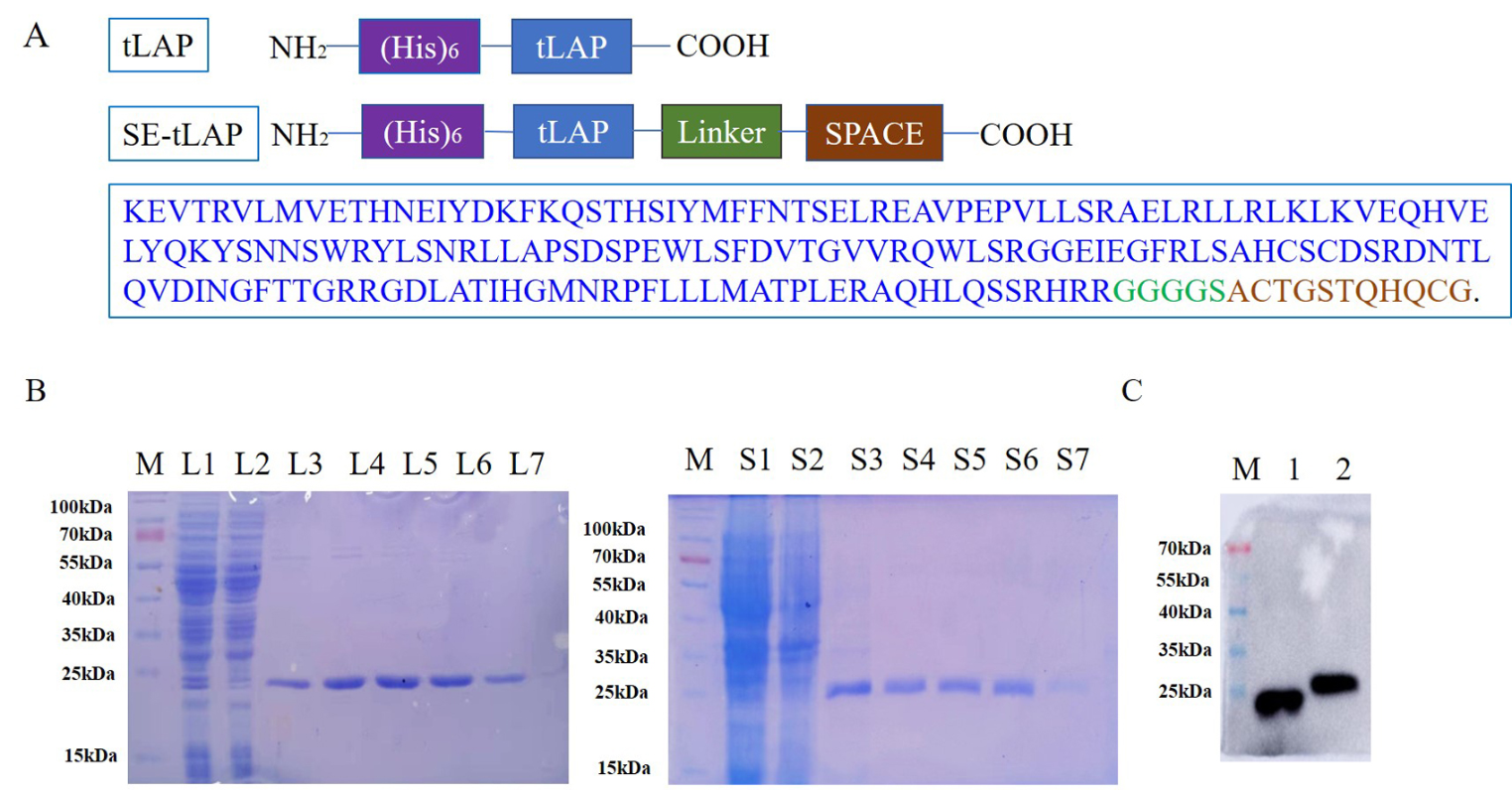
Plasmid construction
The linker (GGGGS) and SPACE peptide (ACTGSTQHQCG) sequences were added to the C-terminal of the tLAP sequence. The SE-tLAP sequences were inserted into the pet28a vector. SE-tLAP/pet28a recombinant plasmid was constructed.
Expression, purification, and identification of SE-tLAP
The SE-tLAP recombinant plasmid was transformed into Rosetta (DE3) competent cells. Peptide expression was induced by IPTG. The bacteria were collected for ultrasonic crushing, and the supernatant was collected after high-speed centrifugation. The recombinant peptides were purified by Ni-agarose affinity columns and detected by SDS-PAGE. Purified SE-tLAP was identified by Western blot. The concentration of the anti-His antibody was 1:1000, and the concentration of the goat anti-mouse IgG antibody was 1:10000.
Cell viability assay
The cell proliferation was investigated by conventional MTT assay. In short, NIH-3T3 cells (1 × 10⁵ cells/well) were inoculated into a 96-well plate. TGF-β1 (10 ng/mL) was added for 12 h. SE-tLAP (30, 60, 90, 120, 150 µg/mL) was added for 24 h. Then, 20µL MTT (5 mg/mL) was added for 4 h. Subsequently, the supernatant was removed and the formazan crystal was dissolved in DMSO (150 µL). The absorbance of 490 nm was detected by an enzyme-labeled instrument.
Wound healing assay
NIH-3T3 cells (15 × 104 cells/well) were inoculated into a 6-well plate and scratched. The cells were treated with TGF-β1 (10 ng/mL) and recombinant peptides. The results were observed by a microscope.
Transwell migration assay
NIH-3T3 cell migration was determined using the Transwell Boyden chamber. The cells were cultured into a 24-well plate with serum-containing medium for 12 h, then treated with TGF-β1 (10 ng/mL) for 12 h, added the tLAP or SE-tLAP for 24 h. The non-migrating cells on the surface of Transwell membrane were washed with PBS, and the migrating cells were stained with 5% cresol violet for 15 min. The results were observed by a microscope.
Real-time PCR
NIH-3T3 cells were inoculated and treated by TGF-β1(10 ng/mL) and recombinant peptides. Total RNA was extracted and reverse-transcribed. The gene expression was detected by the StepOne RT PCR system.
Cell immunofluorescence
The cells were fixed with 4% paraformaldehyde for 20 min. Then the cells were infiltrated with 0.1% Triton X-100 and sealed with 5% BSA for 1 h. The cells were incubated overnight with anti-α-SMA (1:250) and anti-Col-I (1:250) at 4 °C, and then a fluorescent secondary antibody was applied for 1.5 h. Cells were re-stained with DAPI for 15 min. The results were observed using laser confocal microscopy.
Western blot
NIH-3T3 cells were treated with TGF-β1 and polypeptides. The proteins were extracted with a lysate buffer and loaded by SDS-PAGE, and then transferred to PVDF membranes. The membrane was sealed in 5% skim milk for 2 h, rinsed, and incubated with the corresponding antibody at 4℃, followed by the secondary antibody at 37℃ for 1 h. ECL was used for color development.
Enzyme-linked immunosorbent assay (ELISA)
NIH-3T3 cells were treated with TGF-β1 and polypeptides. The cells were digested with trypsin and supernatant was collected. After repeated freeze-thaw, the supernatant was centrifuged. The collagen I content was determined by the ELISA kit.
Preparation of the hyrogels
0.6 g F127 was added into 3mL polypeptide solution and incubated at 4℃ for 12 h until completely dissolved. F127, F127-tLAP, and F127-SE-tLAP hydrogels with a concentration of 20% were prepared and refrigerated at 4℃.
Detection of hydrogel degradation and drug release
1mL PBS was added into 0.5 g F127-tLAP and F127-SE-tLAP hydrogel and incubated at 37℃. The hydrogel weight and polypeptide content in PBS were measured every 4 h.
Hydrogel biocompatibility test
Biocompatibility of the hydrogel was assessed by double staining with Calence AM and PI. NIH-3T3 cells were inoculated in a 6-well plate. 100 µL hydrogel was added to each well at 37 ℃ and 5% CO2 for 24 h. The cells were stained with Calence AM and PI for 15 min. The results were observed by fluorescence microscope.
Animal experiments
The animal experiments were approved by the Institutional Animal Care and Use Committee of Mudanjiang Medical University. ICR mice weighing 23 ± 2 g, were allowed to adapt at a constant temperature (23 ± 2℃) for 7 days before use. The mice were randomly divided into the control group, BLM group, F127 group, F127-tLAP group, and F127-SE-tLAP group. The skin fibrosis model was established by subcutaneous injection of 100µL bleomycin (1 mg/mL). The injection was given every 2 days for 4 weeks. After 2 weeks of injection, 100 µL hydrogel containing 30 µg peptide was applied to the mice once every 2 days for 2 weeks.
Transdermal test of hydrogel
ICR mice were randomly divided into two groups. The hydrogel was prepared after labeling tLAP and SE-tLAP with FITC. 100 µL fluorescent-labeled hydrogel containing 30 ug polypeptide was applied to the back of mice. The skin tissue was embedded in OCT and sliced on a cryostat. Fluorescence was detected using confocal microscopy (Aoki et al. 2019).
Pathological staining and immunohistochemistry
The skin tissues were fixed with 4% formaldehyde, dehydrated, and embedded. Routinely prepared in paraffin sections with a thickness of 5 μm. Paraffin sections were stained and observed by an optical microscope. The paraffin sections for immunohistochemistry were 4 μm thick. Sections were incubated with α-SMA and Col-I antibody at 4℃, followed by the secondary antibody at 37℃ for 1 h. After staining with DAB, the sections were counterstained with hematoxylin. The sections were sealed in a neutral resin and then investigated by an optical microscope.
Detection of hydroxyproline
The skin tissue was weighed and chopped, and PH of its hydrolysate was adjusted to 6.0-6.8. The sample was centrifugally filtered and adjusted concentration with PBS. The absorbance value was detected at 550 nm using a hydroxyproline content detection kit.
Statistical analysis
Experimental data were analyzed by GraphPad Prism 8.3. The results were expressed as mean ± standard deviation (x ± s). Groups were compared by one-way analysis of variance. P < 0.05 represented statistically significant.


留言 (0)