
記住我


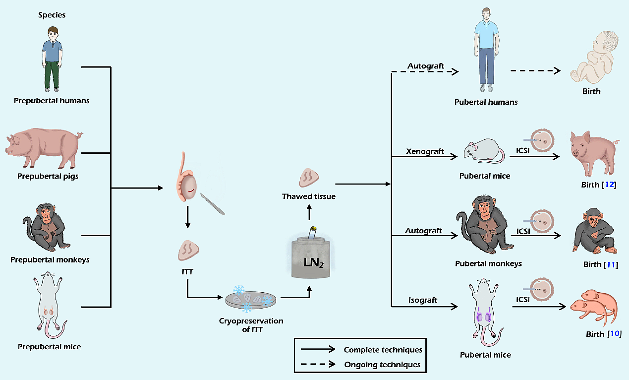
Autologous ITT transplantation is one of the mature techniques to produce sperm and even give birth to healthy offspring. This technique has produced successful spermatogenesis in rodents and also in higher primates [11, 45,46,47]. A summary of autologous testicular tissue grafting in NHPs is provided in Table 2.
Table 2 Summary of autologous testicular tissue grafting in NHPsAs mentioned in Table 2, larger ITT fragments might lead to better tissue survival after transplantation. Nevertheless, a definitive cutoff value to determine the distinction between large and small grafts remains elusive. While smaller grafts, typically with a size smaller than 1mm3, may have better blood supply and a greater propensity to develop new blood vessels, facilitating their integration and survival at the recipient site, the outcomes of autografting were found to be unsatisfactory. There might be a certain risk of graft resorption when using smaller ITT fragments (1 mm3) for autografting [45,46,47]. In the study by Jahnukainen et al. [47], up to 95% of total cryopreserved grafts were lost due to graft resorption. Other studies also showed a poor survival rate of small-size grafts [45, 46]. On the other hand, Fayomi et al. [11]. employed fresh and frozen-thawed ITT fragments of larger sizes (ranging from 9 to 20 mm3) in castrated hosts, resulting in a 100% graft survival rate. The authors hypothesized that the improved outcomes of larger ITT fragments could be attributed to the graft’s ability to generate autocrine/paracrine factors, thereby overcoming ischemic injury. To our best knowledge, in studies related to autologous testicular tissue grafting in NHPs, only this study employed large fragments. Further studies utilizing larger ITT fragments may be necessary to provide additional evidence supporting their superiority over smaller ITT fragments.
Furthermore, the transplantation site is also the key factor that contributes to the success of graft survival. Autotransplantation of testicular tissue in higher primates has been performed at several sites, such as the back skin [11, 45, 46], the scrotal skin [11, 46, 47], the shoulder and the arm [47]. Spermatogenesis was blocked at the spermatocyte stage when grafting occurred in the ectopic site. In contrast, complete spermatogenesis was observed in the scrotal area. One benefit of transplanting to the scrotal site is its more stable temperature compared to ectopic sites. The scrotum’s temperature is closer to the body’s core temperature than peripheral areas like the skin or back. Additionally, in certain species, the scrotum contains muscles that, through the cremasteric reflex, help maintain a cooler temperature within the scrotum. This cooler environment is crucial for protecting the integrity of sperm-producing cells [48]. However, in a study by Fayomi et al. [11] in 2019, all grafts in the back and scrotal skin exhibited complete spermatogenesis. The method employed in this study involved suturing each testicular tissue fragment to the subcutaneous layer of the skin, rather than depositing as a slurry of small pieces in the subcutaneous space. The injury caused by the suture needles or skin flap incision appeared to be adequate in stimulating angiogenic granulation and vascularization of the apposed testicular tissue grafts, ultimately resulting in 100% graft survival and complete spermatogenesis. In conclusion, the scrotal site could become the favored option of graft location. Considering transplantation to an ectopic area may be viable, but it would require a more well-planned and delicate approach.
Xenologous transplantationXenotransplantation of ITT is another technique in which scientists transplant ITT from one species to another. It may have certain disadvantages such as graft rejection, increased risk of zoonotic infections, and potential impairment in producing functional sperm or supporting regular testicular function in the long run. It is important to note that while xenotransplantation of ITT has several disadvantages, it allows for studying testicular development, maturation, and spermatogenesis across species. This technique can deepen our understanding of reproductive biology and contribute to advancements in fertility treatments. For instance, lots of mammalian species (e.g., mouse, rabbit, pig, goat, kitten, horse, cattle, sheep, dog, buffalo, NHPs, etc.) have been reported to complete spermatogenesis by xenografting techniques [10, 49,50,51,52,53,54,55,56]. A summary of xenologous testicular tissue grafting in mammalian species is provided in Table 3.
Table 3 Summary of xenologous testicular tissue grafting in mammalian speciesFrom xenografting experiments in mammalian species, we learned that many parameters may have a strong influence on graft recovery and functionality. As mentioned above, larger fragments might lead to better results. However, most of the xenografting studies in non-human mammal species used graft sizes smaller than 2 mm3. To the best of our knowledge, larger graft sizes have been infrequently employed in xenotransplantation studies, making it uncertain whether smaller grafts would exhibit superior survival rates. In fact, the specific survival rates of small versus large grafts can vary depending on factors such as graft type, grafting technique, and individual characteristics. Therefore, it is important to determine an optimal tissue size for every species to ensure successful outcomes.
As mentioned in Table 3, the back skin of immunodeficient mice is commonly used for grafting ITT. This approach has the advantage of being minimally invasive. Alternatively, grafting the tissue into the testicular parenchyma or the scrotum may provide a more natural environment for the development of sperm, but these kinds of approaches may be more invasive and may require a higher level of operational skills. A study by Ntemou et al. [56] in 2019 demonstrated that the intratesticular transplants led to a higher final graft recovery rate and all recovered transplants showed complete spermatogenesis. The authors hypothesized that the testis was a well vascularized organ that could prevent post-transplantation hypoxia and provide sufficient oxygen for transplant survival. The primary advantage of intratesticular grafting lies in its optimal temperature regulation and the unique hormonal environment within the testes, which are essential for germ cell maturation. This method eliminates the necessity for gonadotrophin stimulation. Consequently, xenotransplantation of ITT into the testicular parenchyma of other species emerges as a promising alternative for ITT grafting.
Moreover, donor age has been shown to influence the efficiency of testis xenografting in several studies. The study conducted by Abrishami et al. [54] in 2010 demonstrated notable outcomes in terms of graft recovery rate, graft weight, seminiferous tubule count, and successful spermatid generation when employing testicular xenografts from prepubertal and pubertal donors. These findings underscore the potential of utilizing immature donors instead of mature donors for testicular tissue xenografting. The authors assumed that the more advanced spermatogenesis occurred in the donor testis tissue, the worse the recovery rate in the graft. Therefore, the higher spermatogenic activity in adult testis tissue would lead to graft degeneration. In addition, adult testis tissue might be more sensitive to ischemic injury before re-vascularization, but the vascularization efficiency of testis tissue xenografting between immature and adult remains disputable.
The impact of supplementation with hormones and other factorsSeveral studies have investigated the potential of supplementation with hormones or other factors to enhance graft outcomes, yielding varied and controversial results. For instance, in mice with infant rhesus monkeys ITT xenografts, the administration of exogenous human chorionic gonadotropin (hCG) improved the growth of xenografted ITT, resulting in complete spermatogenesis [57, 58]. However, supplementing human slow-frozen ITT xenografts with N-acetylcysteine (NAC) or testosterone did not demonstrate a clear impact on germ cell survival or inhibition of apoptosis in the grafted ITT [59]. Additionally, in prepubertal human testis grafts, the exogenous administration of a combination regimen of gonadotrophins, hCG, and follicle-stimulating hormone (FSH) failed to improve germ cell survival and differentiation [60]. Given the uncertain results from administering hormones and other factors post-transplantation, it is logical to consider the site of transplantation and the transplantation technique as more critical factors in improving graft outcomes.
Monitoring viable cell fate in ITT transplantationIn a series of previous studies by Chen [13, 16, 61,62,63,64], bioluminescence imaging (BLI) was employed to track germ cells, ovarian tissues, and ITT across more than a decade. BLI allows for the tracking of tissue-specific luciferase expression in transgenic mice, facilitating the real-time observation of various biological processes, including signaling events and protein-protein interactions within transplanted tissues in vivo [65]. Moreover, BLI empowers researchers to conduct longitudinal studies, enabling the continuous monitoring of germ cell fate over an extended duration. This capability proves invaluable in the assessment of the long-term impacts of ITT and the potential restoration of fertility in pre-pubertal males as they progress towards sexual maturity. Consequently, BLI was introduced as a valuable tool for the evaluation of germ cells and ITT in pre-pubertal male mice.
Biomaterial scaffolds applied for transplantationAs mentioned in the previous part of the paper, there are still obstacles to overcome in ITT transplantation strategies. It has been proven that tissue engineering holds great potential to enhance various aspects of ITT transplantation. For instance, tissue engineering can contribute to the development of suitable scaffolds that mimic the natural architecture of testicular tissue. These scaffolds can provide structural support and guide the formation of functional testicular tissue.
Biomaterial scaffolds can be made from various materials, including natural polymers, synthetic polymers, or a combination of both. Natural polymers commonly used in scaffold fabrication include collagen, alginate, fibrin, and hyaluronic acid which are derived from natural sources such as animals or plants. Synthetic polymers such as poly-L-lactic acid (PLLA), polyglycolic acid (PGA), and poly (lactic-co-glycolic acid) (PLGA) are also widely employed due to their controllable properties and degradation rates.
Currently, numerous research groups are focused on enhancing composite materials and fabrication processes to develop superior scaffolds. Prior to transplantation of the scaffold with an implantable tissue construct into an organism, it is essential to thoroughly evaluate several key factors, including biocompatibility, biodegradability, mechanical properties, scaffold architecture, and manufacturing technology [66]. First, any scaffold for tissue engineering must be biocompatible so that it allows tissues to integrate and grow within the scaffold structure. Second, scaffolds cannot last for a long time, hence, they must be biodegradable, and degradation should not produce toxins. Third, the mechanical properties of the scaffold should be identical to the anatomical site into which it is going to be implanted. Fourth, scaffolds need to maintain a high porosity to achieve effective vascularization. The architecture of scaffolds should have an interconnected pore structure which is of vital importance for the discharge of wastes from the scaffold. Finally, the scaffold manufacturing process should be cost effective, and the final product should be available immediately for clinical use.
Chan et al. summarized four scaffolding approaches over the past few decades [67]. In this paper, we aim to review two major scaffolding approaches for tissue engineering, including tissue fragment encapsulation in hydrogel matrix and premade synthetic porous scaffolds. We also discuss the evolution and prospects of ITT transplantation (Fig. 2).
Fig. 2
Schematic overview of two major scaffolding approaches for tissue engineering. The approaches include tissue fragment encapsulation in hydrogel matrix and premade synthetic porous scaffolds. Additionally, the figure highlights potential outcomes, such as the generation of offspring through ITT transplantation
Hydrogels for tissue fragment encapsulationHydrogels are potential biomaterials for many medical applications, such as delivery systems for bioactive peptides and anticancer drugs [68, 69]. Hydrogels exhibit unique properties, notably their high water content, which is particularly beneficial for fragile or sensitive tissues that require a moist environment for optimal viability and function. In a study by Shojarazavi et al. [70] in 2021, they developed an innovative injectable hydrogel for cartilage tissue engineering by incorporating silk fibroin nanofibers into an alginate/cartilage extracellular matrix (ECM)-based formulation The study provided evidence that this specific hydrogel formulation holds promise in effectively repairing articular cartilage defects by successfully mimicking the natural cartilage environment. In addition, hydrogels can be designed with controlled porosity and permeability, which promote cell interactions by bidirectional diffusion of nutrients, oxygen, and waste [71, 72]. Overall, hydrogels provide an attractive platform for tissue fragment encapsulation due to their hydration capacity and permeability. These advantages make hydrogels an ideal candidate for tissue engineering and regenerative medicine.
Hydrogels can be classified into two categories based on their origin: natural or synthetic polymers. Natural polymers include elastin, collagen, gelatin, keratin, hyaluronic acid, chitosan, heparin, alginate, and fibrin. Synthetic polymers include polyethylene glycol (PEG), poly(N-isopropylacrylamide) (PNIPAAm), and polyacrylic acid (PAA). The main distinction between natural and synthetic hydrogels lies in their properties: natural hydrogels often exhibit excellent biocompatibility and bioactivity due to their resemblance to the native tissue environment, while synthetic hydrogels offer customizable properties and controlled degradation rates, making them suitable for specific applications. Taking natural polymers as an example, alginate is a notable anionic mucoadhesive polymer due to its terminal carboxyl groups [73]. Alginate offers several advantages for male fertility preservation. Poels et al. demonstrated its potential in cryopreservation by encapsulating ITT within alginate hydrogel, effectively improving tissue engraftment, and hence spermatogonial survival [74]. Alginate’s ability to mimic the extracellular matrices plays a pivotal role in supporting stemness potential during the cell cryopreservation process and initiating spermatogenesis following transplantation. Fibrin is another biomaterial used in countless surgical and endoscopic procedures such as improving the efficiency of hemostasis and wound healing [75]. Fibrin has been utilized not only as a matrix for cell growth and differentiation, but also as an excellent delivery system to achieve sustained drug release [76].
The selection of an appropriate hydrogel material that aligns with the specific requirements of the target tissue or cell type is crucial. The process of tissue encapsulation within hydrogels typically involves several key steps, including hydrogel selection, hydrogel preparation, tissue seeding, gelation or cross-linking, culture and maturation, and subsequent assessment of the viability, proliferation, and functionality of the encapsulated tissue within the hydrogel [18]. It is worth noting that the encapsulation process may necessitate optimization depending on the particular tissue type, hydrogel material, and intended application. Factors such as cell density, hydrogel concentration, gelation kinetics, and culture conditions must be carefully controlled to achieve the desired outcomes in tissue encapsulation.
The application of premade synthetic porous scaffoldsSynthetic porous scaffolds are made by incorporating porogens in solid materials so that cells within the tissue can attach easily [77]. They can be designed with tunable properties to match the specific requirements of different tissues or applications. The scaffold’s pore size, porosity, and mechanical strength can be adjusted to create an optimal microenvironment for nutrient diffusion within the tissue. In addition, many synthetic porous scaffolds are designed to be biodegradable, meaning they can be gradually broken down and absorbed by the body as new tissue forms. Typically, the composition of porous scaffolds can be divided into three groups, ceramics, synthetic polymers and natural polymers. Synthetic polymers, such as polycaprolactone (PCL), PLLA, PLGA, or PEG, are commonly used due to their tunable properties and ability to support tissue development. Indeed, PLLA has been widely used as a biomaterial scaffold in the field of human reproduction due to its distinguished mechanical properties [61, 78]. A study conducted by Eslahi et al. [78] in 2013 investigated the application of a PLLA nanofiber scaffold on frozen-thawed neonatal mouse SSCs and testis tissues, resulting in a significant increase in the in vitro formation of neonatal spermatogonial cell clusters. It is noteworthy that while the use of PLLA nanofiber scaffolds may induce SSC differentiation rather than preserving clonogenic and proliferation potential during cell culture, it still demonstrates a substantial impact on germ cells in both clinical and tissue engineering applications.
Another study by Lu et al. [61] in 2022, used electrospun PLLA scaffolds in the transplantation of ITT from transgenic donors, to wild-type recipient mice (both 3-weeks-old) to evaluate ITT spermatogenesis compared with those without a PLLA scaffold. The main finding of the study demonstrated that the PLLA scaffold groups consisted of high-density sperm and a relatively high sperm count in comparison with control groups because of the similarity to normal natural testicular tissue ECM. This result reaffirmed that graft survival was enhanced and spermatogenesis in ITT was also improved by using a PLLA scaffold.
Additionally, it is crucial to take into account the size of PLLA scaffolds. Research conducted by Chen et al. highlighted that ITT from 4-week-old mice exhibited the highest potential for spermatogenesis when combined with fine PLLA electrospun scaffolds [16]. This favorable outcome can be attributed to the fact that the fibrous network structure of the fine PLLA electrospun scaffolds closely resembled the native structure of decellularized testicular albuginea, with fiber fineness measuring approximately 150 nm. This similarity not only mimics the natural microenvironment of the testicular tissue but also promotes high permeability, facilitating the timely delivery of blood flow, nutrients, and other essential biomolecules.
Beyond considering the physical properties of the initial PLLA scaffolds, the degradation behavior of PLLA plays a crucial role. As these polymers break down and are metabolized, they create vacant spaces within the tissue. These spaces serve as opportunities for regenerated cells and interstitial tissue to gradually fill, ultimately restoring structural integrity. In summary, degradable PLLA contributes to stabilizing the microenvironment through the process of recrystallization.
In summary, biomaterial scaffolds play a pivotal role in advancing innovative solutions, potentially leading to the development of artificial testis made by 3D printing of ECM. By employing 3D printing to fabricate biomaterial scaffolds using ECM-derived materials or by integrating ECM components into the scaffold, researchers can establish environments that closely mimic the natural testicular ECM. These meticulously engineered scaffolds can then be populated with testicular tissues. This comprehensive approach holds substantial promise for advancing our understanding of male reproductive biology and effectively addressing challenges associated with male fertility.
Adjuvant agent: VEGF, PDGF, NECINHRecently, researchers have continued to explore advanced techniques and strategies to enhance tissue encapsulation within hydrogels, such as incorporating bioactive molecules, and creating spatially patterned cell distributions. Tissue engineering can utilize bioactive factors to create a favorable microenvironment for transplanted ITTs. These factors can promote cell survival, proliferation, and differentiation, leading to improved graft integration and functionality. The survival of the transplants is influenced by the vascular connection between the graft and host. Sufficient blood supply results in cell survival through the availability of oxygen, along with abundant nutrients and factors. Therefore, during transplantation, the addition of adjuvant agents is employed to promote angiogenesis. Among these agents, vascular endothelial growth factor (VEGF), platelet-derived growth factor (PDGF), and necrosis inhibitor (NECINH) are commonly used. By encapsulating these factors within hydrogels, it becomes possible to regulate the release kinetics of the biomaterial factors. This controlled release mechanism enables sustained, localized, or targeted delivery of the factors to specific sites. Additionally, the hydrogel matrix serves to protect the factors from premature degradation, clearance, or rapid diffusion, thereby extending their presence at the desired target site.
VEGF is described as an angiogenic factor that induces endothelial cell proliferation and migration [79, 80]. Various studies have been conducted on the delivery of VEGF to promote angiogenesis, such as bone regeneration in femur defects [81], stimulation of wound healing in diabetic mice [82], and promotion of neovascularization in the infarcted heart [83]. Many studies have also evaluated the beneficial impact of the incorporation of VEGF microspheres into hydrogels, which promote the formation of blood vessels and facilitate the tissue healing process [84,85,86]. Sustained release of VEGF is therefore of critical importance for accelerating vascularization, and reducing the occurrence of poor endothelialization and incomplete vascularization [87]. Poels et al. aimed to improve spermatogonial recovery of ITT autografts in mice by encapsulating VEGF-nanoparticles (VEGF-NPs) in two types of hydrogels [74]. One was made of 1% alginate, and the other was made of fibrin (formed by fibrinogen and thrombin at a ratio of 1:1). The main finding in this study was that the viability of mouse spermatogonial germ cells was significantly improved (2-fold) as alginate hydrogel existed, regardless of VEGF-NP supplementation, compared to other grafted groups. This observation highlighted the superiority of alginate in promoting the viability of mouse spermatogonial germ cells. This could be explained by variations in structure between the two matrices. The 1% alginate used in this study had a pore size of 200 μm. In fact, pores over 100 μm in diameter were recommended for angiogenesis [88]. Furthermore, the antioxidant properties of alginate have been shown to mitigate the production of reactive oxygen species following hypoxia [89], thereby enhancing the survival of spermatogonia after transplantation.
However, newly generated blood vessels became leaky and prone to rupture [80]. Therefore, PDGF was added to the hydrogels in order to optimize angiogenesis. PDGF played an important role in the stabilization of neo-vascularization by modulating the proliferation and recruitment of perivascular cells [90]. In one study, Del Vento et al. observed that the graft vascular surface was significantly increased when combining VEGF-NPs with PDGF-nanoparticles (PDGF-NPs) after 21 days of transplantation. In addition, vascular maturity improved after 5 days of transplantation when PDGF was added simultaneously, which suggested that PDGF-NPs made revascularization in grafts occur more quickly [91].
Alginate hydrogel loaded with VEGF-NPs supported short-term angiogenesis due to short exposure to hypoxia, but this beneficial effect disappeared in the long run. Several studies revealed that localized delivery of a NECINH could be a potential candidate to improve ITT graft outcomes [91, 92]. Del Vento et al. selected NecroX-5™ to scavenge reactive oxygen and nitrogen species generated by mitochondria due to its significant protective effect against hypoxia during ITT transplantation procedures. The results showed that incorporation of testicular tissue into alginate hydrogels loaded with NECINH-nanoparticles (NECINH-NPs) markedly improved tissue integrity and spermatogonial survival in comparison with the alginate-encapsulated group, indicating the advantages of promising prepubertal fertility restoration with ITT transplantation. However, nanoparticles containing VEGF, PDGF and NECINH simultaneously did not show a positive effect on seminiferous tubule integrity. The interactions between these three molecules are open to dispute.
Advances in nanoparticles loaded with adjuvant agents provide a certain benefit to tissue engineering. Nanoparticles possess several advantageous features, including low toxicity, distinct drug properties (such as high drug loading capacity), potential for targeted delivery, tunable physicochemical characteristics, and precise control of behavior. These attributes make nanoparticles promising tools for enhancing the outcome of ITT transplantation [93,94,95,
留言 (0)