
記住我


All of the experimental procedures were approved by the Ethical Committee and the Institutional Animal Care and Use Committee (20,232,078).
Reagents and antibodiesCisplatin (HY-17,394) was purchased from MCE (New Jersey, USA). The TUNEL ApoGreen Detection Kit (40307ES20), the 1st Strand cDNA Synthesis Kit (11141ES60) and Hieff qPCR SYBR Green Master Mix (11201ES08) were purchased from YEASEN Biotechnology (Shanghai, China). The following primary antibodies were used in this study: anti-Bax (380,709), anti-collagen I (501,352), anti-α-SMA (380,653), anti-NOX4 (380,874), anti-TGF-β1(346,599), anti-p-smad2 (R26361), anti-p-smad3 (380,775), anti-smad2 (200,790), anti-smad3 (R25743). anti-beta tubulin (380,628), and anti-GAPDH (380,626), which were purchased from Zen BioScience (Chengdu, China). Rabbit anti-PCNA polyclonal antibody (bs-2007R) was obtained from Bioss (Beijing, China). Masson (G1340) and Sirus red stain (G1472) were obtained from Solarbio (Beijing, China). 8-OHdG (bs-1278R), 4HNE (bs-6313R), and 3NIT (bs-8551R) were purchased from Bioss (Beijing, China). Endogenous peroxidase blocker was obtained from Zhongshan Jinqiao (Beijing, China). Orthofluorescence microscope was purchased from ZEISS (Germany).
AnimalsA total of 42 SPF female ICR mice (weight of 30 ± 2 g and age of 7–8 weeks) were used in this study and were obtained from the Experimental Animal Center of Anhui Medical University. All of the animals were housed in a relatively stable environment with a cycle of lights on at 8 a.m. and off at 8 p.m., as well as maintained room temperature (21–25 ℃) with water and food available ad libitum. For adaptation to the new environment, the mice were handled for a one-week acclimatization period before the experiment.
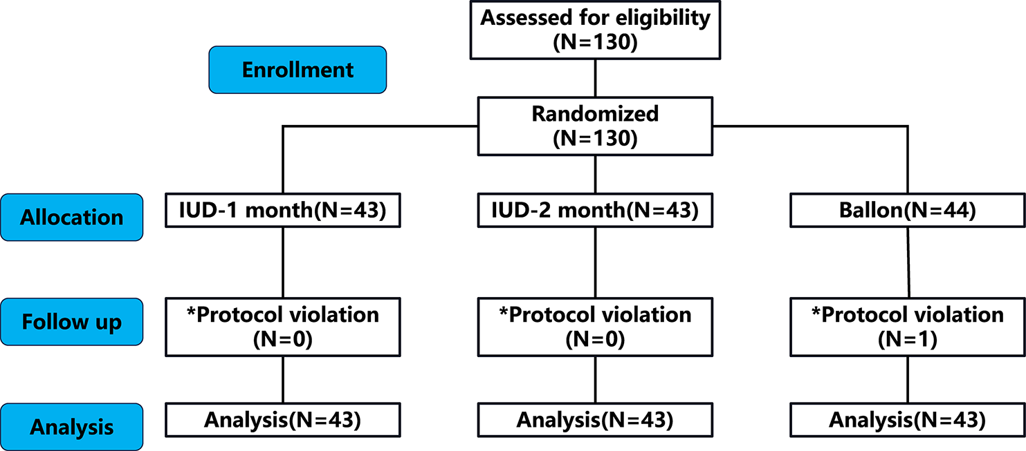
POF model establishmentCisplatin is often used as an inducer for modelling animals with chemotherapy-induced premature ovarian failure, and we also adopted cisplatin to establish the model. Mice were randomly divided into three groups: Cisplatin + LIPUS group (n = 14), Cisplatin group (n = 14) and Control group (n = 14). To better simulate the chemotherapy process, the mice in the Cisplatin group and Cisplatin + LIPUS group were intraperitoneally injected with cisplatin (2 mg.kg− 1) every other day for a total of 10 injections, and the mice in the Control group received an equal volume of saline.
Protocol of LIPUS treatmentThe LIPUS device (Chongqing Haifu Medical Technology Co. LTD, Chongqing, China, RH2022D08) was used in this study. The following parameters were used in this study: the area of the transducer was 5 mm², the frequency of the transducer was 0.5 MHz, the duty cycle was 20%, and the spatial average temporal average intensity (ISATA) was 30 mW/cm2 [17]. Before the LIPUS treatment, the skin of the lower back and the bilateral ovaries were depilated, disinfected, and fully covered with the ultrasonic coupling agent. To better simulate ovarian protection, we performed ultrasound treatment throughout the course of chemotherapy. For the mice in the Cisplatin + LIPUS group, ultrasound treatment was performed on the second day after each injection, and each ovary was irradiated for 20 min until the next day after the last injection. The other two groups were handled in the same manner as the Cisplatin + LIPUS group except for a lack of energy output from LIPUS, the day after the last LIPUS treatment, the mice were sacrificed via cervical dislocation. (Fig. 1).
Fig. 1
Flow chart of the experiment
Haematoxylin and eosin (H&E) staining and follicle countThe day after the last LIPUS treatment, the mice were sacrificed via cervical dislocation, and their bilateral ovaries were completely removed. The ovarian tissue was fixed and embedded in paraffin solution. A microtome was used to make consecutive sections with a thickness of 5 μm, and the slides were dewaxed. Subsequently, the structure of the ovarian tissue was identified by using haematoxylin and eosin staining. Subsequently, the slides were observed under an optical microscope. Follicle stages were classified according to Pederson’s standard. Specifically, an oocyte surrounded by a single layer of flattened or cubical granulosa cells was defined as a primordial or primary follicle; an oocyte surrounded by more than one layer of cuboidal granulosa cells without a visible antrum was defined as a secondary follicle; and a follicle possessing a clear antral space containing follicular fluid was defined as an antral follicle. For the follicle number, we counted every other fifth slice (e.g., 1, 6, 11). To avoid double counting, only nucleated follicles were counted.
TdT-mediated dUTP Nick-End labeling (TUNEL) assayThe ovarian tissue sections were dewaxed, washed once with PBS, and then treated with 20 µg/ml protease K for cell penetration at room temperature for 10 min. The protease K was washed with PBS, and the TUNEL test solution prepared with 50 µl was added, incubated for 60 min at 37 °C away from light, and washed with PBS 3 times, after which the slices were sealed with anti-fluorescence quenching sealing solution and observed under a fluorescence microscope. At least 3 biological experiments were repeated.
Masson and sirus red stainingMasson and Sirus red staining were used to detect fibrosis, and three slices from different ovaries were randomly selected to repeat the experiment. The percentage of positive signal area was analysed via Image J software, and statistical bar charts were drawn. Masson staining: the sections were dewaxed, successively stained with Weigert haematoxylin core, lichen red acid red solution, phosphomolybdic acid differentiation, and aniline blue drop staining, after which the slices were dipped, washed, dried and sealed. Sirus red stain: After dewaxing, the slices were stained successively with weigert hemithylin, differentiated with acidic differentiation liquid, stained with azure scarlet stain, washed and dried and sealed.
Quantitative real-time reverse transcriptase PCR (RT‒qPCR)Inflammatory cytokine (IL-1β, NF-κB, Ccl2,IL-10 and Tnf) mRNA expression levels were assessed by using quantitative real-time reverse transcriptase PCR (RT‒qPCR). After the mice were sacrificed via cervical dislocation, the ovarian tissues were removed, and the TRleasyTM total RNA extraction kit was used in combination with isopropyl alcohol, 75% alcohol and gasform to extract RNA samples. A NANODROP 2000 was used for the quantitative analysis of RNA purity, and Hifairll 1stStrand cDNA Synthesis SuperMix for qPCR (gDNAdigester plus) Kit was used according to the manufacturer’s instructions. Subsequently, the RNA was reverse-transcribed into cDNA, and the obtained cDNA was diluted to an appropriate concentration by using the Hieff qPCR SYBR Green MasterMix (No Rox) kit according to the manufacturer’s instructions. For RT-qPCR, a real-time quantitative Lightcycler480 ll was utilized for light signal detection.
Immunohistochemistry (IHC)IHC for the detection of Bax, Bcl-2, α-SMA, Collagen I, TGF-β1, p-smad2, p-smad3, 4HNE, 8OHdG, and 3NIT was performed by using the peroxidase-labelled streptavidin-biotin method. Specifically, the slides were dewaxed and boiled in citrate buffer solution for 10 min for antigen retrieval. After cooling at room temperature, the sections were incubated with primary polyclonal rabbit antibodies against Bax, Bcl-2, α-SMA, Collagen I, TGF-β1, p-smad2, p-smad3, 4HNE, 8OHdG, and 3NIT. Each antibody was used at a dilution of 1:200, and antibodies were incubated for 2 h at room temperature. Immunostaining was completed by using diaminobenzidine (DAB) as a chromogen, and nuclear counterstaining was performed with Mayer’s haematoxylin. All of the slides were visualized by using a light microscope. At least three slices of unilateral ovarian tissue from different mice in each group were randomly selected to repeat the experiment.
Western blot analysis (WB)After the rats were euthanized in each group, the ovary tissues were rapidly excised and washed in a cold sterile 0.9% NaCl solution. Proteins were extracted and separated via 12% sodium dodecyl sulfate‒polyacrylamide gel electrophoresis, and the product was transferred to a polyvinylidene fluoride membrane, which was blocked with nonfat powdered milk for 1 h and subsequently incubated overnight at 4 °C with primary antibodies. After 4 washes for 10 min each with Tris-buffered saline Tween, secondary antibodies were added to the membrane and incubated for 1 h at room temperature. WB bands were evaluated by using the imaging system, and the grey values of the target bands were analysed by using ImageJ software.
ImmunofluorescenceAfter the slices were dewaxed, high-temperature antigen repair was performed. After natural cooling, endogenous peroxidase blocker was added and incubated for 10 min. Subsequently, the blocker was washed with PBS, and the primary antibody was added and incubated at 4 °C overnight. The next day, the primary antibody was discarded. After washing with PBS, the fluorescent rabbit secondary antibody was added and incubated for 1 h in the dark. After washing, the secondary antibodies were removed, the nuclei were stained with Hoechst, and the stained tissues were photographed under an orthofluorescence microscope. ImageJ software was used for image processing and analysis.
Transcriptome sequencingTotal RNA quality detection, mRNA enrichment, mRNA interruption, double-stranded cDNA synthesis, terminal repair, additions of A and splice, fragment selection, PCR amplification, library quality assessment, and sequencing by Illumina were performed in different biological samples of each group. Quantitative analysis of the gene expression level was performed for each sample, and statistical analysis was conducted after the quantitative analysis was completed to screen the significantly differentially expressed genes in the samples under different states. Mapping of volcano plots and Venn diagrams based on upregulated and downregulated genes were also performed. The differential genes of all of the groups were collected and combined as differential gene sets. Moreover, the FPKM values of genes were analysed via hierarchical clustering, and a heatmap was drawn. GO analysis software GO-seq was used for the enrichment analysis to draw the bar graphs.
Statistical analysisAt least three biological replicates were used to compute the means and SD. To compare significant differences between the groups, one-way analysis of variance using GraphPad Prism 8 (GraphPad Software Inc., San Diego, CA) was performed. To control for type I errors, all results were corrected using the Bonferroni method. P < 0.05 was considered to be statistically significant.
留言 (0)