
記住我

Cardiac magnetic resonance (CMR) provides information on morpho-functional abnormalities and myocardial tissue characterisation. Appropriate indications for CMR in athletes are uncertain.
ObjectiveTo analyse the CMR performed at our Institute to evaluate variables associated with pathologic findings in a large cohort of athletes presenting with different clinical conditions.
MethodsAll the CMR performed at our Institute in athletes aged > 14 years were recruited. CMR indications were investigated. CMR was categorised as “positive” or “negative” based on the presence of morphological and/or functional abnormalities and/or the presence of late gadolinium enhancement (excluding the right ventricular insertion point), fat infiltration, or oedema. Variables associated with “positive” CMR were explored.
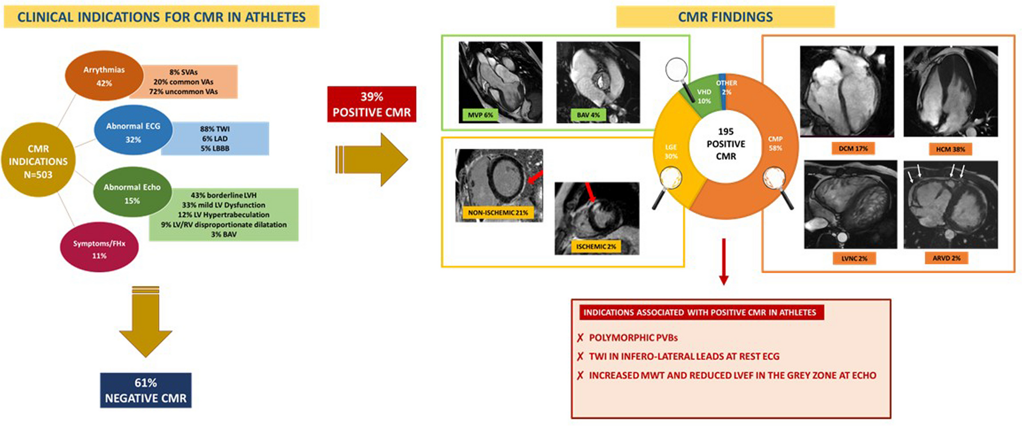
ResultsA total of 503 CMR were included in the analysis. “Negative” and “positive” CMR were 61% and 39%, respectively. Uncommon ventricular arrhythmias (VAs) were the most frequent indications for CMR, but the proportion of positive results was low (37%), and only polymorphic ventricular patterns were associated with positive CMR (p = 0.006). T-wave inversion at 12-lead ECG, particularly on lateral and inferolateral leads, was associated with positive CMR in 34% of athletes (p = 0.05). Echocardiography abnormalities resulted in a large proportion (58%) of positive CMR, mostly cardiomyopathies.
ConclusionCMR is more efficient in identifying a pathologic cardiac substrate in athletes in case of VAs (i.e., polymorphic beats), abnormal ECG repolarisation (negative T-waves in inferolateral leads), and borderline echocardiographic findings (LV hypertrophy, mildly depressed LV function). On the other hand, CMR is associated with a large proportion of negative results. Therefore, a careful clinical selection is needed to indicate CMR in athletes appropriately.
Graphical AbstractClinical indications for CMR and positive CMR findings.
The left panel shows clinical indications for CMR in our athletic cohort and the percentage of negative CMRs. In contrast, the right one shows the percentages of positive CMR findings with some examples. The white arrows in the right panel show an ARVD with a dilated right ventricle and multiple aneurysms of its free wall. The red arrows show the distribution of LGE in the left ventricle. The red panel below shows indications associated with positive CMR findings. ARVD, arrhythmogenic right ventricular disease; BAV, bicuspid aortic valve; CMP, cardiomyopathies; CMR, cardiac magnetic resonance; DCM, dilated cardiomyopathy; ECG, electrocardiogram; Echo, echocardiogram; FHx, family history; HCM, hypertrophic cardiomyopathy; LAD, left axis deviation; LBBB, left bundle branch block; LGE, late gadolinium enhancement; LV, left ventricle; LVH, left ventricular hypertrophy; LVNC, left ventricular non-compaction; MVP, mitral valve prolapse; RV, right ventricle; SVAs, supraventricular arrhythmias; VAs, ventricular arrhythmias; VHD, valvular heart diseases; TWI, T-wave inversion.

留言 (0)