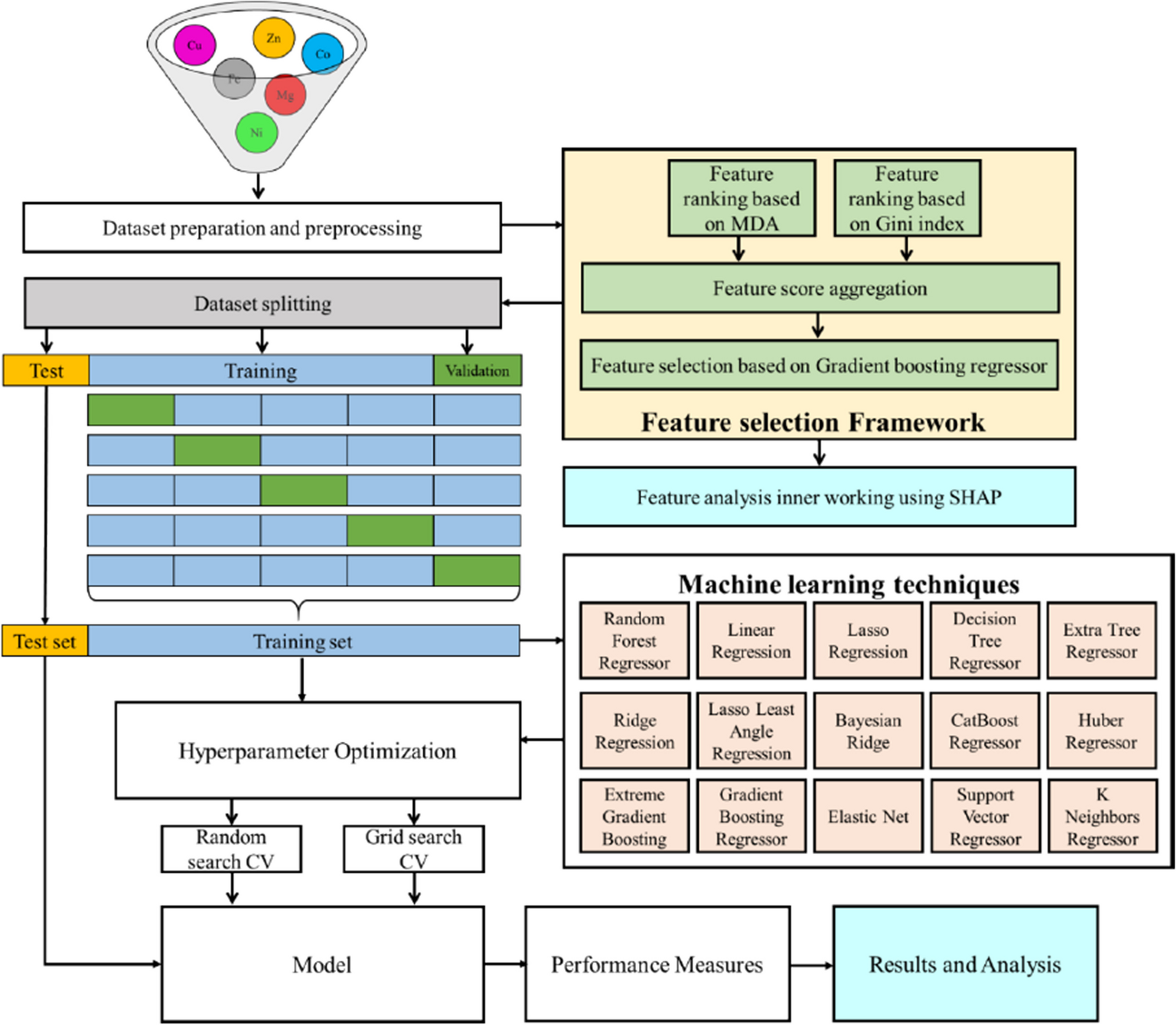
GTPase activity of UreG
UreG (GenBank ID: MN660252) is an accessory protein from the predominant urease obtained from the rumen microorganisms of dairy cows, the preparation of UreG, and the measurement of UreG GTPase activity as described previously (Zhang et al. 2020a). Briefly, the gene of UreG was cloned into pASK-IBA5C plasmid and subsequently transformed into Escherichia coli BL21 (DE3) cells for protein expression, in which the expression plasmid was induced by anhydrotetracycline. The final UreG (purity > 95%) was obtained by purifying crude cell lysates using Strep-Tactin beads (Beaver, Suzhou, China). UreG GTPase activity was measured through the Malachite Green Phosphate Assay Kit according to the manufacturer’s instructions (Sigma-Aldrich, MI, USA). The inhibition rate of alkaloids (Target Molecule Corp., MA, USA) to inhibit UreG GTPase activity was calculated by the following formula:
$$\mathrm\left(\%\right)=\left(1-\frac\right)\times 100$$
where A represents the absorbance value of UreG GTPase activity with alkaloid, B represents the absorbance value of GTPase activity of inactivated UreG with alkaloid, C represents the absorbance value of UreG GTPase activity without alkaloid, and D represents the absorbance value of GTPase activity of inactivated UreG without alkaloid. Each measurement was performed in triplicate.
IC50 value for alkaloid against UreG GTPase activity was calculated using the GraphPad Prism (v.8.0.1) software (GraphPad Software Inc., CA, USA), in which various concentrations of alkaloids (0, 6.25, 12.5, 25, 50, and 100 µM) and standards were prepared at the same reaction conditions, the GTPase activity of inactivated UreG without alkaloid was set as 0, and the GTPase activity of UreG without alkaloid was set at 100%.
Kinetic studies
The kinetics assay of UreG by alkaloid was carried out by Michaelis–Menten equation, and the values of Michaelis–Menten constants (Km and Vmax) were calculated from the Lineweaver–Burk plots through the software of GraphPad Prism. The inhibition with various concentrations of alkaloids (0, 15.6, 62.5, and 250 µM) was measured at different concentrations of GTP substrate (0, 12.5, 25, 50, 100, and 200 µM). Furthermore, the type of enzyme inhibition, including competitive inhibition, uncompetitive inhibition, non-competitive inhibition, and mixed inhibition, was revealed based the trend of Km and Vmax changing with the concentration of alkaloid.
Isothermal titration calorimetry (ITC) measurements and circular dichroism (CD) spectroscopy
The inhibition mechanism of UreG by alkaloid was explored using ITC and CD spectroscopy, to reveal the effects of alkaloid on the combination of nickel towards UreG and the secondary structure of UreG. ITC measurements were carried out with an AutoITC200 microcalorimeter (GE, MA, USA) as described previously (Zhang et al. 2020a). Briefly, 27 µM UreG was titrated with 1500 µM NiSO4 in the present of 238 µM alkaloid. The titration curve was fitted with the one-site binding model, and the final data was analyzed by ITC Analysis Module in the software of Origin 7.0 (OriginLab, MA, USA).
The CD spectra of UreG were recorded using Chirascan Plus spectrometer (Applied Photophysics Ltd., Surrey, UK) equipped with a 1-mm quartz cell, at measurement range between 190 and 260 nm, with 1 nm data pitch and bandwidth. To reduce interference from the background solution, 233 µg/mL UreG and 88 µM alkaloid were prepared using distilled water. The final CD spectrum of UreG was obtained by subtracting the background spectrum (distilled water or alkaloid) from the sample spectrum. The secondary structure proportions (α-helix, β-sheet, β-turn, and random coil) in UreG were calculated using CDNN (v.2.1) software (Applied Photophysics Ltd., Leatherhead, UK).
Molecular docking studies
Binding characteristics of UreG and alkaloid were analyzed performing molecular docking studies. UreG were modeled using the SWISS-MODEL server, and KpUreG (PDB ID: 5XKT) was chosen as the template because of high sequence identity of 77.39%. The 3D structures of berberine chloride and epiberberine were downloaded from PubChem website (https://pubchem.ncbi.nlm.nih.gov/). Molecular docking of UreG and alkaloid was performed using AutoDockTools (v.1.5.6) (Scripps, CA, USA), in which the UreG was considered as a rigid structure receptor, and alkaloid was considered as a flexible molecule. The steps and parameters during molecular docking were consistent with those of previous article (Zhang et al. 2021). A total of 10 poses were obtained for each alkaloid, and the best docking pose with the lowest energy was further analyzed using PyMol molecular graphics system.
Urease activity
Ruminal microbial crude protein was collected by ultrasonication and centrifugation following a previously established method (Zhang et al. 2020b). The urease activity of ruminal microbial crude protein was determined based on the amount of ammonia decomposed by urea through a modified phenol/hypochlorite reaction method (Krallmann-Wenzel 1985), in which the urease activity without alkaloid and urea was set at 0, and the urease activity without alkaloid was set at 100%. The inhibition rate of alkaloid to inhibit urease activity was calculated by the following formula:
$$\mathrm\left(\%\right)=\left(1-\frac\right)\times 100$$
where A represents the absorbance value of urease activity with alkaloid, B represents the absorbance value of urease activity with alkaloid but without urea, C represents the absorbance value of urease activity without alkaloid, and D represents the absorbance value of urease activity without alkaloid and urea. Each measurement was performed in triplicate.
Ruminal microbial fermentation
The study of ruminal microbial fermentation consisted of a control group and 3 epiberberine treatment groups, each containing 70 mL fresh rumen fluid and 140 mL anaerobic medium (Zhang et al. 2020c). Epiberberine treatment groups with the final concentration of 2, 20, and 200 μM were pre-incubated in an anaerobic chamber at 39 °C for 15 min. Immediately after inoculation, 10 mL aliquots of mixed solution were transferred into Hungate tubes, urea with a final concentration of 15 mM was added into each tube. Three tubes were immediately collected from each group as 0 h samples; the others were sealed with rubber stoppers and cultured in an incubator. Samples were collected at 1, 2, 4, 8, and 12 h of incubation, with 3 tubes in each group.
The collected tubes were immediately placed on ice to slow down fermentation; 400 μL of sample solution was mixed with 800 μL metaphosphoric acid solution (25% w/v) and centrifuged at 12,000 × g for 5 min. The supernatant was collected for the detection of NH3 and urea. NH3 content was measured by a modified phenol/hypochlorite reaction method. Urea concentration was determined using the diacetyl monoxime method kit following the manufacturer’s instructions (Jiancheng Bioengineering, Nanjing, China).
Statistical analysis
The data of urease activity and the binding nickel to the UreG were analyzed by one-way analysis of variance (ANOVA) in SPSS v.25.0 software (IBM, NY, USA). Statistical significance P < 0.05 was accepted.


留言 (0)