
記住我
About 55% of patients with advanced cancer experience pain.34 Because of the diversity of tissues involved and the contribution of undergoing anticancer treatments, the clinical presentation of cancer pain can be very variable, with rather complex and not yet well understood pathophysiology.20,27,33 The contribution of a neuropathic component to cancer pain, arising because of a lesion of the somatosensory nervous system,18 should be distinguished from antineoplastic treatment pain.3 The pathophysiology of pain directly because of cancer in these patients can be complex, frequently involving a combination of different mechanisms such as inflammatory, neuropathic, and others that develop as the tumor progresses.19,26,38 With these limitations, it has been estimated that neuropathic cancer pain (NcP) prevalence varies from a conservative estimate of 20% to a liberal estimate of 40%.3 Because cancer pain treatment guidelines8,13 recommend specific adjuvant treatments for NcP, the lack of a standardized assessment method for its diagnosis is certainly clinically relevant.11,25,32
The criteria used for the diagnosis of neuropathic pain proposed by the Neuropathic Special Interest Group (NeuPSIG) of the International Association for the Study of Pain (IASP) follow the clinical method of history taking, examination, and confirmatory tests.14 These approaches have not been widely used in assessing cancer pain, especially because of limitations such as uncertainties on the relative significance of confirmatory tests, especially sensory signs assessment.12 A Delphi expert survey study proposed a modified European Association for palliative care (EAPC)/IASP algorithm7 on the application of the NeuPSIG criteria for cancer pain assessment. To the best of our knowledge, no experience on the use of the EAPC/IASP algorithm has been published to date.
Quantitative sensory testing (QST) is a psychophysical method that uses controlled stimuli to quantify the somatosensory function and presence of sensory positive or negative signs.1 Although not a stand-alone diagnostic test, its utility has been confirmed for the assessment and monitoring of sensory neuropathies and somatosensory deficits, particularly in diabetic and small-fiber neuropathies.1,2,28,37 Yet, few studies have shed light on the function of the somatosensory system in patients with cancer, and little is known on QST discriminative role in NcP.23,24,31
The aim of this study was to estimate the accuracy of the EAPC/IASP algorithm in assessing NcP, comparing it with a gold standard based upon the application of the NeuPSIG grading system. To better analyze the somatosensory profile of patients and assess the presence of positive or negative sensory signs, the objective examination was complemented by the use of QST.1
2. Methods 2.1. Study design and populationA cross-sectional observational study was conducted from August 2020 to March 2023 at the Palliative Care and Pain Outpatient Clinic of a comprehensive cancer center. In predefined days, consecutive patients attending the clinic were screened for eligibility and were included in the study, if they had a cancer diagnosis and had cancer pain (directly because of tissue damage caused by the tumor or its metastases) according to a pain syndrome checklist classification, which allows to classify pain diagnosis into different pain syndromes associated with tumor lesions or antineoplastic treatment9 with a self-reported average pain intensity score ≥3 on a 0 to 10 numerical rating scale (NRS; 0 = no pain and 10 = worst pain imaginable) in both the previous 7 days and 24 hours. Patients had to be able to provide informed consent and be older than 18 years. They were excluded if they had confirmed or suspected pain associated with cancer treatment (ie, chemotherapy-induced peripheral neuropathy [CIPN] or postsurgical pain), unable to poor clinical conditions, were unable to complete the questionnaires required and/or to sit through 60-minute assessment procedure with regular breaks or to understand the information presented in the information sheet had neurological conditions or diseases that could interfere with the examination, and had local skin inflammation or open lesions at the pain site.
2.2. Recruitment and informed consentEligible patients were given the necessary information describing the study steps using a patient's information leaflet and were asked to sign a consent form. The study protocol and supporting documentation was approved by the INT Research Ethics Committee (INT 200/19) and was conducted in accordance with the Declaration of the World Medical Association.
2.3. AssessmentsAll assessments were performed on the same day. The following data were extracted from patients' records: age, sex, primary cancer diagnosis, presence of locally advanced or metastatic disease and site/s of metastasis, pain site, pain syndrome according to the checklist in use at our palliative and pain clinic,9,33 pain flares, and current analgesic and antineoplastic treatments.
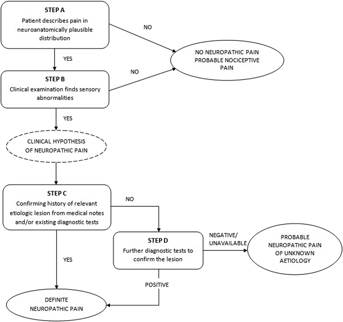
2.4. Pain evaluation and neuropathic cancer pain diagnosis 2.4.1. Pain etiologyPain etiology was identified by applying the codified pain syndrome checklist,9 combining clinical findings with the results of available diagnostic imaging. This classification was used by clinicians to fulfill the algorithm criteria on “relevant etiologic lesion and diagnostic test” (EAPC/IASP algorithm) or “history of relevant lesion or disease” (NeuPSIG criteria) (Figs. 1 and 2).
 Figure 1.:
 EAPC/IASP algorithm for the diagnosis of neuropathic cancer pain.
 Figure 2.:
 NeuPSIG criteria. ENMG, electroneuromyography; NeuPSIG, Neuropathic Special Interest Group.
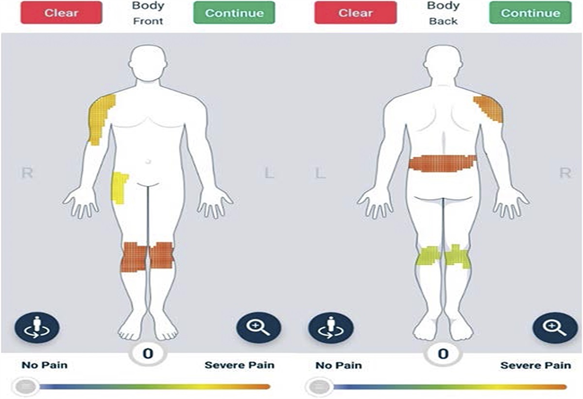
2.4.2. Definition of test and control sitesBased on the patient's reported information and examination, the site of pain (“test site”) was defined. If the patient had more than one painful area, the region with the most intense pain was selected as test site. A nonpainful control site was identified on the contralateral body region, or if this was painful or the test site was located on the midline, a painless ipsilateral control site above/below the test site was selected also free of neurological findings.
2.4.3. Patient-reported outcome measuresPatients reported pain intensity on the pain site (“test site”) using an NRS from 0 to 10 and completed the self-reported Leeds Assessment of Neuropathic Signs and Symptoms (S-LANSS).5
The S-LANSS has 7 questions used to screen for symptoms suggesting the presence of a pain of neuropathic origin. The total score ranges from 0 to 24 points, and scores ≥12 points suggest neuropathic pain.
2.4.4. Neuropathic cancer pain diagnosisClinical assessment and NcP diagnosis were performed by 2 independent investigators: the treating physician and a clinical researcher, as described below.
2.4.4.1. Application of the EAPC/IASP algorithmThe EAPC/IASP algorithm7 was applied by the palliative care treating physician during the scheduled visit. All palliative care physicians had undergone a brief training session on neuropathic pain assessment and were introduced to the EAPC/IASP algorithm (Fig. 1). Indications and checklists were provided for each of the steps included in the algorithm as described in Appendix A, https://links.lww.com/PR9/A221. Based on the answer provided for each step of the algorithm applied at the pain site, pain was considered neuropathic if there was a clinical hypothesis of probable neuropathic pain.
The clinical examination is described in Appendix A, https://links.lww.com/PR9/A221.
2.4.4.2. Application of the Neuropathic Special Interest Group criteriaAfter the EAPC/IASP algorithm application, the patient was assessed by another investigator, blind to the previous assessment, who reviewed the electronic medical records, including available diagnostic tests and examined the patient using also a detailed standardized bedside examination for assessment of sensory signs (Fig. 2). It included the following assessments performed in the pain site, comparing with the control site:
(1) Cold and warm sensitivity, assessed using a thermo-roller (Somedic Roll Temp II). For cold, a thermo-roller at 25°C was rolled over a 2-cm length of skin at a rate of 2-second on, 1-second off. For warm, the thermo-roller at 40°C was rolled over a 2-cm length of skin at a rate of 2 second on, 1 second off; (2) Light touch, assessed using an Owen Mumford Neuropen 10-g monofilament gently pressed against the patient's skin 3 times at a rate of 1 second on, 1 second off; (3) Pinprick sensation was assessed using an Owen Mumford Neurotip, a disposable needle with a defined pressure of 40 g, pressed against the skin. The Neurotip was placed against the patient's skin 3 times at a rate of 1 second on, 1 second off; (4) Dynamic mechanical allodynia (DMA), assessed using a cotton bud gently stroked over a 2-cm area of skin. The cotton bud was stroked over the participants pain site 3 times at a rate of one second on, one second off; and (5) Blunt pressure, assessed using the examiner's thumb pressed against the pain and control sites exerting approximately 4 kg of pressure applied at a rate of 4 kg per second for 4 seconds.Responses to each test were judged by the investigator, in comparison with the control site, and recorded as “normal”: The participant could feel the stimulus as expected with no flinch or withdrawal of body part; “hypersensitivity”: The sensation was perceived to be more intense than expected; and “hyposensitivity”: The sensation was not felt at all or was less intense than expected.
The bedside examination was considered positive if at least one of the above tests, except for the blunt pressure alone, was abnormal.
Based on the clinical evaluation, available diagnostic tests, and the above bedside examination, the investigator was asked to apply the NeuPSIG criteria.14 Pain was considered neuropathic in case of a clinical hypothesis of probable or definite neuropathic pain.
2.4.5. Quantitative sensory testingThen, the same investigator performed the QST. Each QST session was performed in a quiet room maintained at 22 ± 2°C temperature. Patients were familiarized with the QST procedure by demonstrating the stimuli in a body site that was not the test or control site. A QST battery of tests was adapted based on the standardized protocol of the German Research Network on Neuropathic Pain (DFNS)29,30 and was performed at the test (painful) site and then at the control site (Appendix B, https://links.lww.com/PR9/A221). The following parameters were assessed: cold detection threshold (CDT), warm detection threshold (WDT), cold pain threshold (CPT), heat pain threshold (HPT), mechanical detection threshold (MDT), mechanical pain threshold (MPT), pressure pain threshold (PPT), and dynamic mechanical allodynia (DMA).
2.5. Statistical analysisAssuming a prevalence of NcP from 20% to 40% (as per Bennett et al. 2012, PAIN3), and sensitivity values ranging from 60% to 90%, we calculated that a sample size of 95 patients would allow to estimate a 95% CI half width ranging from 21% to 9%. Frequencies and percentages were used to describe categorical variables, whereas mean values and SDs were used for continuous ones. Point and interval estimates (95% confidence intervals [CIs]) for the prevalence of NcP according to different methods of assessment were calculated. Accuracy of the EAPC/IASP diagnostic algorithm in comparison with the application of the NeuPSIG criteria (gold standard) was assessed by sensitivity and specificity, defined as the chance of a true-positive and true-negative test, respectively,6 and estimate precision was measured by 95% CIs. For the QST data, the mean of each parameter was calculated as by the DFNS protocol recommendations.29 The within-subject differences between the test and control site for each patient were analyzed. This choice was based on the lack of normative values for some of the body areas analyzed, on the specific characteristics of patients with cancer and on previous evidence of significant sensitivity of such differences.30 Within-subject differences were calculated either as a difference of test–control or control–test site, to obtain positive values indicating gain of function and negative values indicating loss of function, depending on the QST variable assessed. To test for the ability of QST to discriminate between patients with and without NcP, unequal variance independent-samples t tests were applied to the normally distributed within-subject differences raw data of the QST variables, namely the HPT and CPT. To account for non-normality in the distribution of the rest of the QST variables, nonparametric Kruskal–Wallis tests were applied as sensitivity analysis. P-values <0.05 were regarded as significant. This comparison was made only for patients for whom an agreement was found among both evaluations under study. All data analysis was performed using STATA IC 16.35
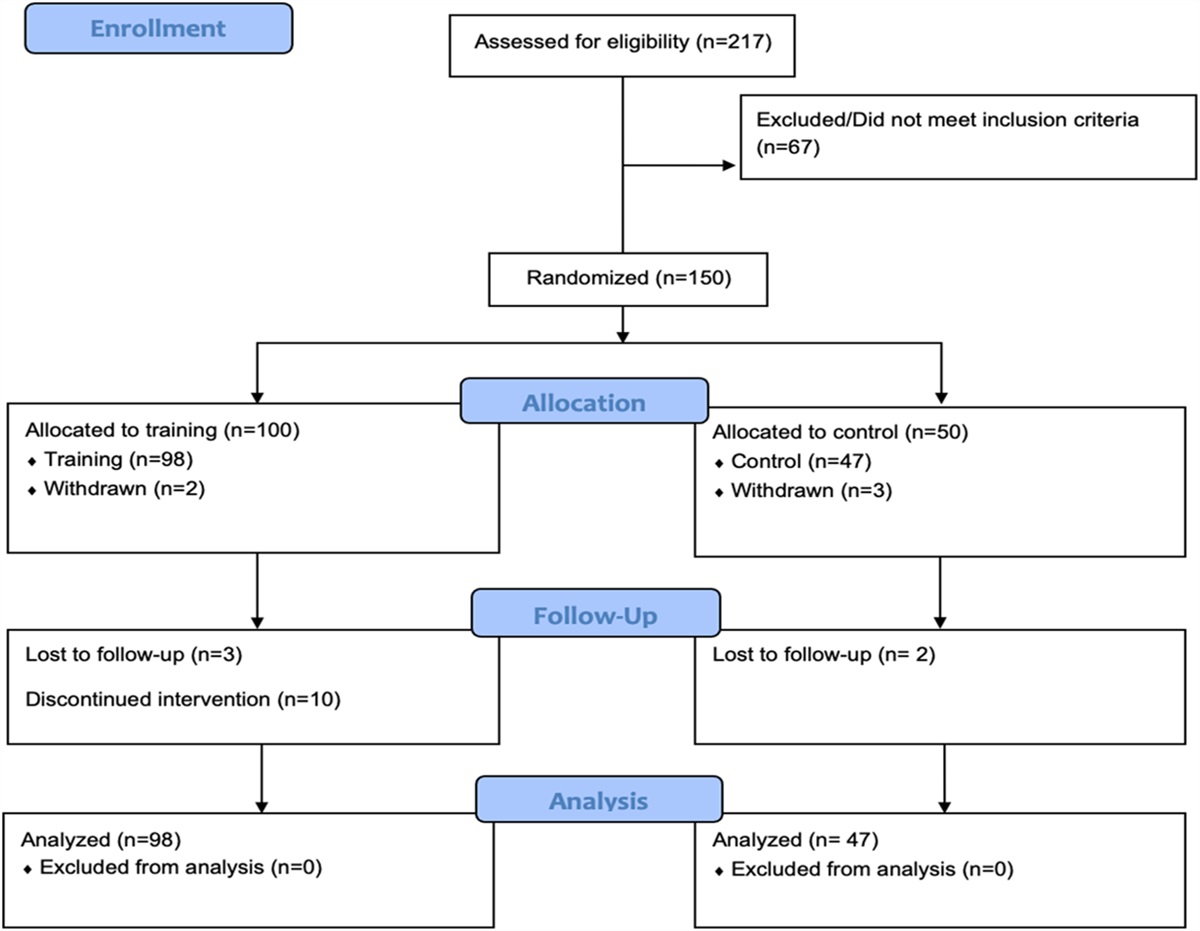
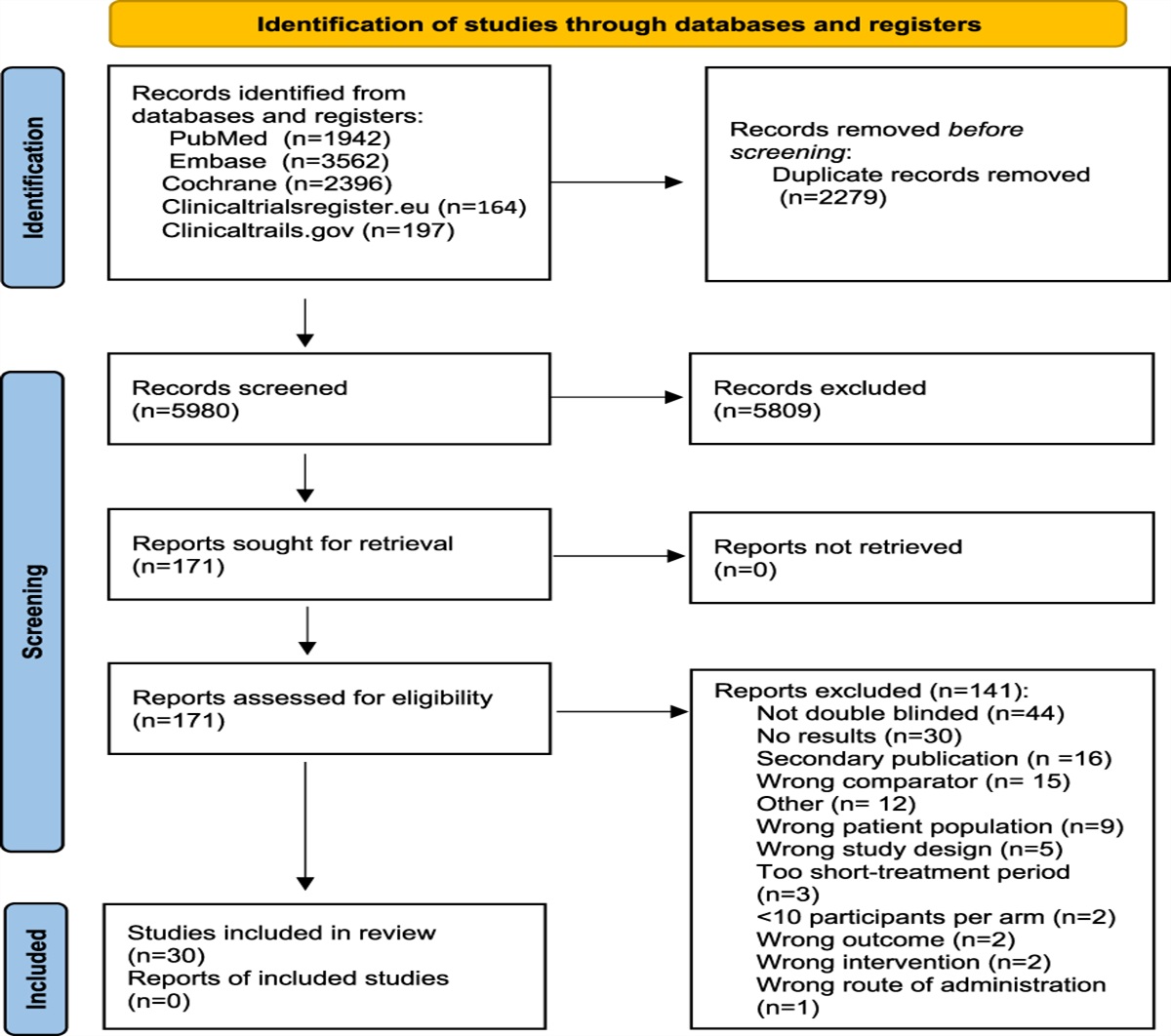
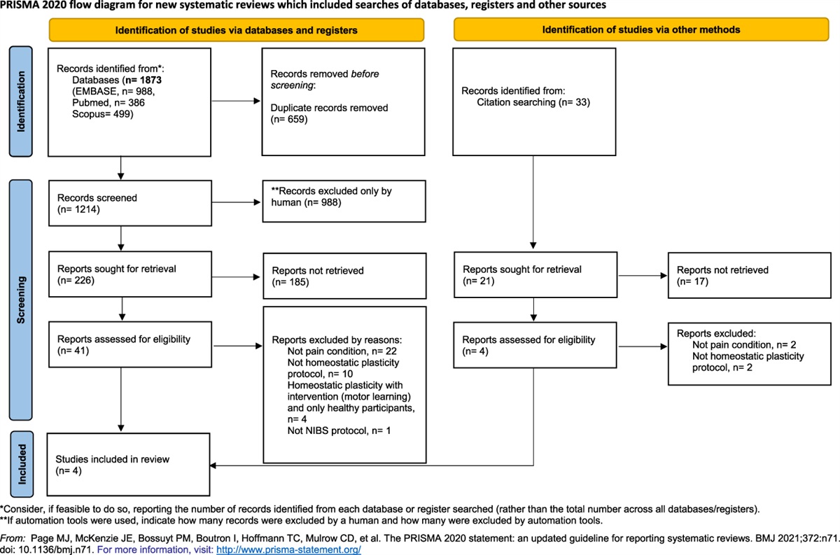
3. ResultsNinety-eight patients were enrolled and included in the final analysis (Fig. 3). Patients and disease characteristics are reported in Table 1. Mean age was 62.5 years, with 57.1% of patients being female and 42.9% male. The most frequent diagnosis was breast (26.5%) and lung cancer (21.4%). Of the enrolled patients, 47.9% had locally advanced disease and 90.8% metastatic disease. The most frequent metastasis sites were bone (66.3%), lymph nodes (38.8%), liver (25.5%), lung (21.4%), and pleura (9.2%). Regarding antineoplastic treatments, 37.8% were receiving none, whereas 37.8% were receiving chemotherapies, 14.4% hormone therapy, 14.4% immunotherapy, and 16.5% biological and target therapies.
 Figure 3.:
 Flowchart of screening and eligibility.
Table 1 - Patients and disease characteristics (98 patients). Characteristic Age, mean (SD), y 62.5 (13.5) Sex, N (%) Female 56 (57.1) Male 42 (42.9) Diagnosis, N (%) Breast 26 (26.5) Lung/bronchial 21 (21.4) Colon/rectum 7 (7.1) Prostate 7 (7.1) Sarcomas 6 (6.1) Renal 6 (6.1) Pancreatic 4 (4.0) Head and neck 2 (2.0) Uterine 2 (2.0) Esophagus 1 (1.1) Thyroid 1 (1.1) Melanoma 1 (1.1) Other 14 (14.4) Locally advanced tumor, N (%) Yes 46 (47.9) No 50 (52.1) Presence of metastasis, N (%) Yes 89 (90.8) No 9 (9.2) Metastasis location*, N (%) Bone 65 (66.3) Lymph nodes 38 (38.8) Liver 25 (25.5) Lung 21 (21.4) Pleura 9 (9.2) Brain 7 (7.1) Peritoneum 5 (5.1) Soft tissues 5 (5.1) Adrenal gland 5 (5.1) Other 8 (8.2) Karnofsky performance status mean (SD) 73.6 (5.8) Antineoplastic treatment*, N (%) Chemotherapy 37 (37.8) Hormone therapy 14 (14.4) Immunotherapy 14 (14.4) Other (biological and target therapies) 16 (16.5) None 37 (37.8)*More than one is possible.
In Table 2, pain and pain treatment characteristics are described. The mean average pain intensity in the past 24 hours and past 7 days was 6.2 (NRS 0–10) for both measurements, and mean pain duration was 7.1 months. The most frequent pain sites were the back (35.7%) and the pelvis (17.3%), followed by lower limbs (13.3%), abdomen (13.3%), chest (12.2%), and upper limbs (8.2%). Summarizing pain syndromes according to tissue involvement, 68 patients (69.4%) had bone pain, 40 (40.7%) pain because of nervous tissue damage (17 a peripheral nerve syndrome because of soft-tissue or bony tumor, 11 a radiculopathy or cauda equina syndrome because of vertebral lesion, 10 a peripheral nerve syndrome because of chest wall mass, one a brachial plexopathy, and one a cervical plexopathy), 33 (33.7%) pain because of soft-tissue damage, and only 15 (15.3%) patients had visceral pain (Table 2). Of these, 46 patients (46.9%) had just one tissue involvement, and the others had a combination of 2 or more tissue etiologies. Forty patients (40.8%) presented pain flares. The most frequent around-the-clock (ATC) analgesic treatments were fentanyl (33.7%) and oxycodone (30.6%), whereas 7 patients (7.1%) were not receiving any ATC treatments. Adjuvant analgesics included acetaminophen (36.7%), anticonvulsants (35.7%), corticosteroids (32.7%), and NSAIDs (16.3%), whereas 23.5% of the patients were not receiving any adjuvants. Thirty patients (30.6%) had received radiotherapy at the pain site before study enrollment. Mean S-LANSS score for the whole group of enrolled patients was 5.5.
Table 2 - Pain and pain treatment characteristics (98 patients). Characteristics Average pain intensity in the past 7 d (NRS 0–10), mean (±SD) 6.2 (±1.6) Average pain intensity in the past 24 h, mean (±SD) 6.2 (±1.7) Pain duration, mean (SD), mo 7.1 (9.3) Presence of pain flares, N (%) Yes 40 (40.8) No 58 (59.2) Pain location, N (%) Back 35 (35.7) Pelvis 17 (17.3) Lower limbs 13 (13.3) Abdomen 13 (13.3) Chest 12 (12.2) Upper limbs 8 (8.2) Pain syndrome*, N(%) Bone pain 68 (69.4) Pain because of nervous tissue damage 40 (40.7) Peripheral nerve syndrome because of soft-tissue or bony tumor 17 (17.3%) Radiculopathy or cauda equina syndrome because of vertebral lesion 11 (11.2%) Peripheral nerve syndrome because of chest wall mass 10 (10.2%) Brachial plexopathy because of tumor invasion 1 (1%) Cervical plexopathy because of tumor invasion 1 (1%) Pain because of soft-tissue damage 33 (33.7) Visceral pain 15 (15.3) Around-the-clock analgesic treatments*, N(%) Fentanyl 33 (33.7) Oxycodone 30 (30.6) Codeine 12 (12.2) Acetaminophen 10 (10.2) Tapentadol 7 (7.1) Tramadol 5 (5.1) NSAIDs 5 (5.1) Buprenorphine 4 (4.1) Morphine 1 (1.1) None 7 (7.1) Adjuvant analgesics*, N(%) Acetaminophen 36 (36.7) Anticonvulsants 35 (35.7) Corticosteroids 32 (32.7) NSAIDs 16 (16.3) None 23 (23.5) Radiotherapy at the pain site, N(%) Yes 30 (30.6) No 68 (69.4) No. of days passed since RT treatment at the time of the visit, mean (±SD) 320 (±78) S-LANSS score, mean (SD) 5.5 (5.9)*More than one is possible.
NRS, numerical rating scale; NSAID, nonsteroidal anti-inflammatory drug; RT, radiotherapy.
Prevalence of NcP as estimated by the EAPC/IASP algorithm, NeuPSIG criteria, and S-LANSS was 35.7% (95% CI 26.1–45.4), 40.8% (95% CI 30.9–50.7), and 18.4% (95% CI 10.6–26.2), respectively. Of the 35 patients classified as having NcP by the EAPC/IASP, 34 had a definite and only one a probable diagnosis. However, of the 40 patients classified as having NcP based on the NeuPSIG criteria, 36 had a definite and 4 a probable diagnosis. Sensitivity and specificity of the EAPC/IASP algorithm compared with the gold standard were 85% (95% CI 70.2–94.3) and 98.3% (95% CI 90.8–100), respectively. The lesion considered to cause NcP was a combination of neurological tissue involvement and either bone (51%), visceral tissue (1%), or soft tissue (48%) in the group diagnosed with the EAPC/IASP algorithm and neurological tissue involvement with bone (55%), visceral tissue (0.8%), or soft tissue (44.2%) in the group diagnosed with the gold standard.
3.1. Quantitative sensory testing findingsA complete QST profile was available for 93 patients. Five patients had an incomplete profile with at least one of the QST variables available. Quantitative sensory testing examination sensory findings are presented in Table 3. The analysis of the within-subject differences, comparing the painful with the nonpainful site, demonstrated sensory abnormalities in the overall group, with an increased threshold in thermal detection both for cold (CDT) and warm sensation (WDT) (P = 0.000) and for heat pain (HPT) (P = 0.0369), demonstrating presence of hyposensitivity. Pressure pain threshold (PPT) was decreased (hypersensitivity) (P = 0.000), and allodynia was found in some patients (P = 0.0001) (Table 3).
Table 3 - Quantitative sensory testing parameters for the whole group. CDTCDT, cold detection threshold; CPT, cold pain threshold; DMA, dynamic mechanical allodynia; HPT, heat pain threshold; MDT, mechanical detection threshold; MPT, mechanical pain threshold; NRS, numerical rating scale; PPT, pressure pain threshold; WDT, warm detection threshold.
Comparing patients with and without NcP, significant differences were found for those with NcP, characterized by hyposensitivity to cold and warm with a reduction of CDT and WDT, hypersensitivity to pressure, and hyperalgesia to cold with a reduction of PPT and CPT; the presence of DMA which was found only in patients with NcP (14 of 34) as seen in Figure 4. Mechanical pain threshold, mechanical detection threshold, and heat pain threshold did not show statistically significant differences. Both the unequal variance independent-samples t tests and Kruskal–Wallis test for the WSD of each QST variable in the group of patients with and without NcP revealed significant differences for CDT (P = 0.0032), WDT (P = 0.0018), CPT(P = 0.02), PPT (P = 0.02), and DMA (0.0001).
 Figure 4.:
 Mean within-subject differences for all QST variables (pain vs control site) of the patients classified as having neuropathic cancer pain (black) and not (gray) (Positive values indicate gain of function, whereas negative ones indicate loss of function). Data are presented as raw mean value of within-subject differences. Negative values indicate loss, whereas positive values indicate gain of function. The graph shows a predominant loss of sensory function (hypoesthesia) in terms of CDT and WDT and gain of function for CPT. Gain of function in the form of hyperalgesia to blunt pressure (PPT) is present and DMA (*P < 0.05, **P < 0.005). CDT, cold detection threshold; CPT, cold pain threshold; DMA, dynamic mechanical allodynia; HPT, heat pain threshold; MDT, mechanical detection threshold; MPT, mechanical pain threshold; PPT, pressure pain threshold; QAT, quantitative sensory testing; WDT, warm detection threshold.
3.2. Characteristics of classifications' disagreement casesOnly 1 patient had a positive EAPC/IASP algorithm diagnosis with a negative gold standard classification, whereas 6 patients were negative at the EAPC/IASP algorithm evaluation and had a positive gold standard (Table 4).
Table 4 - Characteristics of disagreements between the two methods used for neuropathic cancer pain assessment. EAPC/IASP NeuPSIG criteria Distribution History Diagnostic test Simple bedside Standardized bedside Sensory alteration (QST) S-LANSS score NP adjuvant drugs Step A Criterion 1 Step C Criterion 2 Step C Criterion 4 Step B Criterion 3 1 Yes NP No NP Y Y Y N N N Y Y Mechanical hypersensitivity 0 N 2 No NP Yes NP Y Y Y Y Y Y N Y Thermal hyposensitivity and mechanical hypersensitivity 10 Y 3 No NP Yes NP Y Y Y Y Y Y N N Thermal hyposensitivity 5 N 4 No NP Yes NP Y Y Y Y Y Y N Y Thermal hyposensitivity and mechanical hypersensitivity 6 Y 5 No NP Yes NP Y Y Y Y Y Y N Y Mechanical hypersensitivity 1 Y 6 No NP Yes NP Y Y Y Y Y Y N Y Mechanical hypersensitivity 19 N 7 No NP Yes NP Y Y Y Y Y Y N Y Hyposensitivity to thermal stimuli 6 YDisagreements in bold.
NeuPSIG, Neuropathic Special Interest Group; NP, neuropathic pain; QST, quantitative sensory testing; S-LANSS, self-reported Leeds Assessment of Neuropathic Signs and Symptoms.
The positive EAPC/IASP algorithm with a negative gold standard was due to the different evaluation made by the treating physician and the investigator; the first considered the history of the patient suggestive for the presence an etiological lesion, whereas the second did not. This patient had an S-LANSS score of 0. Six patients had a negative EAPC/IASP algorithm and positive gold standard assessment. In 5 patients, this was due to the results of the objective examination step: In 2 of them, thermal perception alone, which was assessed only within the gold standard, was abnormal; in 2 cases, mechanical perception alone was abnormal, whereas in 2 other cases, it was abnormal together with thermal perception. In the last case, the disagreement was due to the lack of objective sensory signs, which are required by the EAPC/IASP algorithm, whereas the NeuPSIG criteria allow for a probable diagnosis of NcP even without sensory signs.
Only one of these 6 patients had a positive S-LANSS score, whereas for the remaining 5 patients, the mean S-LANSS score was 5.5. Four of the 6 patients were receiving adjuvant drugs for NcP.
4. DiscussionIn this study, we show that the EAPC/IASP algorithm7 has good sensitivity and specificity compared with the NeuPSIG criteria. The frequency of neuropathic pain obtained by using the 2 diagnostic algorithms was comparable 35.7% (95% CI 26.1%−45.3%) and 40.8% (95% CI 30.9%−50.7%) according to, respectively, the EAPC/IASP and NeuPSIG criteria. This finding compares with the upper limits of the prevalence previously reported.3 Although the patients were enrolled consecutively, a bias of selection might have occurred as also reflected by the rather high level of pain reported (average NRS in the past 24 hours and past 7 days = 6.2). NcP prevalence obtained by the S-LANSS was lower (18.4% [95% CI 10.6–26.2]), confirming previous evidence on the underestimation of NcP when using screening questionnaires.11,25,32,36
We found satisfactory sensitivity (85% [95% CI 70.2–94.3]) and specificity (98.3% [95% CI 90.8–100]) for the EAPC/IASP algorithm. The relatively lower sensitivity is explained by 6 false-positive cases (6.1%), which are mainly due to the disagreement on the identification of objective sensory findings (Table 4). In fact, in 4 cases, thermal hyposensitivity to cold stimuli was detected by applying the NeuPSIG algorithm, which, in our protocol, included the evaluation of thermal sensation, whereas it was not tested when applying the EAPC/IASP algorithm. The only case of false positives depended on a true disagreement in the interpretation by the 2 independent investigators of patients' history and, therefore, pain etiology. From the above results, we can say that the relatively lower sensitivity of the EAPC/IASP algorithm is likely to depend on the more accurate neurological examination used in applying the NeuPSIG criteria in this study.
The QST profiling showed sensory impairments in the painful sites in the overall group, characterized by hyposensitivity in the detection of thermal stimuli, both for cold and warm (P < 0.001). Hyperalgesia to hot pain stimuli (P = 0.0369), pressure pain stimuli (P = 0.000), and p
留言 (0)