
記住我
Adhesive capsulitis (AC), more commonly known as “frozen shoulder,” is characterized by chronic pain and night pain, affecting approximately 2% to 5% of the global population.31 This condition has a large impact on an individual's health and economic status, with a duration ranging between 1 and 3 years.23,26 Typically, the pain associated with AC has been attributed to peripheral origins, stemming from the local inflammation and fibrosis of capsular tissue.8 However, recent research has shown that central pain mechanisms also have a crucial role in patients with AC.4,24,25 Furthermore, patients with AC are at an increased risk of poor mental health due to persistent pain.5 Research indicated that individuals enduring prolonged pain frequently manifest symptoms of depression, with as many as 85% of those grappling with chronic pain being affected by severe depressive symptoms.9 Furthermore, chronic pain patients with psychological comorbidities such as depression are also likely to have a worse outlook than those who only experience chronic pain.2,5,7,9,11,35
Over the past decades, research has demonstrated the bidirectional relationship between pain and depression in individuals experiencing chronic pain. Kroenke et al.17 conducted a longitudinal study that showed that pain was a powerful predictor of future depression severity, and depression was equivalently strong in predicting future pain intensities. To further investigate this issue, recent studies have conducted neuroimaging techniques to investigate the neural correlates underlying the mutual association between pain and depression. These studies have shown a considerable amount of overlap between brain functional and morphological changes in neuroplasticity and neurobiological mechanisms caused by pain and depression.10,14,33 The demonstrated significance lies in the shared brain plasticity contributing to the concurrent onset and advancement of chronic pain alongside depression. For example, research has revealed a substantial overlap of brain regions involved in mood regulation and pain modulation, such as the prefrontal cortex, thalamus, insular cortex, anterior cingulate, amygdala, and hippocampus. This structural basis supports the coexistence of pain and depression.10,14,16,21,22,30 Moreover, studies focused on the molecular mechanisms associated with chronic pain, and depression-induced neural plasticity changes have also identified the shared molecular changes in both pain and depression. These results demonstrated that the neuroplasticity associated with the cooccurrence and development of chronic pain and depression may involve the same brain structures, neurotransmitters, and signaling pathways.10,14 However, the precise neural mechanisms underlying the contribution of cortical plasticity to AC remain largely unknown. Furthermore, the association between these neural plasticity changes and pain-associated emotional comorbidities has not been extensively investigated.
Therefore, in this study, we employed a combination of psychophysics, structural MRI, and functional MRI techniques to examine the brain's structural and functional changes in AC. We also examined the association between these brain alterations and the duration of pain, as well as pain-related emotional comorbidities assessed using the hospital anxiety and depression scale (HADS). By conducting these investigations, our study aims to enhance our comprehension of the underlying mechanisms involved in AC pathogenesis, thereby establishing a robust foundation for the mechanism-based diagnosis of patients with AC.
2. Methods 2.1. SubjectsThe Institutional Review Board of Xi'an Honghui Hospital approved this study, and each participant was provided with written informed consent before each procedure. During 2022 to 2023, a total of 54 individuals with a primary diagnosis of adhesive capsulitis, along with 52 healthy controls (HC) were included.
To validate the reproducibility of our results, we used an external data set using the same inclusion and exclusion criteria. The external validation set included part of the data of an ongoing prospective clinical trial registered in the China Clinical Trial Registry (ChiCTR-2100047183). A total of 31 patients with AC and 32 HC were included in the Tianjin Medical University General Hospital.
The inclusion criteria for patients with adhesive capsulitis were as follows: (1) the external rotation of the affected shoulder was reduced by 50%, in comparison to the unaffected shoulder, which was below 30°; (2) the range of movements were reduced by 25% in at least 2 planes when compared with the unaffected shoulder; (3) the duration of the symptoms was more than 3 months; and (4) normal X-rays of shoulder. Exclusion criterion included the following: (1) patients with secondary adhesive capsulitis and other shoulder conditions (eg, injury, infection, arthritis, cervical radiculopathy, previous surgery, etc.); (2) other chronic pain issues such as fibromyalgia, rheumatic diseases, and the like; (3) other comorbidities include cardiovascular, neurological, and cognitive diseases; (4) patients who were on antidepressants and/or anticonvulsants; and (5) cannot complete the fMRI scan.
The same exclusion criteria for both data sets were applied to healthy participants except for the diagnosis of adhesive capsulitis.
2.2. Clinical assessmentFor both data sets, all patients with AC were assessed within 1 week before their fMRI scan and were clearly instructed to indicate the mean level of pain intensity (for the last month) with a numerical rating scale (with values ranging from 0 to 100; 0 indicating no pain, 20 indicating threshold pain, and 100 indicating intolerable pain), as well as to report the duration of their pain in months. Both patients with AC and HC were evaluated using the HADS within 1 week before their fMRI scan. This study used the Chinese version of HADS, which is a self-report measure containing 7 items to measure anxiety (HADS-A subscore) and 7 items to measure depression, each scored from 0 to 3 (HADS-D subscore).20
2.3. Data acquisition and preprocessing steps 2.3.1. Data acquisition for discovery data setA 64-channel phase-array head-neck coil was used with a 3T MR scanner (MAGNETOM Prisma, Siemens, Erlangen, Germany) for acquiring fMRI data. To reduce unconscious movement, participants were provided with a sponge pad for head support and directed to remain still. Furthermore, patients were asked to close their eyes and maintain an impartial mental state, while staying alert. The fMRI scanning was conducted with a simultaneous multislice gradient-echo echo-planar imaging (EPI) sequence with a repetition time (TR) of 800 milliseconds, an echo time (TE) of 30 milliseconds, an in-plane resolution of 3 mm × 3 mm, a field of view (FOV) of 222 mm × 222 mm, a flip angle (FA) of 54°, a matrix of 74 × 74, a slice thickness of 3 mm, several slices of 48, a bandwidth of 1690 Hz/pixel, no gap, parallel acquisition technique mode, slice orientation of transversal, a slice acceleration factor of 4, and a phase encoding acceleration factor of 2. Four hundred fifty functional images were acquired in a total time of 6 minutes. The 2-inversion contrast magnetization prepared rapid gradient echo sequence (MP2RAGE) was employed to acquire a high-resolution 3D T1 structural image in 6 minutes and 42 seconds. The parameters used included an inversion time (TI1/TI2) of 700 milliseconds/2110 milliseconds, TR/TE of 4000 milliseconds/3.41 milliseconds, matrix of 256 × 240, FA1/FA2 of 4°/5°, FOV of 256 mm × 240 mm, slice thickness of 1 mm, number of slices of 192, and slice orientation of sagittal with an in-plane resolution of 1 mm × 1 mm.
2.3.2. Data acquisition for external validation data setData were acquired using a 3.0T magnetic resonance scanner (Discovery MR750, General Electric, German) with a 20-channel phased-array head coil. Functional images were collected using a gradient echo-planar pulse imaging (EPI) sequence with the following parameters: repetition time = 2000 milliseconds, echo time = 30 milliseconds, flip angle = 90°, field of view = 240 mm × 240 mm, matrix = 64 × 64, 38 slices, and slice thickness = 3.0 mm. A total of 180 images were collected in 6 minutes. Structural images were collected using a 3D T1-weighted image (3D T1WI) for coregistration and normalization of functional images with the following parameters: sagittal acquisition, repetition time: 7.8 milliseconds, echo time: 3.0 milliseconds, inversion time: 450 milliseconds, flip angle: 13°, field of view: 256 mm × 256 mm, matrix: 256 × 256, 180 slices, and slice thickness: 1.0 mm.
2.3.3. Preprocessing stepsData preprocessing in this study was done with the Data Processing Assistant for rs-fMRI (DPARSF; http://www.restfmri.net/forum/DPARSF) toolbox. The preprocessing steps included the following: (1) 10 time points from each scan were excluded due to magnetization stabilization and acclimation; (2) Motion correction was done through the realign function embedded in the DPARSFA toolbox to eliminate the influence of head movement; (3) the T1 images were coregistered to functional images and normalized to the MNI template; (4) the voxels of functional images were resampled to 3 × 3 × 3 mm3; (5) linear drift, mean global signal, white matter signal, CSF signal, and friston-24 parameters were regressed out as covariates; (6) the scrubbing process was conducted on time points with high motion; and (7) a filter step was performed across 0.01∼0.08 Hz to eliminate high-frequency noise. The data obtained were then used for further analysis.
2.4. Brain functional and structural metrics 2.4.1. Grey matter volumeThe T1-weighted images were used to conduct voxel-based morphometry (VBM) analyses to obtain whole brain grey matter volume for both patients with AC and HC. The preprocessing steps for the high-resolution T1-weighted images by means of the Computational Anatomy Toolbox (CAT12) and SPM12 included (1) manually aligning the images to the anterior commissure orientation after inspecting for any artifacts or gross anatomical abnormalities for better registration; (2) splitting them into the cerebrospinal fluid (CSF), white matter (WM), and gray matter (GM) after adjusting for inhomogeneities in the signal; (3) through DARTEL with nonlinear registration to a standard space, followed by a linear and nonlinear intensity adjustment based on the Jacobian determinant from the deformation fields; (4) finally summing the volumes of the segmented CSF, WM, and GM compartments to calculate the intracranial volume. Finally, the resultant brain maps were z-scored for further analysis.
2.4.2. Regional homogeneityThe regional homogeneity (ReHo) value was calculated as follows: The preprocessed data were used to calculate the similarity between a single voxel and the surrounding 27 voxels through Kendall coefficient of concordance (KCC) based on ReHo, to measure the similarity of the time series in the functional cluster. Subsequently, the individual's ReHo image was normalized by dividing it by the mean ReHo value for the entire brain of all participants in the group. Finally, the ReHo brain map was then spatially smoothed using a 5-mm smooth core and z-scored for further analysis. Regional homogeneity reflects the synchronization of the fMRI BOLD signal between neighboring voxels, providing a measure of local connectivity. By contrast, grey matter volume (GMV) reflects structural volume differences, not function. Although GMV shows structural abnormalities, ReHo can reveal additional insights into functional changes.
2.5. Functional connectivityFollowing the between-group comparison of ReHo and GMV to identify significant regions, a post hoc functional connectivity (FC) analysis was performed on 2 regions of interest (ROIs). Significant clusters were analyzed by drawing spheres with a radius of 5 mm. Two ROIs were then defined, namely, the right ventral part of medial prefrontal cortices (vmPFC) (x, y, z: 3, 63, 0) and left amygdala (x, y, z: −18, −3, −12). The average temporal series across all the voxels present in the ROIs was extracted, and the voxelwise functional connectivity was calculated. This resulted in ROI to whole brain FC maps, which were then converted to z-scores for further statistical analysis.
2.6. Statistical analysesThe differences in ReHo, FC, and GMV were investigated using voxelwise 2-sample t-tests between AC patient's HCs within a gray matter mask. False discovery rates (FDR) correction was performed to address multiple comparison issues. The voxel-level significance threshold was set to P < 0.001, and the cluster-level significance threshold was set to P < 0.05 using SPM12 (http://www.fil.ion.ucl.ac.uk/spm). Pearson correlation analyses were performed to reveal the association among clinical measures and were also performed to assess the relationship between brain alterations in patients with AC and clinical measures. Bonferroni correction was performed for multiple comparison correction, and the significant threshold was set to P < (0.05/number of correlation performed for a given analyses).
Furthermore, to investigate the path association among pain duration, HADS-D subscore, and brain abnormalities in patients with AC (eg, altered GMV within vmPFC identified by voxelwise comparison), mediation analyses were performed. Considering there is no empirical evidence suggesting the directional association between pain duration and HADS-D score, 2 models were tested: (1) in which GMV of vmPFC was the mediator, pain duration was the independent variable, and HADS-D subscore was the dependent variable; and (2) GMV of vmPFC was the mediator, HADS-D subscore was the independent variable, and pain duration was the dependent variable. The mediatory association was investigated through a bootstrapped mediation analysis to determine whether the total effect (path c) and the direct effect (path c′) that involve the mediator (M) show a substantial distinction. The PROCESS macro (www.processmacro.org, version 2.16.3) in SPSS (IBM, version 23.0) was used with 2000 bootstrap samples to identify 95% confidence intervals for the components of the model. Statistically significant mediation is present when the upper and lower 95% confidence intervals (CIs) determined through bootstrapping do not contain zero.
All analyses were performed in both the discovery data set and external validation data set, respectively, to validate our results. It should be noted that due to the relatively small sample size of the validation set, we performed the statistical analyses within preidentified brain regions (eg, GMV, ReHo comparison) obtained in the discovery set.
3. Results 3.1. Demographic dataThe demographic information and clinical assessments for both discovery and external validation data sets can be found in Table 1. There is no significant difference between patients with AC and HCs regarding age, sex, or education years (P > 0.05).
Table 1 - Demographic data of the discovery and external validation data set. Discovery data set Validation data set AC HC P AC HC P Age 48.1 ± 10.2 47.3 ± 7.5 0.64 47.4 ± 11.3 47.8 ± 7.2 0.87 Gender (F/M) 26/26 26/26 1 15/16 14/16 0.89 Education (y) 8.9 ± 5.2 9.4 ± 7.8 0.70 7.8 ± 6.4 7.6 ± 5.2 0.89 Trait pain intensity 53.4 ± 11.1 N/A N/A 57.4 ± 13.2 N/A N/A PD (mo) 9.7 ± 2.3 N/A N/A 9.9 ± 3.0 N/A N/A HADS-D 8.5 ± 2.3 4.2 ± 1.5 <0.001 9.2 ± 1.5 3.8 ± 1.7 <0.001 HADS-A 7.1 ± 2.2 4.0 ± 1.4 <0.001 7.3 ± 2.2 4.1 ± 1.3 <0.001AC, patients with adhesive capsulitis; F, female; HADS-A, anxiety subscale of hospital anxiety and depression scale; HADS-D, depression subscale of hospital anxiety and depression scale; HC, healthy controls; M, male; PD, pain duration; PT, pain threshold; trait pain intensity, measure by numerous rating scale range from 0 to 100.
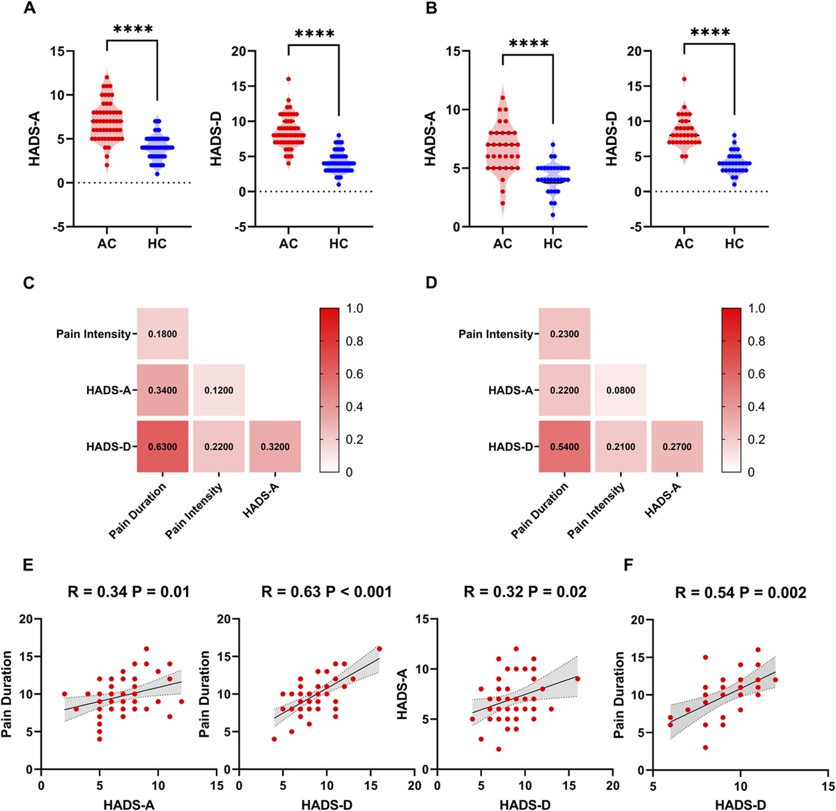
Relative to HCs, AC patients exhibited a significantly higher level of depression and anxiety score measured by HADS for both the discovery and validation set (Table 1, Fig. 1A, B). The association among clinical measures in AC patients for both the discovery and validation sets was illustrated in heat maps (Fig. 1C, D). Specifically, a mild positive association was observed between HADS-A and HADS-D scores (discovery set: R = 0.32, P = 0.02; validation set: R = 0.27, P = 0.13), and a positive relationship was observed between HADS-D and pain duration in AC patients (discovery set: R = 0.63, P < 0.001; validation set: R = 0.54, P = 0.002, survived Bonferroni correction). The scatter plots were also illustrated in Figure 1E, F.
 Figure 1.:
 (A). Differences in depression and anxiety subscale of hospital anxiety and depression scale (HADS) between adhesive capsulitis (AC) and healthy controls (HC) using discovery data. ****P < 0.001. (B). Differences in HADS subscore between AC and HC using validation data. ****P < 0.001. (C). The heat map for association among HADS-D, HADS-A, pain duration, and pain intensity (NRS) in patients with AC using discovery data. (D). The heat map for association among HADS-D, HADS-A, pain duration, and pain intensity in patients with AC using validation data. (E). The scatter plots for the significant correlation between HADS-D and pain duration, between HADS-A and pain duration, and between HADS-D and HADS-A in patients with AC using discovery data. (F). The scatter plots for the significant correlation between HADS-D and pain duration in patients with AC using validation data.
3.3. Grey matter volume, regional homogeneity, and Region of Interest-to-whole-brain functional connectivityIn this study, we observed significantly lower GMV within the left amygdala and right vmPFC in AC patients relative to HCs (Fig. 2A; Table 2). Compared with HCs, AC patients also exhibited a significantly lower ReHo within the right vmPFC (Fig. 2B, Table 3). Furthermore, these 2 significant clusters within vmPFC identified by GMV and ReHo analyses were largely overlapped (Fig. 2C).
 Figure 2.:
 (A). The brain regions exhibited lower grey matter volume (GMV) in patients with adhesive capsulitis (AC) relative to healthy controls (HC) in discovery data. (B). The brain regions exhibited lower ReHo in patients with AC relative to HC in discovery data. (C). The intersection for structural and functional alterations in the ventral part of medial prefrontal cortices (vmPFC). The intersection was used for functional connectivity (FC) analysis. (D). The brain regions exhibited higher FC in patients with AC relative to HC. (E). The brain regions exhibited lower GMV and higher FC in patients with AC relative to HC using validation data. **P < 0.01. ReHo, regional homogeneity.
Table 2 - Grey matter volume differences between patients with adhesive capsulitis and healthy controls. Brain regions Peak MNI (x, y, z) Voxels T value Left amygdala −18 −3 −12 204 −8.7 Right mPFC 3 63 0 323 −9.4The clusters exhibited significant between-group differences in GMV between AC and HC revealed by voxelwise 2-sample t test.
AC, adhesive capsulitis; GMV, Grey matter volume; HC, healthy controls; mPFC, medial prefrontal cortices.
The clusters exhibited significant between-group differences in ALFF between AC and HC revealed by voxelwise 2-sample t test.
AC, adhesive capsulitis; ALFF, amplitude of low-frequency fluctuation; HC, healthy controls; mPFC, medial prefrontal cortices.
For ROI-to-whole-brain FC, we found that the FC between right vmPFC and right ACC (vmPFC-ACC) was significantly higher in patients with AC (Fig. 2D, Table 4). Lower GMV within the left amygdala and right vmPFC and higher FC between the right vmPFC and right ACC were also observed in patients with AC relative to HCs in the validation set (Fig. 2E). Furthermore, the HADS-D score positively correlated with the GMV within vmPFC in patients with AC (discovery set: R = 0.58, P < 0.001, survived Bonferroni correction, Fig. 3A; validation set: R = 0.61, P < 0.001, Fig. 3E; survived Bonferroni correction). Furthermore, the duration of pain also positively correlated with the vmPFC GMV in patients with AC (discovery set: R = 0.47, P = 0.004, survived Bonferroni correction, Fig. 3B; validation set: R = 0.36, P = 0.03, Fig. 3F).
Table 4 - Medial prefrontal cortices-to-cortical functional connectivity differences between patients with adhesive capsulitis and healthy controls; adhesive capsulitis > healthy controls. Brain regions Peak MNI (x, y, z) Voxels T value Right ACC 3 54 15 79 7.8The clusters exhibited significant between-group differences in FC between AC and HC revealed by voxelwise 2-sample t test.
AC, adhesive capsulitis; ACC, anterior cingulate cortex; FC, functional connectivity; HC, healthy controls.
 Figure 3.:
 The association between brain alterations and clinical measurements in patients with adhesive capsulitis. (A). The scatter plot for the positive association between vmPFC grey matter volume (GMV) and HADS-D. (B). The scatter plot for the positive association between vmPFC GMV and pain duration. (C). The scatter plot for a positive association between vmPFC GMV and regional homogeneity. (D). The scatter plot for the positive association between vmPFC-ACC FC and pain intensity. (E). The scatter plot for the positive association between vmPFC- GMV and HADS-D. (F). The scatter plot for the positive association between vmPFC- GMV and pain duration. ACC, anterior cingulate cortices; HADS-D, depression subscale of hospital anxiety and depression scale; vmPFC, ventral part of medial prefrontal cortices.
There was no significant association between ReHo and pain intensity, between ReHo and pain duration. Regional homogeneity positively correlated with GMV (discovery set: R = 0.28, P = 0.04, Fig. 3C). For ROI-to-whole-brain FC, we found that the FC between right vmPFC and right ACC (vmPFC-ACC) correlated with pain intensity in patients with AC (discovery set: R = 0.44, P = 0.005, survived Bonferroni correction, Fig. 3D; validation set: R = 0.24, P = 0.21), whereas no significant association between pain duration and vmPFC-ACC FC, and between HADS-D and vmPFC-ACC FC was observed.
3.4. Mediation analysesIn this study, the mediation analyses revealed that the impact of pain duration on HADS-D score was mediated by vmPFC GMV in patients with AC (direct effect = 0.44, P = 0.002, indirect effect = 0.16, 95% confidence interval: [0.04, 0.36]) (Fig. 4A). Furthermore, such mediatory effect could be replicated by our validation data (direct effect = 0.18, P = 0.022, indirect effect = 0.08, 95% confidence interval: [0.09, 0.21]) (Fig. 4B).
 Figure 4.:
 (A). The medication analyses in discovery data: The effect of pain duration on HADS-D was mediated by vmPFC GMV in patients with AC. (B). The medication analyses in validation data. (C). The medication analyses using discovery data: The effect of HADS-D on pain duration was not mediated by vmPFC GMV. (D). The medication analyses using validation data: The effect of HADS-D on pain duration was not mediated by vmPFC GMV. *P < 0.05. AC, adhesive capsulitis; GMV, grey matter volume; HADS-D, depression subscore for hospital anxiety and depression scale (HADS); vmPFC, ventral part of medial prefrontal cortex.
By contrast, the effect of the HADS-D score on pain duration was not mediated by the vmPFC GMV (direct effect = 0.56, P = 0.051; indirect effect = 0.09, 95% confidence interval: [−0.03, 0.29]) (Fig. 4C). Similar results were also obtained in validation data (direct effect = 1.06, P = 0.013, indirect effect = 0.22, 95% confidence interval: [−0.31, 0.53]) (Fig. 4D).
4. DiscussionIn this study, 4 main findings were observed: (1) patients with AC exhibited a higher level of HADS-D score, which correlated with the GM atrophy within right vmPFC compared with HCs; (2) relative to HCs, patients with AC exhibited lower level of ReHo within right vmPFC, which largely overlapped with the structural abnormalities compared with HCs; (3) relative to HCs, patients with AC exhibited significantly higher vmPFC-ACC FC, which correlated with pain intensity; (4) the impact of pain duration on HADS-D score was mediated by GMV of the right vmPFC in patients with AC.
4.1. Relative to healthy control, patients with adhesive capsulitis exhibited a significantly higher level of hospital anxiety and depression scale score, which is associated with brain morphological alterationOur current findings were in line with previous studies that negative emotions, such as depressed status and anxiety, are associated with a heightened perception of pain in AC.9 It is considered that depression and anxiety may contribute to a distorted perception, exaggerating the perceived impairment beyond what is objectively measured, leading to more difficulty in adapting to and dealing with problems of the upper extremity; moreover, depression and anxiety could have an impact on the efficacy of treatment.35 Such phenomenon has also been reported in other chronic pain conditions and the association between depression and chronic pain has been demonstrated to be bidirectional, with both of them being potential risk factors for one another.3 Studies have found that depression and anxiety can intensify the pain, whereas prolonged pain can also trigger mood disturbances (eg, depression and anxiety).3,15,18
Brain structural analyses showed that patients with depression had notably diminished gray matter volumes in the medial prefrontal cortex (PFC) and hippocampus, which were linked to the severity of the depression. Moreover, researchers have elucidated the medial part of prefrontal areas in the restraint of pain through the descending pain modulation pathway.1,13,29 Evidence has shown that the prefrontal cortex communicates with the periaqueductal gray matter and is then connected to the rostral ventromedial medulla, which sends direct signals to the spinal cord, and this pathway is closely linked to pain relief.13 In summary, chronic pain and depression may be based on common neuroplasticity changes, which are a potentially important route for the onset and aggravation of chronic pain and depression.
4.2. Functional alterations in the ventral medial prefrontal cortex overlapped with structural abnormalitiesIn this study, we observed altered GMV and ReHo within the ventral part of mPFC, as well as higher vmPFC-ACC FC in patients with AC compared with HC. It has been shown in previous studies that more anxiety symptoms in fibromyalgia correlated with higher levels of connectivity strength between the vmPFC and right sensorimotor cortex, suggesting the potential role of vmPFC in pain and negative emotions.36 Evidence from anxiety-like behaviors in mouse models of chronic pain highlighted the crucial role of nNOS-expressing neurons in the ventromedial prefrontal cortex (vmPFC) in the manifestation of pain-induced anxiety19; furthermore, these neural alterations were not confined solely to anxiety (eg, pathological or physiological stress-related responses).12,32 Taken together, neuroplasticity plays a significant role in the development and manifestation of chronic pain and negative emotions, likely involving similar brain structures, neurotransmitters, and pathways. Having said this, an alternative explanation also exists—it has also been shown by recent studies that the altered FCs between patients with chronic pain and HC were associated with the timing of assessment, specifically the presence or absence of pain at that moment of MR scanning.6 Therefore, it is plausible that functional or structural alterations precede the onset of pain and may serve as predisposing factors rather than mere consequences.
4.3. Structural alterations within ventral part of medial prefrontal cortices mediated the influence of pain duration on depression severity in patients with adhesive capsulitisIt is well established that there is a bidirectional relationship between pain and depression in patients with chronic pain. Kroenke et al.17 conducted a longitudinal study that showed that pain was a powerful predictor of future depression severity, and depression was equivalently strong in predicting future pain intensities. This study found that the impact of pain duration on HADS-D score in patients with AC was mediated by the structural abnormalities of the vmPFC in patients with AC. Furthermore, the alternative path, where vmPFC GMV mediated the effect of HADS-D on pain duration, was not statistically significant in this study. This finding implies that although there is a bidirectional relationship between pain and depression, reciprocal causal effects on one another can be initiated through distinct neural systems. Research is needed to determine the exact neural mechanisms that mediate causal effects. Moreover, the GMV abnormalities within the amygdala observed in patients with chronic pain have been linked to negative affective disorders by past research and can predict the chronification of pain.27,28,34 Regarding clinical significance, our present findings demonstrated that AC is not only generated peripherally but also associated with psychological comorbidities such as depression and anxiety. Furthermore, our results revealed the potential route for depression-induced pain in AC and provided insights into possible analgesic targets that could break the vicious circle between pain and depression in patients with AC.
4.4. LimitationsIt is noteworthy that our study has several limitations. First, the participants have all received analgesics before enrolling in this study. There is a possibility that this could affect our results. Second, the vmPFC is composed of various distinct subregions, and in this study, we have not explored the functional and structural alterations in subregions of vmPFC. This limited the illustration of detailed mechanisms. Third, are the functional and structural changes that occur a characteristic “brain signature” of AC, or do they overlap with other chronic pain situations? This question remains unsolved. It remains unclear whether pain is sensitized at the spinal cord level, which can be clarified with spinal cord functional magnetic resonance imaging.
5. ConclusionIn summary, our current findings suggest that vmPFC alterations correlate with both the pain duration and the emotional comorbidities experienced by patients with AC. Our research provides an enhanced comprehension of the underlying mechanisms of AC, thereby facilitating the development of more effective treatment approaches for AC.
DisclosuresThe authors have no conflict of interest to declare.
AcknowledgementsAuthor contributions statement: J.L.: Conceptualization; Data curation; Formal analysis; Investigation; Methodology; Roles/Writing—original draft. R.Z.: Conceptualization; Data curation; Formal analysis; Software; Roles/Writing—original draft. C.W.: Data curation; Formal analysis; Methodology. J.S.: Data curation; Software. X.G.: Formal analysis. Y.G.: Software. X.C.: Funding acquisition; Project administration; Resources; Supervision; Writing—review and editing. All authors agree to be accountable for all aspects of the work.
Data availability statement: The data used in this study can be availed upon reasonable request.
This research did not receive any specific grant from funding agencies in the public, commercial, or not-for-profit sectors.
References [1]. Apkarian AV, Bushnell MC, Treede RD, Zubieta JK. Human brain mechanisms of pain perception and regulation in health and disease. Eur J Pain 2005;9:463–84. [2]. Bagheri F, Ebrahimzadeh MH, Moradi A, Bidgoli HF. Factors associated with pain, disability and quality of life in patients suffering from frozen shoulder. Arch Bone Joint Surg 2016;4:243–7. [3]. Bair MJ, Robinson RL, Katon W, Kroenke K. Depression and pain comorbidity: a literature review. Arch Intern Med 2003;163:2433–45. [4]. Balasch-Bernat M, Dueñas L, Aguilar-Rodríguez M, Falla D, Schneebeli A, Navarro-Bosch M, Lluch E, Barbero M. The spatial extent of pain is associated with pain intensity, catastrophizing and some measures of central sensitization in people with frozen shoulder. J Clin Med 2021;11:154. [5]. Brindisino F, Silvestri E, Gallo C, Venturin D, Di Giacomo G, Peebles AM, Provencher MT, Innocenti T. Depression and anxiety are associated with worse subjective and functional baseline scores in patients with frozen shoulder contracture syndrome: a systematic review. Arthrosc Sports Med Rehabil 2022;4:e1219–34. [6]. Čeko M, Frangos E, Gracely J, Richards E, Wang B, Schweinhardt P, Catherine Bushnell M. Default mode network changes in fibromyalgia patients are largely dependent on current clinical pain. Neuroimage 2020;216:116877. [7]. Debeer P, Commeyne O, De Cupere I, Tijskens D, Verhaegen F, Dankaerts W, Claes L, Kiekens G. The outcome of hydrodilation in frozen shoulder patients and the relationship with kinesiophobia, depression, and anxiety. J Exp Orthop 2021;8:85. [8]. Dias R, Cutts S, Massoud S. Frozen shoulder. BMJ 2005;331:1453–6. [9]. Ding H, Tang Y, Xue Y, Yang Z, Li Z, He D, Zhao Y, Zong Y. A report on the prevalence of depression and anxiety in patients with frozen shoulder and their relations to disease status. Psychol Health Med 2014;19:730–7. [10]. Doan L, Manders T, Wang J. Neuroplasticity underlying the comorbidity of pain and depression. Neural Plasticity 2015;2015:504691. [11]. Ebrahimzadeh MH, Moradi A, Bidgoli HF, Zarei B. The relationship between depression or anxiety symptoms and objective and subjective symptoms of patients with frozen shoulder. Int J Prev Med 2019;10:38. [12]. Hsieh JC, Stone-Elander S, Ingvar M. Anticipatory coping of pain expressed in the human anterior cingulate cortex: a positron emission tomography study. Neurosci Lett 1999;262:61–4. [13]. Huang J, Gadotti VM, Chen L, Souza IA, Huang S, Wang D, Ramakrishnan C, Deisseroth K, Zhang Z, Zamponi GW. A neuronal circuit for activating descending modulation of neuropathic pain. Nat Neurosci 2019;22:1659–68. [14]. Humo M, Lu H, Yalcin I. The molecular neurobiology of chronic pain-induced depression. Cell Tissue Res 2019;377:21–43. [15]. IsHak WW, Wen RY, Naghdechi L, Vanle B, Dang J, Knosp M, Dascal J, Marcia L, Gohar Y, Eskander L, Yadegar J, Hanna S, Sadek A, Aguilar-Hernandez L, Danovitch I, Louy C. Pain and depression: a systematic review. Harv Rev Psychiatry 2018;26:352–63. [16]. Koenigs M, Grafman J. The functional neuroanatomy of depression: distinct roles for ventromedial and dorsolateral prefrontal cortex. Behav Brain Res 2009;201:239–43. [17]. Kroenke K, Wu J, Bair MJ, Krebs EE, Damush TM, Tu W. Reciprocal relationship between pain and depression: a 12-month longitudinal analysis in primary care. J Pain 2011;12:964–73. [18]. Leonard BE. Pain, depression and inflammation: are interconnected causative factors involved? Mod Trends Pharmacopsychiatry 2015;30:22–35. [19]. Liang HY, Chen ZJ, Xiao H, Lin YH, Hu YY, Chang L, Wu HY, Wang P, Lu W, Zhu DY, Luo CX. nNOS-expressing neurons in the vmPFC transform pPVT-derived chronic pain signals into anxiety behaviors. Nat Commun 2020;11:2501. [20]. Lin X, Chen Z, Jin L, Gao W, Qu B, Zuo Y, Liu R, Yu M. Rasch analysis of the hospital anxiety and depression scale among Chinese cataract patients. PLoS One 2017;12:e0185287. [21]. Liu W, Ge T, Leng Y, Pan Z, Fan J, Yang W, Cui R. The role of neural plasticity in depression: from Hippocampus to prefrontal cortex. Neural plasticity 2017;2017:1–11. [22]. Malfliet A, Coppieters I, Van Wilgen P, Kregel J, De Pauw R, Dolphens M, Ickmans K. Brain changes associated with cognitive and emotional factors in chronic pain: a systematic review. Eur J Pain 2017;21:769–86. [23]. Mamarelis G, Moris D. Frozen shoulder. Lancet 2021;397:372. [24]. Mena-Del Horno S, Dueñas L, Lluch E, Louw A, Luque-Suarez A, Mertens MG, Fuentes-Aparicio L, Balasch-Bernat M. A central nervous system focused treatment program for people with frozen shoulder: a feasibility study. Int J Environ Res Public Health 2022;19:2628. [25]. Mertens MG, Struyf F, Lluch Girbes E, Dueñas L, Verborgt O, Meeus M. Autonomic nervous system function and central pain processing in people with frozen shoulder: a case-control study. Clin J Pain 2022;38:659–69. [26]. Millar NL, Meakins A, Struyf F, Willmore E, Campbell AL, Kirwan PD, Akbar M, Moore L, Ronquillo JC, Murrell GAC, Rodeo SA. Frozen shoulder. Nat Rev Dis primers 2022;8:59. [27]. Neugebauer V. Amygdala pain mechanisms. Handbook Exp Pharmacol 2015;227:261–84. [28]. Neugebauer V, Mazzitelli M, Cragg B, Ji G, Navratilova E, Porreca F. Amygdala, neuropeptides, and chronic pain-related affective behaviors. Neuropharmacology 2020;170:108052. [29]. Peng WW, Tang ZY, Zhang FR, Li H, Kong YZ, Iannetti GD, Hu L. Neurobiological mechanisms of TENS-induced analgesia. Neuroimage 2019;195:396–408. [30]. Pizzagalli DA, Roberts AC. Prefrontal cortex and depression. Neuropsychopharmacology 2022;47:225–46. [31]. Robinson CM, Seah KT, Chee YH, Hindle P, Murray IR. Frozen shoulder. J Bone Joint Surg Br 2012;94:1–9. [32]. Shany O, Greental A, Gilam G, Perry D, Bleich-Cohen M, Ovadia M, Cohen A, Raz G. Somatic engagement alters subsequent neurobehavioral correlates of affective mentalizing. Hum Brain Mapp 2021;42:5846–61. [33]. Sheng J, Liu S, Wang Y, Cui R, Zhang X. The link between depression and chronic pain: neural mechanisms in the brain. Neural Plast 2017;2017:9724371. [34]. Thompson JM, Neugebauer V. Amygdala plasticity and pain. Pain Res Manag 2017;2017:8296501. [35]. Toprak M, Erden M. Sleep quality, pain, anxiety, depression and quality of life in patients with frozen shoulder1. J Back Musculoskelet Rehabil 2019;32:287–91.
留言 (0)