
記住我
Therefore, searches on the NCBI (National Center for Biotechnology Information) PubMed database (https://pubmed.ncbi.nlm.nih.gov/) were performed to identify relevant publications dealing with effects of marine biotoxins on CYP expression, induction, and activity. The database was searched for different classes of marine biotoxins, not limited to DSP toxins: dinophysis toxins (incl. OA), pectenotoxins, spirolides, yessotoxins, azaspiracides, brevetoxins, and ciguatoxins (with both, group names as well as individual toxin names, and their commonly used abbreviations, as search terms). Search for marine biotoxins was combined with a search for a mention of CYP enzymes or relevant CYP-regulating xeno-sensing receptors (i.e., mainly the aryl hydrocarbon receptor (AHR), the constitutive androstane receptor (CAR), and the pregnane-X-receptor (PXR)), again used as search terms in full or abbreviated.
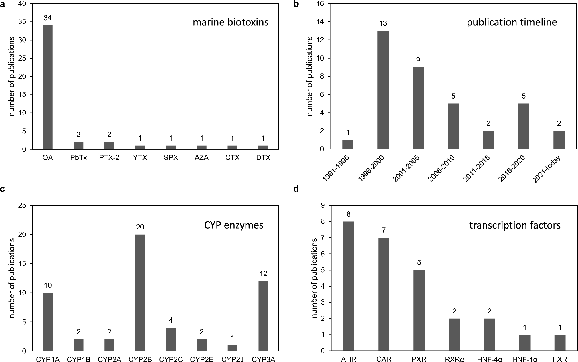
Overview of literature search resultsSearch until May 16, 2023, yielded a total of 37 relevant publications after removal of non-relevant hits, for example papers describing the metabolism of marine biotoxins by CYPs, but not the regulation of CYPs by marine biotoxins. Moreover, results were filtered for mammalian CYPs from CYP subfamilies 1–3, to focus on mammalian drug and xenobiotic metabolism. Papers dealing with other species or CYP families were therefore also excluded. Given the fact that the numbers of publications retrieved with PubMed searches for many research topics are very often three- or four-digit, this gave a first indication that the field had not been subject of extensive research in the past. The first striking observation was that okadaic acid was the toxin of choice in the vast majority of papers (34 out of 37 publications). By contrast, all other toxins, namely AZA1-3, PTX2, DTX1-2, YTX, SPX (spirolide), CTX (ciguatoxin), and PbTX, had been subject of research in only in one or two papers. This is visualized in Fig. 1A. Please note that more than one toxin has been used in several studies. Therefore, the total numbers in the diagram (and subsequent diagrams) may exceed the overall number of papers used as a data source. A publication peak was noted between 1996 and 2005, while the numbers of papers published regarding the regulation of CYPs by marine biotoxins declined somewhat afterwards, with an average of not more than one publication per year or even less since 2011 (Fig. 1B). The CYP subfamily mentioned most in the published literature as being regulated by marine biotoxins (i.e., mainly be okadaic acid), was CYP2B (20 publications), followed by CYP1A (10 publications), CYP3A (12 publications), and CYP2C (4 publications). Other CYP subfamilies were studied less often (Fig. 1C). Among the xenobiotic-sensing receptors, research was rather similarly distributed over the classic xeno-sensors AHR (8 publications), CAR (7 publications), and PXR (5 publications), while some research has also been performed on the nuclear receptors retinoid-X-receptor alpha (RXRα), farnesoid-X-receptor (FXR), and the different hepatocyte nuclear factors (HNFs) (Fig. 1D).
Fig. 1
Results of a literature search on the effects of marine biotoxins on the regulation of CYP family 1–3 enzymes in mammalian cells. a Numbers of publications containing data on effects of individual marine biotoxins on CYP enzymes. b Illustration of publication timeline of relevant papers. c Numbers of papers in which the regulation of specific CYP subfamilies has been studied under the influence of marine biotoxins. d Identity of the different xeno-sensors/nuclear receptors, which have been studied in the context of CYP regulation by marine biotoxins
Most of the work on CYP regulation by marine biotoxins has been done in vitro. To this end, human cells have been used most frequently, followed by cells of mouse and rat origin (Fig. 2A). Research in vitro was equally distributed between the use of primary cells and permanent cell lines (Fig. 2B). Studies involving in vivo research were rarer (6 publications), and were done in rats or mice or (Fig. 2A). With respect to nuclear receptor involvement, a clear preference of researchers for human cells and receptors (10 publications) was evident, whereas nuclear receptors from other species were studied only occasionally (Fig. 2C).
Fig. 2
Overview of species and in vitro/in vivo model systems in research on CYP regulation by marine biotoxins. a Species origin of model systems in vitro and in vivo for research on CYP regulation by marine biotoxins. b Use of primary or permanent cell lines for in vitro research on marine biotoxin effects of CYP regulation. c Species origin of model systems in vitro and in vivo for research on the regulation of CYP-regulating xeno-sensors by marine biotoxins
Regulation of CYP family 1 members by marine biotoxinsCYPs from family 1 are known to metabolize primarily planar and hydrophobic substrates. Accordingly, the AHR, activated for example by dioxins and polycyclic aromatic hydrocarbons, is a major transcriptional regulator of the expression of CYP family 1 members (Abel and Haarmann-Stemmann 2010; Haarmann-Stemmann and Abel 2006). CYP 1 regulation by marine biotoxins was investigated in 10 different papers (Alarcan et al. 2017; Ferron et al. 2016; Hukkanen et al. 2000; Oesch-Bartlomowicz et al. 1997; Posti et al. 1999; Shimoyama et al. 2014; Sidhu and Omiecinski 1997; Tamaki et al. 2005; Wuerger et al. 2022; Wuerger et al. 2023), with a total of 21 different entries for individual CYPs, experimental systems and toxins. A summary of the observations along with the respective references is provided in Table 1. OA was most intensively investigated, while PTX-2 was used in two studies. Analyses of effects of other marine biotoxins on CYP1A regulation have not been published. Data for CYP1A1 regulation by OA almost consistently point towards a downregulation of CYP1A1 in different human and rodent in vitro systems (Table 1). No effect of OA on CYP1A1 has been observed in a single paper, where the toxin did not affect the TCDD-induced levels of CYP1A1 in human A549 lung carcinoma cells (Hukkanen et al. 2000). The remaining studies listed in Table 1 are dealing with hepatic cells from rodents or humans; and based on the available data, a general consistent downregulation of CYP1A1 in human and rodent liver cells by OA can be concluded. This appears to apply to both, basal CYP1A1 expression, as well as CYP1A1 expression activated by ligands of the AHR (Table 1). In one study, the effect of OA on TCDD-induced CYP1A1 levels has additionally been investigated in human intestinal and breast cells, and a downregulation of CYP1A1, similar to what was seen in liver cells, was observed here (Shimoyama et al. 2014). Similar observations were made for OA-dependent regulation of CYP1A2, where also a downregulation of the CYP isoform by OA was observed, both in its basal and AHR activator-induced state (Table 1). CYP1B1 expression alterations by OA have been investigated in only two studies with human in vitro models, where a slight but not statistically significant upregulation of TCDD-induced CYP1B1 levels in A549 cells was observed in one publication (Hukkanen et al. 2000), whereas downregulation was observed in liver cells by others (Shimoyama et al. 2014). In contrast to the CYP-repression effects of OA, the few data available for CYP1 regulation by PTX-2 show consistent upregulation of CYPs 1A1 and 1A2 in human cells by this toxin (Table 1).
Table 1 Effects of marine biotoxins on CYP1 isoformsEffects of marine biotoxins on CYP family 1 members might, in principle, be indirectly caused by interference with the xeno-sensor and key CYP1A/CYP1B regulator AHR. No or only minor effects of OA on AHR expression or activity were observed in some studies with different models (Alarcan et al. 2017; Kurl 1994; Shimoyama et al. 2014); or downregulation of AHR expression in human cells occurred only at OA concentrations much higher than needed for CYP1A1/1A2 repression (Wuerger et al. 2022). This may indicate that a mechanism distinct from simple regulation of the overall cellular amount of the AHR is responsible for the OA-mediated decrease in CYP1A. On the other hand, increased nuclear AHR localization has been observed in a human epidermis cell line after treatment with OA (Ikuta et al. 2004). Moreover, it was observed in Hepa-1 mouse liver cells that OA does not affect AHR DNA binding and basal AHR-dependent transcription, but increased TCDD-dependent transcription mediated by AHR (Li and Dougherty 1997). OA also appears to be able to affect phosphorylation of the AHR dimerization partner ARNT in COS-1 monkey kidney cells (Levine and Perdew 2002). It is, however, not yet clear, whether these observations result in consequences for the AHR/ARNT dimer and its transactivation potential. Shimoyama and co-workers have conducted mechanistic analyses of AHR activation in human cells and discovered an AHR-independent pathway involving the phosphorylation and dephosphorylation of the transcription factor Sp1 at Ser-59 by protein phosphatase 2A (PP2A), thus offering a possible molecular explanation of the inhibitory effect of OA on CYP1A1 via PP2A inhibition and interference with SP1 phosphorylation (Shimoyama et al. 2014).
Regulation of CYP family 2 members by marine biotoxinsThe CYP2 family gathers many isoforms and metabolizes a wide range of drugs, as for example reviewed by (Pelkonen et al. 2008): Key enzymes from this family are CYP2A6, which typically metabolizes smaller planar molecules (e.g., nicotine), CYP2B6, which catalyzes the biotransformation of neutral molecules, CYP2D6 being well known for its important role in drug metabolism and its genetic polymorphisms, or CYP2E1, well-expressed in the liver and mainly involved in the metabolism of small molecules including ethanol. Members of the CYP2C (e.g., CYP2C9, 2C8, and 2C19) metabolize a large number of commonly used drugs such as fluoxetine, fluvastatin, diclofenac, diazepam, mephenytoin or omeprazole. CYP2 regulation by marine biotoxins was investigated in 23 different papers (Abe et al. 2017; Alarcan et al. 2017; Ferron et al. 2016; Gahrs et al. 2013; Ganem et al. 1999; Honkakoski and Negishi 1998; Inoue et al. 2006; Joannard et al. 2000; Kawamoto et al. 1999; Kawamura et al. 1999; Morey et al. 2008; Nirodi et al. 1996; Posti et al. 1999; Pustylnyak et al. 2005; Samudre et al. 2002; Sidhu and Omiecinski 1997; Swales et al. 2005; Tohkin et al. 1996; Wuerger et al. 2022, 2023; Yamasaki et al. 2018; Yoshinari et al. 2003; Zhang et al. 2006), as summarized in Table 2. Thus, CYP2 family members constitute the CYP enzymes which have been most often mentioned in the context of CYP regulation by marine biotoxins. Notably, 6 of those studies are vivo trials, mostly in rats. Similar to studies of the CYP1 family, OA was the most investigated toxin, while only few studies included on PTX-2 or CTX. In short, following a single dose administration of CTX to male C57/BL6 mice, gene expression analysis in the liver revealed upregulation of CYPs 2B9, 2B10, 2B13, 2E1, and 2J9, while CYPs 2J11 and 2J13 were downregulated (Morey et al. 2008). Data for PTX-2 showed an upregulation of CYP2B6, 2C9, and 2C19 following 24 h treatment in human HepaRG liver cells (Alarcan et al. 2017). These findings at the gene expression level were correlated with increases in the enzymatic activity of 2C9 and 2C19 following 72 h incubation in HepaRG cells (Ferron et al. 2016). Contrary to the upregulations observed for CTX and PTX-2, data in regards to CYP modulation by OA mostly points towards a downregulation of CYP2 enzymes (see Table 2). It is important to note that in most studies, OA was used as pre-treatment before incubation with the indirect CAR activator phenobarbital. Examples of CYP downregulation include CYPs 2B1, 2B2, and 2B10, observed both in in vitro studies using mouse or rat cells. These findings were also reported in most of the in vivo studies conducted in rat (4 out of 5). Data in human liver HepaRG cells show not only gene expression downregulation for CYP2C8, CYP2C9, CYP2C19, and CYP2E1, but also decreased CYP activity (Wuerger et al. 2022). In addition, decreases in the activity of CYP2C9 and CYP2C19 following 72 h incubation in HepaRG cells were observed by (Ferron et al. 2016). Conflicting data were reported for CYPB26: while (Wuerger et al. 2022) observed gene downregulation, upregulation was observed by (Swales et al. 2005) (mRNA) and (Inoue et al. 2006) (reporter assay). However, it is important to note that the reported upregulation occurred at most when OA was co-incubated with the CAR activator 1,4-bis [2-(3,5-dichloropyridyloxy)] benzene (TCPOBOP). This outcome points towards the key importance of CAR. Indeed, CYP 2 family enzymes, and especially CYP2B6, are under the transcriptional regulation of CAR. Under physiological conditions, CAR is located in the cytoplasm in an inactive state due to a multi-protein retention complex constituted of heat-shock protein (HSP) 90 and CAR cytoplasmic retention protein (CCRP). HSP70 has also been shown to stabilize this complex in the inactive state (Timsit and Negishi 2014; Yoshinari et al. 2003).
Table 2 Effects of marine biotoxins on CYP2 isoformsRegulation of CYP family 3 members by marine biotoxinsCYP3 family members engaged in xenobiotic metabolism in humans or rodents are the different isoforms of sub-family CYP3A, which is mainly regulated by PXR; for example, see the review by (Tompkins and Wallace 2007). Key isoforms in humans include CYP3A4, CYP3A5 and CYP3A7 (Pelkonen et al. 2008). CYP3A regulation by marine biotoxins has been studied in 12 different publications (Alarcan et al. 2017; Ding and Staudinger 2005; Ferron et al. 2016; Gahrs et al. 2013; Joannard et al. 2000; Morey et al. 2008; Swales et al. 2005; Wuerger et al. 2022, 2023; Yamasaki et al. 2018; Yokobori et al. 2019; Zhang et al. 2006), and again OA was the main biotoxin studied, with PTX-2 being investigated as a second toxin (Table 3). Data univocally show a downregulation of CYP3A enzymes, in cell line models of mostly human, more rarely of rodent origin, as well as in rodent in vivo studies (Table 3). Moreover, results also show decreased CYP3A4 activity following 24 h (Wuerger et al. 2022) or 72 h incubation in HepaRG cells (Ferron et al. 2016). Administration of CTX to male C57/BL6 mice led to CYP3A44 downregulation in the liver (Morey et al. 2008). In contrast to the generally rather CYP-repressive effects of OA, available data show no effect or very slight upregulation of CYPs 3A4 and 3A5 by PTX-2 in human liver cells (Table 1). In a HepG2-hPXR luciferase reporter assay, OA induced luciferase activity, suggesting a PXR-dependent activation of the luciferase reporter construct through the XREM sequence of the human CYP3A4 promoter (Ferron et al. 2016). Conversely, in transfected HEK-T cells, OA provoked strong decreases in firefly luciferase signals in human PXR and RXRα transactivation assays (Wuerger et al. 2023).
Table 3 Effects of marin
留言 (0)