In this study, the objective was to construct a model for predicting the probability of recurrent implantation failure (RIF) after assisted reproductive technology (ART) treatment based on the clinical characteristics and routine laboratory test data of infertile patients. The results revealed that increased infertility duration, uterine cavity abnormalities, low AMH levels, insulin resistance, ANA positivity, and A-β2-GPI Ab positivity were associated with an increased risk of RIF. In subsequent ART cycles for RIF patients, advanced age increases the risk of no live births, and blastocyst transplantation is more conducive to achieving a live birth.
With the development of ART, clinical treatment protocols are becoming increasingly effective, and the success rate of IVF has improved; however, there are still some families who undergo multiple high-quality embryo transfers for fertility treatment and are unable to achieve pregnancy. With the development of preimplantation genetic testing (PGT), approximately 70% of embryos are identified as high quality [12]. Thus, maternal autopathological factors are closely related to the occurrence of RIF. In previous studies, aetiological screening for RIF included examination of thrombosis tendency, immunoglobulin levels, lymphocyte subsets, and multiple immune cytokine profiles [13,14,15]. However, the significance of several indicators in the diagnosis and prediction of RIF is still controversial. Patients with RIF suffer great psychological and economic pressure due to multiple implantation failures. Therefore, analysing the clinical characteristics of patients and the results of routine laboratory tests are particularly important for the analysis and prediction of RIF to provide patients with more reasonable clinical management strategies to improve pregnancy outcomes.
With increasing infertility duration, sperm may exhibit decreased acrosomal protein activity and nuclear chromatin immaturity, which increases the possibility of sperm–egg union disorder in infertile couples [16]. This increases the risk of embryo implantation failure. The results of this study suggest that an increase in infertility duration is a risk factor for RIF. Studies have shown that pregnancy rates are closely related to a woman’s age [17]. The proportion of oocytes with chromosomal abnormalities begins to increase after the age of 26 years, and the older the woman is, the greater the probability of chromosomal abnormalities in oocytes [18], which may affect embryonic development potential. Previous studies have shown that primary infertility is associated with IVF-assisted pregnancy failure [19]. In this study, there were significant differences in age and infertility type between the RIF group and the control group, but logistic regression analysis showed that age and infertility type were not risk factors for RIF. In future studies, data from more centres and larger samples are needed for verification.
Embryo implantation requires a good uterine environment, and studies have shown that the incidence of uterine cavity abnormalities in RIF patients can reach 25–50% [20]. Ultrasonography and hysteroscopy are commonly used in clinical monitoring. The most common intrauterine lesions closely related to embryo implantation include intrauterine adhesions, endometritis, endometrial polyps and submucosal myoma. Hysteroscopy is the gold standard for detecting and treating uterine factors and can detect intrauterine lesions that may be missed by other examinations [21]. Most studies suggest that patients with RIF should undergo hysteroscopy before undergoing further assisted reproduction cycles and that pregnancy should be facilitated after ruling out/treating uterine cavity lesions, therefore significantly improving the pregnancy rate [22]. In this study, the proportion of uterine abnormalities in patients with RIF was significantly greater than that in healthy individuals, and logistic regression analysis revealed that uterine abnormalities were a risk factor for RIF.
AMH is a member of the transforming growth factor β (TGF-β) superfamily and is secreted by ovarian granulosa cells [23]. The serum AMH concentration is widely used to evaluate and predict ovarian function, COH, embryo quality and pregnancy outcomes [24, 25]. Previous studies have shown that the AMH concentration is an important clinical predictor of ART cycle outcomes [26]. In a study based on single-dominant follicles and in vitro fertilization (IVF), AMH levels in follicular fluid, but not in serum, were correlated with embryo implantation potential [27]. In this study, the AMH concentration in the RIF group was significantly lower than that in the control group, and logistic regression analysis revealed that a low AMH concentration was a risk factor for the occurrence of RIF. Low level of AMH may affect the quality of embryos and thus the implantation of embryos.
Studies have shown that IR can affect oocyte meiosis in PCOS patients and delay oocyte maturation, thereby reducing the number of mature oocytes [28]. A study in a mouse model of insulin resistance showed that IR increased oxidative stress and mitochondrial dysfunction in mouse oocytes, resulting in poor oocyte quality and reduced fertility [29]. A prospective clinical study showed that the implantation and pregnancy rates of PCOS patients with IR were significantly lower than those of PCOS patients without IR, and there was no significant difference in embryo quality between the two groups. It is speculated that IR may reduce the embryo implantation rate by affecting the function of the endometrium in patients [30]. In this study, the HOMA-IR score was significantly greater in RIF patients than in control individuals. Logistic regression analysis revealed that a high IR was a risk factor for the occurrence of RIF.
Previous studies have shown that autoimmune coordination is an important condition for a successful pregnancy [31, 32]. ANA is a screening antibody for autoimmune diseases. ANA positivity can reduce oocyte quality, affect embryo development, and reduce the embryo implantation and pregnancy rates, resulting in repeated pregnancy loss [33,34,35]. Anti-phospholipid antibodies (APAs), such as anti-cardiolipin (aCL) and A-β2-GPI Abs, can affect pregnancy outcomes by interfering with oocyte development, embryo morphology, uterine contractions, and appropriate decidua and are potential causes of hypofertility [36]. In this study, the percentages of patients who tested positive for thyroid antibodies, ANAs and A-β2-GPI Abs in the RIF group were significantly greater than those in the control group. Logistic regression analysis revealed that ANA and A-β2-GPI Ab positivity were risk factors for the occurrence of RIF.
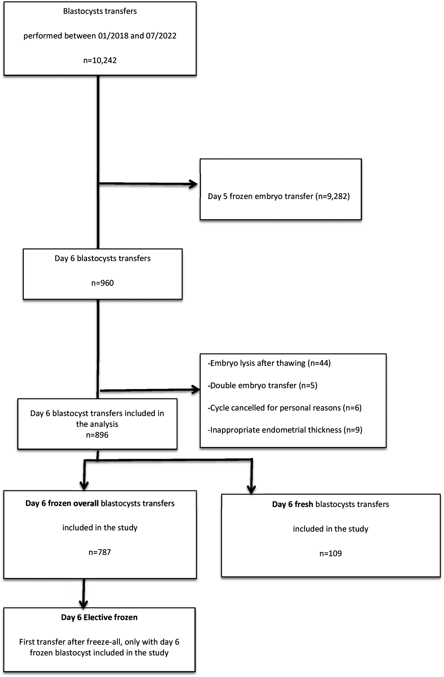
Aetiological screening was carried out for RIF patients, and assisted reproduction therapy was continued after symptomatic treatment. This study further analysed RIF patients with and without live births after the subsequent first assisted reproduction cycle. Logistic regression analysis revealed that advanced age was a risk factor for no live births after subsequent assisted reproduction cycles in RIF patients, and blastocyst transfer improved the live birth rate after subsequent assisted reproduction cycles in RIF patients. Advanced age leads to a decrease in the number of transferable embryos and strongly affects the quality of embryos; in particular, an increase in the number of aneuploid embryos significantly reduces the pregnancy rate [37, 38]. It has been reported that with increasing age, the asynchronism of embryo-intima development increases. In women aged 35 years, the increase in asynchronism of the embryo intima can lead to a significant decrease in the embryo implantation rate and a significant increase in the biochemical pregnancy rate [39]. Therefore, for RIF patients undergoing subsequent assisted reproduction cycles, the appropriate length of time should be determined to improve the live birth rate. A prospective cohort study showed that implantation rates were significantly greater in patients who underwent blastocyst transfer than in patients who underwent cleavage-stage embryo transfer [40]. Studies have shown that blastocyst transfer can improve the clinical pregnancy rate in patients with RIF [41]. Therefore, for the subsequent assisted pregnancy cycle of RIF patients, blastocyst transfer should be performed as much as possible, and assisted reproduction treatment should be continued as soon as possible through reasonable cycle management to minimize the adverse effects of age on assisted pregnancy outcomes.
Limitations
This was a single-centre regression study, for which the results need to be evaluated and verified by prospective large-scale randomized controlled studies. The small sample size for the analysis of factors influencing pregnancy outcomes in subsequent assisted reproduction cycles for RIF patients resulted in the inclusion of fewer covariates, and future studies with larger samples and the inclusion of more factors are needed for assessment and validation.


留言 (0)