
記住我


SILu™Lite SigmaMAb infliximab Monoclonal Antibody, Tris(2-carboxyethyl) phosphine hydrochloride (TCEP), and Iodoacetamide (IAA) was purchased from Sigma-Aldrich (St. Louis, US). Trypsin Gold (trypsin 1), Platinum (trypsin 2) and sequencing grade (trypsin 3) were purchased from Promega (Madison, US). Trypsin sequencing grade (trypsin 4) Trypsin MS approved (trypsin 5) was purchased from Serva Electrophoresis (Heidelberg, Germany). Recombinant trypsin (trypsin 6) trypsin bovine (trypsin 7) purchased from Sigma-Aldrich (St. Louis, US). Oasis HLB µElution Plate 30 μm was purchased from Waters (Eschborn, Germany). All solvents used for chromatography were purchased from Biosolve (Biosolve BV, Valkenswaard, Netherlands) with LC-MS-grade quality.
Patient cohort and sample materialAnonymous leftover serum samples were collected from the MVZ Laboratory Dr. Limbach after routine analysis. The handling of specimens for this laboratory-based analytical study was in accordance to the prerequisites that have been defined by the Central Ethics Committee of the German Medical AssociationFootnote 1. In particular, the cohort consists of human blood serum from 8 patients who are being treated with the monoclonal antibody infliximab. Samples were aliquoted, snap frozen using liquid nitrogen, and stored at -80 °C until used. In addition, for each serum sample corresponding ELISA measurement values were available.
Calculation of digest yield of the antibody infliximab peptidesTo assess the most suitable hydrolase in terms of digest yield and reproducibility, two distinct experiments were conducted. To ensure a fair comparison of digestion efficiencies among different hydrolases in an exploratory experiment, the same sample matrix (1 µl NIST plasma) with a fixed digestion time of 16 h was used for each analyis. Moreover, the digestion process adhered consistently to the same protocol, maintaining an identical protein:trypsin ratio of 20:1. To determine the digestion yield, 500 µg SILuTMLite SigmaMAb infliximab Monoclonal Antibody (infliximab Standard, Sigma-Aldrich, USA) was first rehydrated and then diluted with plasma as matrix to obtain a final concentration of 0.025 µg/µL antibody in solution. Subsequently, 1.25 µg per vial was diluted according to the conventional FASP protocol to finally obtain a concentration of 3.79 µg/ml after elution of the digest from the FASP filter reflecting complete digestion. To determine the experimental yield of the digest, a stable isotope labeled standard of known concentration was added to the digested peptides. The corresponding peak areas of standard to digested peptide reflect the experimentally determined digestion yield in regard to the theoretical amount per well.
To further explore the results of these experiments, we performed a time-resolved study in a second experiment to investigate the effects of different incubation times on digestion of the antibody. For this purpose we used the best performing trypsin from the exploratory study. Specifically, incubation periods of 2 h, 4 h, 6 h, and 16 h were systematically tested using the same experimental setup described above.
Comparison of four common sample preparation methodsIn conjunction with the digestion yield experiments we compared four sample preparation methods: S-Trap 2-hour digestion (as recommended by the manufacturer), S-Trap 16-hour digestion, FASP and in-solution digestion. Each method involved the processing of five NIST plasma samples, with two individuals working on the preparation independent from each other. In total, each preparation was accomplished in 10 technical replicates from a pooled plasma sample to monitor the reproducibility.
S-TrapThe buffer used contained 5% SDS and 100 mM triethylammonium bicarbonate (TEAB), PhosStop and Protease Inhibitor. The binding buffer used subsequently, consisting of 50 ml 1 M TEAB, was adjusted to a pH of 7.2. For plasma, 5 µl of the plasma sample was taken and diluted with 95 µl of a freshly prepared SDS lysis buffer. Of this solution, 30 µl were taken for reduction and alkylation. For reduction, 0.6 µl of a 500 mM TCEP solution was added to the sample, and it was incubated for 30 min at 37 °C with shaking. After the incubation period, 0.9 µl of a 500 mM IAA solution was added. Another incubation step was conducted in the dark at room temperature with shaking for 30 min. Subsequently, 3.5 µl of phosphoric acid (12%) and 210 µl binding buffer were added to each sample. Sample clean up and proteolysis were carried out using the S-Trap protocol (Protifi) for S-Trap Mini cartridges using a protein to trypsin ratio of 10:1. Samples were incubated either 2 h at 47 °C or 16 h at 37 °C. After desalting, the samples were dried down and reconstituted at a concentration of 3.3 µg/µl.
FASPFor the preparation and digestion of plasma according to FASP, the plasma sample underwent alkylation. Therefore, 30 µl of a 50 mM ABC buffer (pH 8.5) and 7 µl of a 10% DOC (sodium deoxycholate) solution were added to 5 µl of the sample, followed by shaking for five seconds. Subsequently, 1 µl of 500 mM TCEP was added, and the mixture was incubated at 37 °C for 30 min. After the incubation period, a 500 mM IAA solution was added until a final concentration of 15 mM was reached. Another incubation step was carried out in the dark at room temperature with shaking for 30 min. The FASP filter was conditioned with 100 µl of the 8 M urea solution and centrifuged at 13,800 rcf for 5 min. The eluate was discarded in the same manner as for the sample loading and washing steps. The alkylated sample was mixed with the 8 M urea solution on the filter and centrifuged at 13,800 rcf for 20 min. Washing steps were performed three times with 100 µl of 8 M urea solution and three times with 100 µl of 50 mM ABC solution. Centrifugation followed each washing step. For each sample, 100 µl digestion buffer (100 µg trypsin in 1 ml 50 mM ammonium bicarbonate (ABC), 2 µl 1 M CaCl2 solution) was added (trypsin to substrate ratio of 1:10), treated for 5 min at 37 °C with shaking, and then incubated for 16 h at 37 °C. Subsequently, 50 µl 50 mM ABC were pipetted onto the filter and centrifuged. Finally, 50 µl ultrapure water was added and centrifuged, completing the elution and enabling the disposal of the filter. To achieve a pH value of < 3.0, 20 µl of 10% TFA was added to the sample solution and checked with pH paper. The sample was frozen in this dissolved state at -80 °C.
In solution digestionFor in-solution digestion, 5 µl of plasma (NIST) or serum (patients) was initially added to 10 µl of the urea buffer in a 96-well plate. Subsequently, 3.6 µl of 50 mM TCEP was added and incubated for 30 min at 37 °C. For alkylation, 1.4 µl of a 250 mM IAA solution was added to the wells, mixed, and allowed to stand for an additional 30 min at room temperature, protected from light. A volume of 6.1 µl of the sample was then transferred to a new reaction vessel. To initiate digestion, 100 µg of trypsin was dissolved in 450 µl of ABC buffer, and 0.9 µl of CaCl2 solution was added. Subsequently, 45 µl of each was added to the samples to achieve a final trypsin-to-substrate ratio of 1:10. The mixture was then incubated at 37 °C for approximately 16 h. Digestion was halted by adding 5.1 µl of 10% formic acid (FA). Purification was carried out simultaneously for both the FASP and in-solution samples using the same protocol. All subsequent steps were executed with the semi-automated sample preparation device Resolvex A200 from Tecan (Männedorf, Switzerland). To prepare the 96-well filter plates, they were conditioned twice with 100 µl of ACN with 0.1% TFA and twice with 100 µl of 0.1% TFA in water. The samples were then transferred to the filters. Subsequent washing steps were performed three times with 100 µl of 0.1% TFA, followed by elution twice with 50 µl of 60% ACN containing 0.1% FA. The plate was dried and frozen at -80 °C until further analysis.
Quality control of prepared samplesFollowing the desalting process, the entirety of the proteolytic digests underwent meticulous evaluation for complete digestion using monolithic column separation (PepSwift monolithic PS-DVB PL-CAP200-PM, Dionex) integrated into an inert Ultimate 3000 HPLC system (Dionex, Germering, Germany). This assessment involved the direct injection of a 1 µg sample. Utilizing a binary gradient (solvent A: 0.1% TFA, solvent B: 0.08% TFA, 84% ACN) with a progression from 5 to 12% B within 5 min and subsequently from 12 to 50% B over 15 min, the flow rate was maintained at 2.2 µl/min, and the temperature was set at 60 °C. UV traces were recorded at 214 nm throughout this procedure [11].
Data independent-acquisition LC-MS/MSAll samples were analyzed using an UltiMate 3000 RSLC nano UHPLC coupled to a QExactive HF mass spectrometer, and 1 µg of peptide mix was applied. Samples were first transferred to a 75 μm × 2 cm, 100 Å, C18 precolumn at a flow rate of 10 µl/min for 20 min., followed by separation on the 75 μm × 50 cm, 100 Å, C18 main column with a flow rate of 250 nl/min and a linear gradient consisting of solution A (99.9% water, 0.1% formic acid) and solution B (84% acetonitrile, 15.9% water, 0.1% formic acid), with a pure gradient length of 120 min (3–45% solution B). The gradient was applied as follows: 3% B for 5 min, 3–35% for 120 min, followed by 3 wash steps, each reaching 95% buffer B for 2 min. After the last wash step, the instrument was allowed to equilibrate for 20 min. MS data acquisition was performed in DIA (data independent acquisition) mode using an in-house generated spectral library. Each analyzed sample was mixed with an appropriate amount of iRT standard (Biognosys). Full MS scans were acquired from 300 to 2,000 m/z at a resolution of 60,000 (Orbitrap) using the polysiloxane ion at 445.12002 m/z as the lock mass. The automatic gain control (AGC) was set to 3E6 and the maximum injection time was set to 20 milliseconds. The full MS scans were followed by 23 DIA windows, each covering a range of 28 m/z with an overlap of 1 m/z starting at 400 m/z and acquired at a resolution of 30,000 (Orbitrap) with an AGC of 3E6 and an nCE of 27 (CID). For analysis of samples acquired by nanoscale liquid chromatography coupled to mass spectrometry (LC-MS/MS) in DIA mode, data were imported into Spectronaut software (Biognosys) and analyzed using a library-based search. The search and extraction settings were kept as default (BGS Factory settings). Human proteome data from UniProt (www.uniprot.org) with 20,374 entries were selected as the proteome background. For reliable label-free quantification, only proteins identified with ≥ 2 unique peptides were considered for further analysis. Next, the average normalized abundance (obtained by Spectronaut) for each protein were calculated and used to determine the ratios of patient muscle samples with their respective controls. Finally, a log2 transformation of the generated ratios and Student’s t-test p-values were calculated for each protein using MS Excel.
Setup and optimizing a targeted assay for infliximab by LC–MS/MS using MRMThe peptides suitable for quantification were synthesized in-house by use of 15N and 13C stable isotope-labeled arginine and lysine (Fig. 1). The selection of peptides was limited to tryptic peptides. In addition, only peptides from the variable range of monoclonal antibodies were selected. This ensured that the peptides subsequently identified by mass spectrometer were antibody-specific. Verification was carried out by localizing the peptide in the amino acid sequence of the antibody. The BLAST algorithm (Basic Local Alignment Search Tool) was used for this purpose [12]. Furthermore, each of the peptides was also available in a natural, non-labeled version for usage as internal standard at a later point.
Fig. 1
Schematic structure of infliximab and its amino acid sequence depicting Fab heavy and light chains. Blue regions represent conserved peptide sequence, red shows unique amino acid sequences and green the hypervariable regions and interaction points with TNFa. The Peptide sequences selected for quantification are marked with bold and underlined
The fragment ions were selected in the Skyline software using the Prosit neural network architecture [13]. Five fragment ions were determined for each previously selected precursor ion. The preliminary selection of fragment ions was based on the top 5 most intense fragment ions calculated by Prosit in silico. An overview of the selected transitions can be found in Table 1. These fragments were then verified by comparing the spectra calculated by Prosit with experimentally determined spectra. The top 5 most intense fragment ions per precursor ion determined in this way were then saved in Skyline for all further measurements.
All MRM measurements were performed with a Vanquish Horizon UHPLC coupled to a TSQ Altis Triple-Quad mass spectrometer (all Thermo Fisher Scientific, Germany) using a binary reversed phase gradient (Waters Acquity UPLC Peptide CSH C18; 1 mm x 150 mm), equipped with a respective VanGuard column at a flow rate of 100 µL/min (0–18 min, 3–35%, 18–20 min, 35–95%, 20–24 min 95%, 24–25 min 3%, 25–26 min 95%, 26–34 min 3%). First, an equimolar mix of 3.47 pmol/µl was prepared from the SIL peptides in 0.1% TFA. Transitions were monitored in positive ion mode using dynamic MRM acquisition with a detection window of 2.5 min, 3 s cycle time, Q1 resolution (FWHM) of 0.7, Q3 resolution (FWHM) of 1.2, and a dwell time of at least 290 ms. All MRM raw data were processed and inspected using the Skyline software (version 23.1.0.268).
For scheduling, 3.47 pmol per peptide was injected to the HPLC and the retention windows of the fragment ions were determined using the Skyline software [14]. All measurements were carried out in scheduled MRM mode with a retention time window of 2.5 min. The best-responding transitions were monitored by MRM-MS for each peptide in all subsequent assay development experiments. A summary of the developed targeted assay used for quantitation can be found in Table 1.
Table 1 Summary of the chosen Infliximab peptides with their corresponding precursor and product m/z values and their optimized collision energyThe optimal CE was calculated using Skyline based on the manufacturer’s specifications for the slope and the y-intercept of the respective charge states. For optimization of the CE, collision energy is ramped stepwise (step size 1.0 V). The experimentally determined optimum CE for each fragment ion was selected based on the signal intensities. These were stored in the method and automatically selected by Skyline for all further measurements. Therefore, raw data was imported into Skyline and the quantification of the monoclonal antibody was carried out using a calibration curve calculated by Skyline. To establish the calibration curves, different amounts of SIS peptides range from 0.25 to 50 µg antibody /ml plasma were spiked into 10 µg of tryptic digested serum as the matrix for the calibration curves in the experiment with addition of corresponding unlabeled peptides with a concentration of 12.5 µg antibody /ml for normalization. Technical triplicates of the individual samples were injected in order of lowest to highest concentration. The calibration line was created using the ratio of the area under the curve of native to isotope-labeled peptides and analyzed without weighting to determine the assay’s linear range and LLOQ. Peptide Settings for quantification and figures of merit were set here as followed: linear regression fit, no regression weighting, maximum LOQ bias 20%, maximum LOQ CV 20%, LOD calculated on blank plus 3*standard deviation.
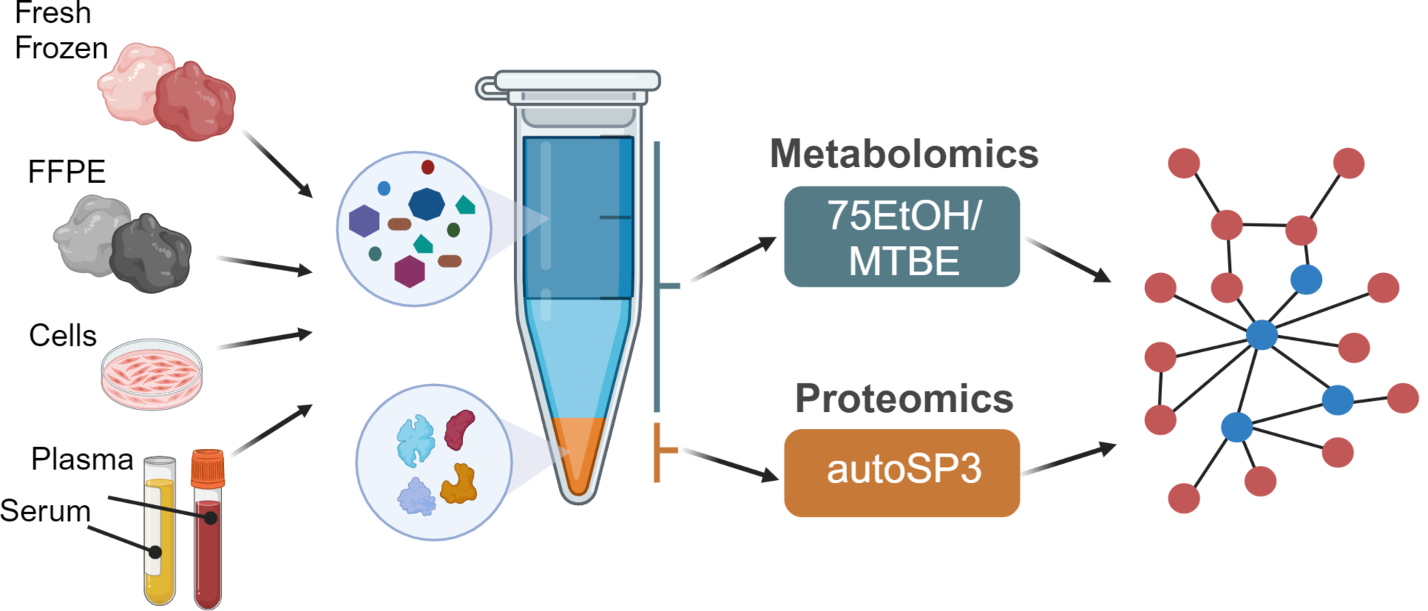
Semi-automated sample processing and trypsin digestionFinally, 5 µL patient serum were added to 10 µL 9 M urea and reduced using 10 mM TCEP for 30 min at 37 °C and further alkylated in 30 mM of IAA for 30 min at room temperature in the dark. The remaining IAA was quenched with 20 mM dithiothreitol (DTT) for a further 15 min at room temperature. Subsequently, samples were digested using trypsin (Serva, ratio [protease:protein] 1:10 in 50 ammonium bicarbonate buffer supplemented with 2 mM CaCl2) at 37 °C for 2 h according to our results of the time-resolved antibody digestion experiment. Digestion was stopped by acidification using 10% formic acid. Finally, peptides were de-salted in a semi-automated way using a Waters OASIS HLB µElution plate, ending up after lyophilisation and re-constitution of the peptides in 95.7 µL 0.1% trifluoroacetic acid (TFA). A comprehensive overview of the final semi-automated workflow for Infliximab quantitation in human serum samples is shown in Fig. 2.
Fig. 2
Semi-automated proteomic workflow: To enhance reproducibility in the sample preparation of TDM samples, an automated 96-well plate FASP process was employed. This allowed for the concurrent preparation of over 90 samples, minimizing manual pipetting steps to reduce inaccuracies and save time. (Figure was created using BioRender)
留言 (0)