
記住我


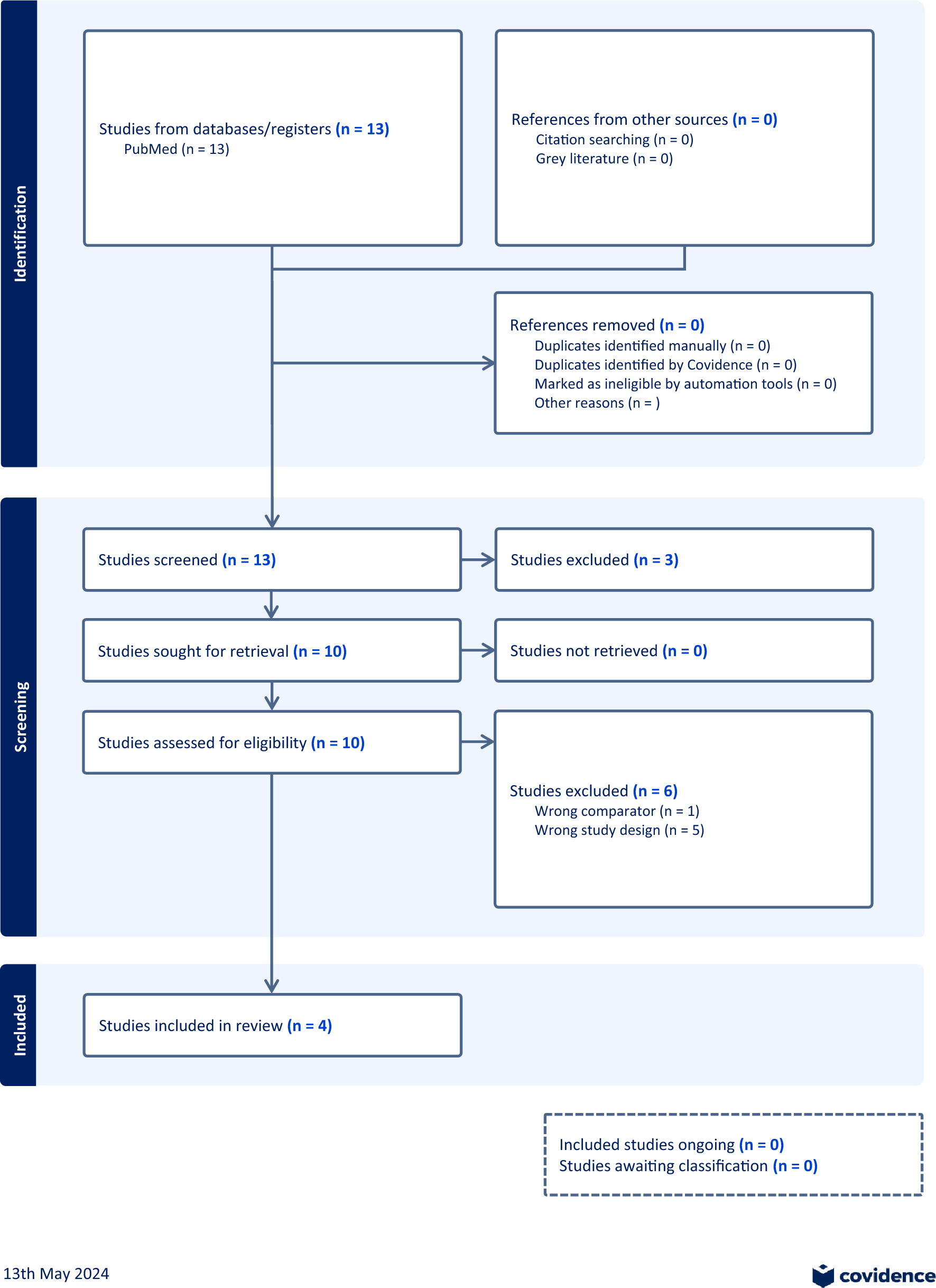


A total of 43 infant-mother dyads were identified through diagnostic codes on the basis of the infant being exposed to lithium through mother’s own milk. Of these, 30 consented to be included in the follow-up study on effects of breastfeeding during lithium therapy (Heinonen et al. 2022). All but three infants were also exposed to lithium in utero, and for 25 of them, the serum lithium concentration measured in the umbilical cord or at the neonatal screening was available. These 25 infants were included in the analyses on the effects of intrauterine and breast milk exposure during the first weeks of life (Fig. 1).
Fig. 1
A flow chart of the study participants and exposure groups. 1 Heinonen et al. 2022. Lithium use during breastfeeding was safe in healthy full-term infants under strict monitoring (Heinonen et al. 2022). 2 Infant serum lithium concentration was measured in cord blood in all but three infants, for whom the lithium concentration measured at two days of age was used instead
Infant characteristicsThe 25 infants had a median (range) GA of 39 + 2 (35 + 2–41 + 0) weeks. The average (SD) birth weight was 3557 (407) g. All infants had APGAR at 5 min of 7 points or more. Seven infants were categorized into the high exposure group (HEG) and 18 to the low exposure group (LEG). One infant in the HEG was born preterm in week 35 + 2, all other infants were term. The same infant was large for gestational age, while all the other infants were appropriate for gestational age. One of the infants in the low exposure group was born with an atrioventricular septal defect. No other malformations were reported. A higher percentage of the infants were exclusively breastfed at follow-up (58.3%) compared with the time of discharge from hospital (45.8%). There was no difference in the level of exclusive breastfeeding between exposure groups. All other infants were partially breastfed. Initially, the mother’s need for sleep was the most common reason for giving additional formula. One infant was given formula due to prematurity and significant weight loss and one after an apparent life-threatening episode (ALTE) and therapeutic lithium concentrations at two days of age.
Maternal characteristicsAll but three mothers were diagnosed with bipolar disorder (Table 1). In 16 women, lithium treatment was initiated before pregnancy, and in seven during pregnancy (data not available for two mothers). Data on psychiatric symptoms at the time of delivery was available for 22 women. Of these, one woman in HEG and two in LEG were experiencing psychiatric symptoms at the time of delivery. The rest were reported to have no or mild symptoms.
Table 1 Background characteristics of the included mother-infant dyadsThe average (SD) dose of lithium sulfate or equivalent was 207 (97) mg/day at delivery and 197 (78) mg/day at follow-up, equaling 1109 (520) and 1055 (418) mg/day in lithium carbonate. The mean lithium dose at delivery was significantly higher in mothers with infants in the HEG than in the LEG (274 (98) vs 180 (85) mg of lithium sulfate a day, equaling 1468 (525) vs 964 (455) mg of lithium carbonate a day, p < 0.05). The difference in maternal lithium dose decreased until follow-up and was no longer statistically significant (222 vs 187 mg/day of lithium sulfate, equaling 1189 vs 1002 mg/day of lithium carbonate, p = 0.27, Table 2). Of the included women, 16 (70%) were also treated with other psychotropic drugs, whereof six with two or more drugs other than lithium. The level of polypharmacy was similar in both exposure groups(Table 1).
Table 2 Comparison of lithium serum concentrations and clinical characteristics between exposure groupsThree months before pregnancy, two (33.4%) women in HEG and five (31.3%) in LEG smoked on a daily basis. Three women (50%) in HEG and nine (60%) in LEG used alcohol. All women had discontinued use of alcohol and all but one in the LEG had discontinued smoking at admission to maternity care in the first trimester (Table 1). No illicit drug use was reported three months prior to or during pregnancy.
Lithium concentrationsMaternal serum lithium concentration at delivery was available for 23 mothers. The median maternal serum lithium concentration at delivery was 0.40 meq/l. The median infant/mother ratio at delivery was 1.0 with no difference between the groups (Table 2). There was a strong correlation between maternal and infant lithium concentrations measured in the umbilical cord, Pearson’s R 0.91, p < 0.05 (Fig. 2). Infant/mother ratio at follow-up was significantly lower than at birth (mean difference 0.89, p < 0.05).
Fig. 2
Scatterplot between infant and maternal lithium serum concentrations measured at delivery. There was a strong correlation between maternal and infant serum lithium concentrations at birth, R = 0.91, p < 0.05
Median infant serum lithium concentration measured in the umbilical cord was 0.45 meq/l, 0.9 meq/l in the HEG and 0.4 meq/l in the LEG, p < 0.05 (Table 2). Ten infants had both the serum lithium concentration analyzed in cord blood as well as a repeat measurement in infant serum at a median age of 48 h. There was no significant difference in the median serum lithium concentration between birth and at two days of age, 0.45 vs 0.40 meq/l, p = 0.20 (Fig. 3).
Fig. 3
Boxplots of infant serum lithium concentrations. Infant serum lithium concentrations measured at birth were <0.6 meq/l in low exposure group (LEG) and ≥ 0.6 meq/l in high exposure group (HEG). The concentrations were measured in the umbilical cord (n = 15 LEG, 7 HEG), at around 2 days of age (n = 9 LEG, 4 HEG) and at the first policlinical follow-up visit at median 20 and 26 days of age in LEG vs HEG (n = 18 LEG, 7 HEG)
The median infant serum lithium concentration at follow-up was 0.10 meq/l. The median lithium concentration was significantly lower at follow-up than at birth, but higher in the HEG than in the LEG (0.20 vs 0.06 meq/l, p < 0.05, Fig. 3). There was no difference in median lithium concentration between exclusively and partially breastfed infants at 48 h of age, 0.40 vs 0.55 meq/l, p = 0.17, or at follow-up, 0.10 vs 0.08 meq/l, p = 0.37. In the LEG, the median serum lithium concentration in the exclusively breastfed infants was 0.07 meq/l, and 0.04 meq/l in the partially breastfed infants at follow-up, p < 0.05.
There was a significant correlation between the maternal dose of lithium sulfate and infant serum lithium concentration at birth, R = 0.41 (p < 0.05), but not at follow-up, R = 0.32 (p = 0.12).
Infant clinical outcomesAt birth, 15 of 24 infants (62.5%) had clinical symptoms, 85.7% of the HEG and 41.2% of the LEG (p = 0.08), presented in Table 3.
Table 3 Neonatal symptoms in infants in high vs low exposure groupsThe most registered symptoms at birth were respiratory symptoms (apnea, labored breathing, need for CPAP and/or ventilation), seen in 25% of the infants with no statistically significant difference between HEG and LEG (Table 3). A fifth of all infants needed neonatal resuscitation within one hour after birth, with continuous positive airway pressure (CPAP) and/or ventilation, 29% in HEG vs 18% in LEG, p = 0.61. The CNS complications seen at birth were jitteriness, agitation and lethargy, seen in 16.7% infants, without a statistically significant difference between HEG and LEG. Thyroid levels were measured in five infants at birth and nine at follow-up. Two infants had elevated levels of thyroid hormone at birth, both normalized without treatment. Plasma creatinine levels were measured in nine infants at birth and 23 at follow-up. One infant had an elevated plasma creatinine level at birth, which was normalized at follow-up. No cases of hypoglycemia were detected.
Four infants (16%) were admitted to neonatal care, two in each exposure group, including the infant with atrioventricular septal defect in the LEG, who was excluded from the analyses of clinical symptoms. The length of stay at neonatal care was 1 day for the infants in the LEG and 0 and 2 days for the infants in the HEG. The median (range) length of stay at maternity ward was 4.5 (3–6) days for LEG and 4 (3–6) days for HEG. There was no statistically significant difference in the separate neonatal morbidities between the exposure groups (Table 3). A detailed description of the infants with possibly severe symptoms and/or high lithium levels is presented in Table 4.
Table 4 Description of infants with possibly severe symptoms and/or high lithium levelsSymptoms at follow-upThe first follow-up visit was at an average (SD) age of 24 (12.5) days, with no statistically significant difference in time to first visit between the exposure groups (Table 2). Four infants were categorized as having clinical symptoms at follow-up. These included poor infant growth (4 infants) and tiredness (2 infants, one from each group). In addition, two infants in the HEG and one in the LEG were recommended to reduce breastfeeding due to high serum lithium concentrations and/or clinical symptoms (Tables 2, 4). There was no difference in clinical symptoms between exclusively and partially breastfed infants at follow-up (14.3 vs 20.0%, p = 1.0). There was a significant reduction in symptoms to the time of follow-up in both groups, to 28.6% symptoms in the HEG, and 11.8% in the LEG, but no statistically significant difference in symptoms between HEG and LEG (p = 0.55).
Symptoms in relation to polypharmacyOf the 15 infants to mothers treated with other psychotropic drugs in addition to lithium, 8 (53.3%) had neonatal symptoms, compared to 4 (57.1%) of the 7 infants to mothers treated with lithium in monotherapy (p = 1.0). At follow-up, all 4 infants with poor growth had mothers treated with other psychotropic drugs as well, equaling 26.7% of the infants of the polypharmacy group, vs 0 infants in lithium only group, p = 0.26.
留言 (0)