
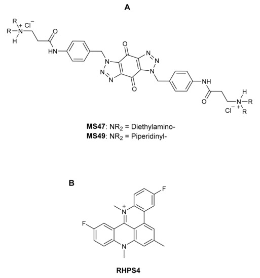
 Ligand MS47 (NSC778438) satisfied the pre-determined criteria for the threshold inhibition in a minimum number of cells in the NCI 60 cell one-dose screening, so it was tested against the panel of sixty tumor cell lines of NCI at five small doses.
 3.4. Cell Cycle AnalysisTo determine whether the MDA-MB-435 cell cycle was perturbed by treatment with MS47 and MS49 and help establish the cell death modality, both a cell cycle analysis and an apoptosis assay were performed [
 3.5. Annexin V-FITC and Propidium Iodide Apoptosis AssayTo investigate the cell death mechanism and test the hypothesis that MS47 and MS49 may initiate apoptosis, human melanoma MDA-MB-435 cells were treated with both ligands (0.5 × GI50, 1 × GI50 and 2 × GI50) for 24, 48 and 72 h, stained with Annexin V-FITC/PI and analyzed by flow cytometry (
記住我


Renal cancer cell lines demonstrate an exceptional resistance to MS47. The renal cancer TK-10 and UO-31 cell lines, ovarian cancer NCI/ADR-RES cell line, and colon cancer HCT-15 cell line have the lowest growth inhibition and highest relative resistance.
Figure 2. The mean graph of the one-dose (10 μM) screen of MS47 (NSC778438) illustrates the sensitivity of the sixty human tumor cell lines to the cytotoxic activity of MS47. Yellow highlights represent the most sensitive cell lines to MS47 cytotoxic (lethal) activity and their PG values in the range of 90%.
Figure 2. The mean graph of the one-dose (10 μM) screen of MS47 (NSC778438) illustrates the sensitivity of the sixty human tumor cell lines to the cytotoxic activity of MS47. Yellow highlights represent the most sensitive cell lines to MS47 cytotoxic (lethal) activity and their PG values in the range of 90%.
The total growth inhibition activity of MS47 (NCS778438) against the NCI 60 human cancer cell lines can be described by the NCI 60 TGI values, which range from 0.26 µM to 38.1 µM. Resistance to MS47 (TGI values > 100 µM) can be found in the leukemia CCRF-CEM, colon cancer HCT-15, ovarian cancer NCI/ADR-RES, and renal cancer 786-0 and CAKI-1 cell lines. MS47 showed the highest potency in total growth inhibition against the ovarian cancer OVCAR-4 cell line (TGI = 0.26 µM), followed by the melanoma MALME-3M cell line (TGI = 0.265 µM), and other cell lines showing sub-micromolar TGI values less than 0.4 µM. While the lowest potency in the total growth inhibitory activity was against the renal cancer UO-31 cell line.
The NCI 60 LC50 values show the lethality and cytotoxicity of MS47 (NCS778438) against the NCI 60 human cancer cell lines. The values range from 0.515 µM to 86.1 µM. The ovarian cancer OVCAR-4 cell line showed the highest sensitivity to MS47 (LC50 = 0.515 µM), followed by the melanoma MDA-MB-435 and MALME-3M, colon cancer COLO 205, and CNS cancer U251 cell lines, with sub-micromolar LC50 values of 0.602, 0.636, 0.644 and 0.694 µM, respectively, and other cell lines with LC50 values of <1 µM. The least lethal activity shown by MS47 was for the renal cancer UO-31 cell line (LC50 = 86.1 µM). All of the leukemia cell lines showed interesting resistance to MS47 cytotoxic activity, in addition to the colon cancer HCT-15, ovarian cancer NCI/ADR-RES, renal cancer 786-0 and CAKI-1, prostate cancer PC-3 and breast cancer HS 578T cell lines, with LC50 values of >100 µM. The resistance and sensitivity shown by the panel of sixty human cancer cell lines reflected a significant selective cytotoxic (lethal) activity for MS47.
Table 1. The anticancer activity of MS47 (NSC778438) against the NCI 60 human cancer cell lines illustrated by its GI50, TGI and LC50 values (µM). Yellow highlights represent the highest potency of MS47, while green highlights represent its lowest potency against NCI 60 panel.
Table 1. The anticancer activity of MS47 (NSC778438) against the NCI 60 human cancer cell lines illustrated by its GI50, TGI and LC50 values (µM). Yellow highlights represent the highest potency of MS47, while green highlights represent its lowest potency against NCI 60 panel.
Panel/Cell LineResponse Parameters of Ligand MS47 (µM)GI50TGILC50Leukaemia CCRF-CEM0.251>100>100HL-60(TB)0.2260.643>100K-5620.59714.4>100MOLT-40.2260.600>100RPMI-82260.6293.86>100SR0.4575.35>100Non-Small Cell Lung Cancer A549/ATCC1.433.177.04EKVX1.543.056.04HOP-621.542.915.52HOP-920.4231.405.88NCI-H2261.563.126.24NCI-H230.2670.79424.8NCI-H322M0.2891.043.45NCI-H4600.3441.386.24NCI-H5220.1940.4814.76Colon Cancer COLO 2050.1740.3350.644HCC-29980.3171.124.49HCT-1160.2831.143.57HCT-1522.9>100>100HT290.3210.90033.8KM120.5442.176.39SW-6200.4071.737.40CNS Cancer SF-2680.3261.456.88SF-2951.362.725.46SF-5390.2020.4200.875SNB-190.1820.3710.755SNB-750.1760.4211.01U2510.1750.3480.694Melanoma LOX IMVI0.1870.4892.62MALME-3M0.1110.2650.636M141.583.387.24MDA-MB-4350.1760.3260.602SK-MEL-20.2190.5538.81SK-MEL-280.9132.466.24SK-MEL-50.2270.7052.70UACC-2570.7112.427.07UACC-621.052.566.26Ovarian Cancer IGROV10.2330.6873.79OVCAR-30.2610.7617.37OVCAR-40.1310.2600.515OVCAR-50.5922.529.07OVCAR-81.563.66-NCI/ADR-RES>100>100>100SK-OV-31.995.3236.9Renal Cancer 786-024.1>100>100A4981.616.0226.0ACHN9.5623.254.6CAKI-1>100>100>100RXF 3933.6015.254.4SN12C0.1850.3710.745TK-1011.130.181.3UO-3116.938.186.1Prostate Cancer PC-32.407.52>100DU-1451.032.234.83Breast Cancer MCF70.1440.3580.894MDA-MB-231/ATCC0.25310.261.4HS 578T2.889.17>100BT-5491.733.165.78MDA-MB-4680.1730.3820.843Figure 3. Dose–response curves of the cytotoxic activity of MS47 (NSC778438) against the panel of sixty human cancer cell lines. Value of 100 for the growth percentage indicates the growth of untreated cells, while a growth percentage value of 0 indicates no net growth throughout the period of the experiment, and a growth percentage value of −100 represents that all of the cells were killed by MS47.
Figure 3. Dose–response curves of the cytotoxic activity of MS47 (NSC778438) against the panel of sixty human cancer cell lines. Value of 100 for the growth percentage indicates the growth of untreated cells, while a growth percentage value of 0 indicates no net growth throughout the period of the experiment, and a growth percentage value of −100 represents that all of the cells were killed by MS47.
The NCI 60 cell five-dose screening results indicated that MS47 (NSC778438) has potent and selective cell growth inhibition and cytotoxic activities against tumor cell lines isolated from distinct organs, making it a promising anticancer drug candidate for further development for the treatment of multiple carcinomas, such as renal, melanoma, ovarian, colon, breast, and CNS cancers.
The cytotoxicity of MS47 indicated by the NCI 60 panel in vitro screen results and the higher selective potent growth inhibitory shown by MS49 indicate that further preclinical evaluations of both ligands are warranted. Specifically notable is the sensitivity of the melanoma MDA-MB-435 cell line (LC50 = 0.602 µM); thus, this cell line was selected for more thorough interrogation of anticancer activity.
3.2. MTT AssayThe growth inhibitory activities of MS47 and MS49 were tested further in vitro using an MTT assay [20,41,42] against the human melanoma MDA-MB-435 cell line, which was chosen in particular for further in vitro anticancer evaluation of both ligands as it is one of the most sensitive cell lines to the lethal effect of MS47, as shown by the NCI 60 panel data.The estimated concentrations at 50% cell growth inhibition (GI50) were determined from the dose–response curves (Figure 4) after 72 h exposure of melanoma MDA-MB-435 cells to MS47 and MS49, and are provided in Table 2. The GI50 values resulting from examination of the growth inhibition by both ligands in human non-tumorigenic MRC-5 (embryonic lung fibroblasts) to evaluate their putative selective cytotoxic activities are also presented in Table 2. In the concentration range (0.01–100 µM), both MS47 and MS49 inhibit the growth of the human melanoma MDA-MB-435 cell line potently. However, ligand MS49 (GI50 value of 75 nM) shows more potent growth inhibitory effect than MS47 (GI50 value of 226 nM). Comparing the GI50 values with those of human normal lung MRC-5 fibroblasts, both ligands demonstrate greater potency in the melanoma cell line than in non-tumorigenic lung fibroblasts, with indicated cancer selectivity indices (SI) of 9.8 for MS47 and 17.7 for MS49, revealing their good selective growth inhibitory effects for cancer cells over normal cells.Table 2. Growth inhibition effects of MS47 and MS49 on human melanoma MDA-MB-435 and human normal lung MRC-5 cell lines. Values of GI50 are presented as mean ± standard deviation of at least three separate experiments (n = 8 per trial). “SI: Selectivity index (GI50 MRC-5/GI50 melanoma cell line)”.
Table 2. Growth inhibition effects of MS47 and MS49 on human melanoma MDA-MB-435 and human normal lung MRC-5 cell lines. Values of GI50 are presented as mean ± standard deviation of at least three separate experiments (n = 8 per trial). “SI: Selectivity index (GI50 MRC-5/GI50 melanoma cell line)”.
LigandGI50 (μM) ± S.D.SIMDA-MB-435 Cell LineMRC-5 Cell Line470.226 ± 0.0572.219 ± 0.0769.8490.075 ± 0.0101.325 ± 0.13717.7Figure 4. Dose–response curves that show the growth inhibiting effects of MS47 and MS49 against melanoma MDA-MB-435 cell line. Values are mean ± SD, n = 8, graphs are representative of experiments performed on at least three separate occasions.
Figure 4. Dose–response curves that show the growth inhibiting effects of MS47 and MS49 against melanoma MDA-MB-435 cell line. Values are mean ± SD, n = 8, graphs are representative of experiments performed on at least three separate occasions.
Figure 6. Statistical analyses of cell cycle phases: G0/G1, S and G2/M in human melanoma MDA-MB-435 cells, treated with 0.5 × GI50, 1 × GI50 and 2 × GI50 of MS47 and MS49 compared to the untreated cells (control group) for (A) 24 h, (B) 48 h and (C) 72 h. MS47 (2 × GI50) evoked significant arrest in the G0/G1 and G2/M phases (* p < 0.05 and ** p < 0.005; experiments were repeated ≥ three times, n = 2). (D) Flow cytometric cell cycle analysis (histograms) of human melanoma MDA-MB-435 cells, treated with MS47 and MS49 of 2 × GI50 concentrations for 72 h compared to the control cells and stained with propidium iodide (PI).
Figure 6. Statistical analyses of cell cycle phases: G0/G1, S and G2/M in human melanoma MDA-MB-435 cells, treated with 0.5 × GI50, 1 × GI50 and 2 × GI50 of MS47 and MS49 compared to the untreated cells (control group) for (A) 24 h, (B) 48 h and (C) 72 h. MS47 (2 × GI50) evoked significant arrest in the G0/G1 and G2/M phases (* p < 0.05 and ** p < 0.005; experiments were repeated ≥ three times, n = 2). (D) Flow cytometric cell cycle analysis (histograms) of human melanoma MDA-MB-435 cells, treated with MS47 and MS49 of 2 × GI50 concentrations for 72 h compared to the control cells and stained with propidium iodide (PI).
Figure 7. Statistical analysis of the percentages of healthy, apoptotic (early and late) and necrotic human melanoma MDA-MB435 cells, treated with MS47 and MS49 (0.5 × GI50, 1 × GI50 and 2 × GI50) compared to the untreated cells (control group) for (A) 24 h, (B) 48 h and (C) 72 h. Both ligands caused significant increase in the percentage and number of the late apoptotic cells (* p < 0.05, ** p < 0.005, *** p < 0.0005; experiments were repeated ≥ three times, n = 2). (D) Flow cytometric analysis (histograms) of cell apoptosis mechanism (apoptosis/necrosis) in human melanoma MDA-MB435 cells, treated with MS47 and MS49 of 2 × GI50 concentrations compared to the untreated cells (control group) and stained with FITC-conjugated annexin V and propidium iodide (PI) for 72 h.
Figure 7. Statistical analysis of the percentages of healthy, apoptotic (early and late) and necrotic human melanoma MDA-MB435 cells, treated with MS47 and MS49 (0.5 × GI50, 1 × GI50 and 2 × GI50) compared to the untreated cells (control group) for (A) 24 h, (B) 48 h and (C) 72 h. Both ligands caused significant increase in the percentage and number of the late apoptotic cells (* p < 0.05, ** p < 0.005, *** p < 0.0005; experiments were repeated ≥ three times, n = 2). (D) Flow cytometric analysis (histograms) of cell apoptosis mechanism (apoptosis/necrosis) in human melanoma MDA-MB435 cells, treated with MS47 and MS49 of 2 × GI50 concentrations compared to the untreated cells (control group) and stained with FITC-conjugated annexin V and propidium iodide (PI) for 72 h.
留言 (0)