1. IntroductionMycotoxin contamination in agricultural and food products is one of the major food safety concerns worldwide [
1,
2]. Penicillium- and Aspergillus-derived ochratoxin A (OTA) [
3] is one of the most toxic mycotoxins that widely contaminate various crops (corn, wheat, beans, and cocoa), resulting in a serious threat to human health and consequential economic losses worldwide [
4]. Many countries and international organizations have set strict limit standards for OTA residues [
5,
6], which are 0.5–10 µg kg−1 in different food commodities according to European Union regulations [
7]. Regarding the universality and severe consequence of mycotoxin pollution, cheap “point of care” (POC) methods without expensive instruments and special technicians play a crucial role in the high-throughput analysis of mycotoxins [
8]. Enzyme-linked immunosorbent assay (ELISA) is the most widely used POC method because of its merits of rapidity, simplicity, cost-effectiveness, and excellent specificity [
9]. However, the conventional competitive ELISA for mycotoxin detection suffers from two major shortcomings [
10,
11]. First, the low-intensity and single-color colorimetric signal of enzymatic reaction-derived products leads to limited sensitivity and poor adaptability of on-site detection based on naked-eye interpretation in resource-constrained areas [
12,
13]. Second, using toxic target mycotoxin and organic reagents for preparing enzyme- or carrier protein-analyte conjugates as competing antigens leads to serious secondary pollution and a nonnegligible occupational hazard [
14]. Hence, highly sensitive and eco-friendly immunoassays for mycotoxin screening must be developed urgently.In recent decades, various colloidal nanoparticles have been used in developing biosensors to enhance or supersede current analytical techniques, making a great impact in research and practice applications [
15,
16]. In particular, plasmonic nanoparticles are interesting because their unique localized surface plasmon resonance (LSPR) enables them to produce intense responses to incident light [
17], which can be linked to the presence of a target analyte to yield extremely sensitive detection [
18,
19]. Gold nanoparticles (AuNPs) are the most widely used plasmonic nanoparticles due to their merits, including easy preparation, excellent colloidal stability, and high accessibility for functionalization [
20,
21]. Highly sensitive AuNP-based sensing methods adopting various output signals, such as colorimetric signals [
12], surface-enhanced Raman scattering [
22], fluorescence [
23], and photothermal signals [
24] have been developed. AuNP-based colorimetric and photothermal sensing methods are particularly well suited for POC applications because of their simple signal readout via the naked eye or a thermometer [
25]. Both colorimetric and photothermal signals generated from AuNPs are far more sensitive than those from conventional ELISAs because of the strong LSPR absorbance and photothermal effect of AuNPs. In recent years, plasmonic and photothermal ELISAs, which integrate LSPR modulation of AuNPs with conventional ELISA platforms, have attracted considerable attention because of their enhanced sensitivity [
26]. The distinct color changes of plasmonic ELISAs are convenient for naked-eye readout without an excitation; the photothermal signal is more suitable than the colorimetric signal for muddy or colored samples [
24]. Therefore, the combination of colorimetric and photothermal signals provides enhanced feasibility in POC applications and enhanced accuracy and reliability because of the mutual verification of the two signals [
27]. Given that the two signals synchronously follow the LSPR modulation, dual modal plasmonic-photothermal ELISAs (ppELISAs) have been developed using AuNPs as signal transducers for the detection of disease biomarkers [
28], nucleic acids [
24], microorganisms [
29], and mycotoxins [
30]. A slight change in the compositions, shapes, sizes, or aggregation states of AuNPs may give rise to a remarkable LSPR variation. Among these modulation strategies, the AuNP aggregation-induced redshift of the LSPR absorbance always results in remarkably contrasting color changes and a distinct photothermal effect with a certain excitation. Previously, we reported a tyramine-mediated AuNP aggregation system based on the horseradish peroxidase (HRP)–catalyzed polymerization of phenolic hydroxyl in tyramine [
31]. On this basis, a direct competitive ppELISA (dc-ppELISA) with enhanced sensitivity, accuracy, and reliability over conventional ELISAs can be promoted for mycotoxin detection.The use of competing antigens of the enzyme-analyte conjugates in conventional direct competitive ELISAs for mycotoxin detection limits the sensitivity because one target molecule can only competitively inhibit the binding of one enzyme molecule. This approach also poses severe secondary pollution problems because of the consumption of toxic target mycotoxin and organic reagents. Therefore, novel competing antigens should be explored, particularly those that are eco-friendly and loaded with enhanced amounts of enzymes. Therefore, the M13 bacteriophage (M13 phage) carrying a mimic antigen epitope and multiple enzymes was proposed as a promising surrogate for competing antigens. The filamentous M13 is noninfectious to humans and composed of 2700 copies of the major p8 proteins and 3–5 copies of minor p3, p6, p7, and p9 proteins at the two ends [
32]. It can be easily modified to mimic various target analytes, such as small molecules or proteins, by integrating mimotopes at the N-terminal of p3 proteins through a gene modification–fusion expression process [
33]. The abundant copies of p8 proteins can be extensively functionalized as a container for high-density loading of signal transducers or regulators (e.g., fluorescent dyes and enzymes), resulting in remarkably enhanced signal intensities of biosensors [
34]. Previously, we adopted an OTA-mimicking M13 phage (M13OTA) integrated with an OTA mimotope at the p3 proteins as an enzyme container to improve the sensitivity of a fluorescent ELISA for OTA detection [
35].
Herein, we developed an eco-friendly dc-ppELISA for the highly sensitive detection of OTA in corn, and a colorimetric–photothermal dual model gave the developed method more applicability in POC screening. A biotinylated M13 phage (bio-M13OTA) was used as a competing antigen and container for glucose oxidase (GOx) enzymes, and citrate-AuNPs were applied as dual-modal signal transducers. The aggregation of citrate-capped AuNPs was induced by the HRP-catalyzed polymerization of tyramine, which was electrostatically adsorbed onto AuNPs in the presence of H2O2 produced from GOx-catalyzed oxidation of glucose. The analytical performance of the proposed dc-ppELISA, including the limit of detection (LOD), 50% competitive inhibition concentration (IC50), accuracy, and reliability, was evaluated and compared with that of conventional HRP-based ELISAs.
4. Materials and Methods 4.1. Materials and Instruments
The following reagents were used: ochratoxin A (OTA), aflatoxin B1 (AFB1), fumonisin B1 (FB1), zearalenone (ZEN), deoxynivalenol (DON) and citrinin (CIT) (Huaan Magnech Bio-Tech., Beijing, China); HRP, GOx, bovine serum albumin (BSA), streptavidin, chloroauric Acid (HAuCl4) and trisodium citrate (Sigma-Aldrich, St. Louis, MO, USA); tyramine (Solarbio, Beijing, China); anti-OTA ascitic fluids (Wuxi Zodoboer Biotech., Wuxi, China); sulfosuccinimidyl 6-(biotinamido) hexanoate (Sulfo-NHS-LC-Biotin, NHS-biotin; Macklin, Shanghai, China); M13 bacteriophage with an OTA-mimicking peptide sequence of GMSWMMA (M13OTA) was given by Prof. Xuelan Chen of Jiangxi Normal University (Nanchang, China). All other analytical-grade chemicals were purchased from Sinopharm Chemical Corp. (Shanghai, China) and applied without further purification. We obtained 96-well plates from Corning, Inc. (New York, NY, USA); 18.2 MΩ cm deionized water was prepared using a Millipore Milli-Q water purification system (Millipore, Milford, MA, USA).
Transmission electron microscopy (TEM) images were obtained with a JEOL JEM-2100 electron microscope (Tokyo, Japan). Ultraviolet–visible (UV-Vis) absorption spectra were recorded using a double-beam UV-vis spectrophotometer (Cintra 10e; GBC, VI, Melbourne, Australia). Average hydrodynamic diameter was determined via dynamic light scattering (DLS) particle size analyzer (Zeta Sizer Nano ZS90, Malvern Instruments Ltd., Worcestershire, UK). The surface plasmon resonance (SPR) signal intensity of the AuNPs was determined using a Multiskan GO multimode reader (Thermo Fisher, Vantaa, Finland). Temperature of the AuNP solution under the excitation of an 808 nm laser was measured by an infrared camera, and all measurements were performed at room temperature.
4.2. Synthesis of Citrate-capped AuNPs
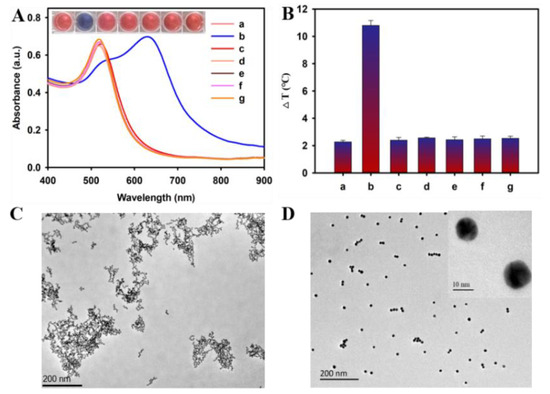
Citrate-capped AuNPs with an average diameter of 13 nm were synthesized via a one-step reduction method. Briefly, 250 mL of 1 mM HAuCl4 solution was heated to a boil under vigorous stirring, and 25 mL of 38.8 mM citrate solution was quickly added followed by 15 min of reaction under a stirring and boiling condition. During this period, the solution color changed immediately from pale yellow to blue and then gradually to dark red. Subsequently, the reaction solution was placed in an ice bath for 15 min, and the resultant AuNP solution was filtered with a 220 nm microporous membrane and stored at 4 °C for later use. The concentration of AuNPs was calculated to be 15.9 nM according to Beer’s Law.
4.3. Propagation of M13OTA Bacteriophage
Escherichia coli ER2738 cells were cultured at 37 °C overnight in tetracycline-containing (10 μg mL−1) Luria–Bertani (LB) medium. Then, 200 μL of the above LB with enriched Escherichia coli cells and 1 μL of M13OTA bacteriophage solution was inoculated into 20 mL of LB solution and cultured at 37 °C for 4~5 h under constant shaking at a speed of 250 rpm. Subsequently, the cell debris was removed via 10 min centrifugation at 5000 rpm, and then the proliferated M13OTA in the supernatant was precipitated at 4 °C overnight after adding a PEG-NaCl solution (2.5 M NaCl, 20% PEG-8000). Thereafter, the precipitates of M13OTA were collected via centrifugation (13,500 rpm, 10 min) and resuspended in 1 mL of phosphate-buffered (PB) solution. The cell debris was removed by spinning for 10 min at 5000 rpm. Then, 200 μL of PEG-NaCl solution was added to the supernatant for 1 h incubation on ice. The M13OTA phage was obtained via centrifugation (13,500 rpm, 10 min) and resuspended with 200 μL of PB buffer. The concentration of amplified M13OTA phage bacteriophages was determined via a plate count method.
4.4. Preparation of Bio-M13OTA and Bio-GOx
Bio-M13OTA and biotinylated GOx (bio-GOx) were obtained via the coupling between the amine groups on p8 proteins of the phage or GOx and the active ester group of NHS-biotin. During the preparation of bio-M13OTA at the different molar ratios, different volumes of NHS-biotin (1 mg mL−1, dissolved in DMF) were added to 1 mL of M13OTA solution (2 × 10⁹ pfu mL−1, pH = 8.6) After 4 h incubation on ice under vigorous stirring, 200 μL of PEG-NaCl solution was added for another 1 h incubation on ice. The bio-M13OTA phage was purified via centrifugation (13,500 rpm, 10 min), resuspended in 1 mL of PB buffer, and stored at 4 °C. Similarly, for the preparation of bio-GOx at the molar ratio of 20:1, 74 μL of NHS-biotin (1 mg mL−1, dissolved in DMF) and 1 mg of GOx powder were mixed in 1 mL PBS solution (0.01 M, pH 8.6) and incubated on ice for 4 h under vigorous stirring. Excess NHS-biotin was removed via dialysis in PBS (0.01 M, pH 7.4) for 72 h. The resultant bio-GOx solution was stored at −20 °C with some glycerin added.
4.5. dc-ppELISA Procedure for OTA
The procedure for the dc-ppELISA using bio-M13OTA as a competing antigen is as follows. First, 100 µL of protein G (25 µg mL−1, 0.01 M PBS pH 8.6) is added into each well of a 96-well microplate and incubated overnight at 4 °C. After washing three times with PBST (PBS containing 0.05% Tween 20) and once with PBS, 100 μL of anti-OTA ascitic fluids (0.75 μg mL−1 in PBS, pH = 8.6) is added to each well and incubated for 2 h at 37 °C. After removing excess anti-OTA ascitic fluids, 300 μL of blocking buffer (1% BSA in 0.01M PBS 7.4) is added for another 2 h incubation at 37 °C. After a repeated washing process, 50 μL of the sample solution and 50 μL of bio-M13OTA solution (2 × 10⁹ pfu mL−1) are added to each well. After incubating for 60 min at 37 °C, the microplate is washed again. Then, 100 μL of streptavidin solution is added and incubated for 30 min at 37 °C. After washing away the unbound streptavidin, 100 μL of bio-GOx is added and incubated for another 30 min at 37 °C. After unreacted bio-GOx is wiped off, 100 μL of D (+)-glucose (1 mg mL−1 in PBS, pH = 7.4) is added for 1 h incubation at 37 °C. Finally, 150 μL of substrate solution containing HRP (50 μL, 5 µg mL−1), tyramine (50 μL, 100 µg mL−1), and AuNPs (50 μL, 15.9 nM) is added into each well. After 5 min incubation, the microplate is photographed to record the color development and the optical density (OD) of each well at 520 and 630 nm are detected by multimode microplate reader. Furthermore, an infrared camera is used to record the temperature of each well after irradiation with an 808 nm laser for another 5 min. The inhibition rates were calculated according to the following formula: inhibition rate (%) = (1 − B/B0) * 100%, where B and B0 represent OD630nm/OD520nm values (colorimetric signal) or temperature changes (∆T, photothermal signal) obtained from detecting OTA-positive and -negative samples.
4.6. Sample Preparation
Corn samples were purchased from grain procurement agencies (Shandong, China), and verified to be OTA-free via high-performance liquid chromatography (HPLC). The HPLC method was according to the Chinese national standard GB 5009.96-2016, which is listed below: (a) chromatographic column: C18 column, column length 150 mm, inner diameter 4.6 mm, particle size 5 μm; (b) mobile phase: acetonitrile water glacial acetic acid (96 + 102 + 2); (c) flow rate: 1.0 mL/min; (d) column temperature: 35 °C; (e) injection volume: 50 μL; (f) detection wavelength: excitation wavelength 333 nm, emission wavelength 460 nm. All samples were thoroughly ground and mixed before use, and the corn samples for assay validation were prepared following a previously reported method and Chinese national standard GB 5009.96-2016. In brief, several portions of finely ground corn samples (5.0 g each portion) were spiked with OTA at different levels (2–100 μg kg−1). Each portion of OTA-spiked corn sample was extracted with 25 mL methanol-H2O solution (80% methanol: 20% H2O) under vigorous shaking for 20 min. After centrifugation at 10,000 rpm for 5 min, the supernatant was then collected and stored at 4 °C for further use.


留言 (0)