
記住我

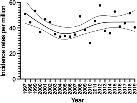
Uterine cancer is the sixth most common cancer in women worldwide with about 417,000 women newly diagnosed in 2020.1 It is also the fourth most common cancer in the United States, with data from the U.S. National Cancer Institute’s Surveillance, Epidemiology, and End Result program indicating that new uterine cancer cases have increased an average 0.5% annually during the last 10 years.2-4 The incidence of uterine cancer in Taiwan was 24.7 per 100,000 women according to the nationwide population-based Taiwan Cancer Registry in 2018.5 In Taiwan, uterine cancer was not in the top 10 cancers prior to 2006,6, 7 but its incidents have been rapidly increasing since 2000.8
Uterine cancers can be categorized as carcinomas, carcinosarcomas, or sarcomas. Approximately 86% of uterine cancers are carcinomas.5, 9 About 80%–90% of endometrial cancers, also called endometrial carcinomas (ECs), are endometrioid type.10 The majority of ECs is diagnosed in the initial stages because the main symptom of abnormal or menopausal vaginal bleeding appears early in the disease and prompts investigation. The endometrioid carcinomas were regarded as type I uterine carcinomas. Uterine papillary serous and clear cell carcinoma were regarded as type II uterine carcinomas which had different treatment strategies and outcomes compared with those type I uterine carcinomas. The International Federation of Gynecology and Obstetrics (FIGO) Committee shifted from clinical to surgical staging of EC in 1988 and revised the staging system in 2009.11 A standard staging procedure, including hysterectomy, bilateral salpingo-oophorectomy, extrauterine tumor excision, and selective pelvic and/or para-aortic lymph node dissectionis routinely recommended for uterine cancer patients.12 Hysterectomy with/without bilateral salpingo-oophorectomy alone would be an alternative for low-risk patients based on the imaging results or those who could not tolerate longer operative time.
Because staging depends on postoperative findings, the estimated stage and risk of extrauterine disease determines the extent of surgery. Extrauterine spread, the depth of myometrial invasion (MI), tumor grade and histological subtype, and lymph node metastases are the factors considered in endometrial cancer prognosis.12 Complete lymph node dissection is associated with morbidity including lymphedema, lymphocele formation, and neuralgia.13 An international consensus conference generated the recommendation that the indication for lymph node dissection should stratify the cases to low, intermediate, or high risk.12 Currently, preoperative magnetic resonance imaging (MRI) and/or computerized tomography (CT), intraoperative frozen section and sentinel lymph node mapping could be used if full staging is needed.14 The GOG 99 and PORTEC trials defined risk factors for those at high to intermediate risk of recurrence, including MI on preoperative imaging and intraoperative surgical findings. Low risk for nodal metastasisis characterized by <50% MI, tumor size <2 cm, and well to moderately differentiated histology.15 One retrospective analysis of low-risk patients without lymphadenectomy reported no significant difference in overall survival and progression-free survival compared to those who underwent lymphadenectomy.16 Evidence on the accuracy of the currently available tools (especially imaging) remain lacking in literature.17-27 Therefore, the optimal selection of patients who can avoid lymph node dissection and the determination of the extent of lymphadenectomy for high- to intermediate-risk patients remain clinical challenges.28, 29
The guidelines recommend MRI for estimating the preoperative stage because of the better resolution of soft tissue contrast for assessing the depth of myometrial or cervical invasion compared to CT.12, 30-32 Ultrasound relies on operator expertise. Positron emission tomography (PET) is not yet widely used for preoperative evaluation in EC due to cost and inaccessibility.
Several studies have reported the assessment of preoperative image reliability, but numbers of patients are limited and not all risk factors were discussed.24-26, 28 So, we conducted a retrospective study to evaluate the diagnostic performance of preoperative MRI in EC staging in routine clinical practice.
2 MATERIALS AND METHODS 2.1 PatientsA total of 1020 patients with EC were identified from the National Taiwan University Hospital covering the period from January 1, 2013, to December 31, 2018. This study was approved by the Institutional Research Ethics Committee at the National Taiwan University Hospital. All of the patients’ data were fully anonymized before we accessed them and the Research Ethics Committee waived the requirement for informed consent. All patients were diagnosed by endometrial biopsy or curettage, with confirmation by hysterectomy. We excluded cases in which patients did not undergo hysterectomy because of personal reasons, were not available for preoperative MRI at our hospital, were not good candidates for surgery, had undergone surgery at another hospital, had incidental cancer such as ovarian cancer after the hysterectomy, or had undergone surveillance at the other hospitals after surgery. We also excluded 66 patients with other histological types, including serous and clear cell carcinoma, adenosarcoma, carcinosarcoma, leiomyosarcoma, and neuroendocrine carcinoma. Data of the remaining 527 patients were eligible for further analysis (Figure S1).
2.2 MRI examinationsAll of the 527 patients underwent abdomino-pelvic MRI to examine upper abdomen. The chest CT scan was only performed when suspected pulmonary metastasis by CxR or clinical symptoms such as cough. MRI examinations were performed using a 1.5-T MRI unit (SignaHDx; GE Healthcare). The pulse sequences for pelvic imaging included T2-weighted fast spin echo (FSE) sequences in the sagittal, coronal oblique, and axial oblique views according to the axis of the uterine body, an axial T2-weighted FSE sequence with fat saturation (FS) of the whole pelvis, an axial T1-weighted gradient-echo (GRE) sequence with FS, and an axial diffusion-weighted echo-planar imaging (DW-EPI) sequence (b-values, 0, and 800 s/mm2) for the whole pelvis. Examinations performed after 2018 also included a sagittal DW-EPI of the uterus. Apparent diffusion coefficient (ADC) maps were derived from the diffusion-weighted sequences, generated by the scanner software. The patients received intravenous gadolinium contrast medium (0.1 mmol/kg of gadoterate meglumine, Dotarem; Guerbet) if there were no contraindications. Post-contrast images include T1-weighted three-dimensional-spoiled GRE sequence with FS in the sagittal, coronal, and axial views. Images of the upper abdomen were also obtained to detect possible metastases. The pulse sequences for upper abdomen include an axial T1-weighted GRE sequence with FS, an axial T2-weighted FSE sequence with FS, and post-contrast T1-weighted three-dimensional-spoiled GRE sequence with FS in the coronal and axial views. The details of these pulse sequences are summarized in Table 1. The MRI examinations were interpreted by total 11 well-experienced and qualified radiologists, who are familiar with abdominal and pelvic imaging. The imaging reports were obtained from the electronic medical record of the hospital. We recorded the following findings from the imaging reports including MI, CI, AM, pelvic and/or para-aortic lymph node metastases, and intra-abdominal metastases. MI was defined as abnormal signal intensity of the tumor extended into the myometrium. CI was defined as disruption of the hypointense cervical stroma by the tumor. AM was defined as abnormal mass involving the adnexa. Lymph nodes with a short axis >1 cm, or with suspicious features including multiple small rounded nodes, irregular contour, abnormal signal intensity similar to that of primary tumor, or presence of necrosis, were considered to be nodal metastasis.32, 33 The definition of intra-abdominal metastasis was tumor lesions which were not included in the other five parameters including para-aortic lymphadenopathy above the renal vessels, peritoneal metastasis such as enhancing omental or peritoneal nodules, or hepatic metastasis as hepatic nodules with mild hyperintensity on T2-weighted images with hypoenhancement on post-contrast images.32
TABLE 1. The relevant methods and conditions of MRI examination Plane Repetition time (TR) (ms) Echo time (TE) (ms) Flip angle (°) Slice thickness (mm) Matrix Field of view (mm) Pelvis T2-weighted FSESagittal
Coronal oblique
Axial oblique
3500–5500 80–100 — 3–4 256 × 192 240–250 T2-weighted FSE with FS Axial 3500–5500 80–100 — 5–6 288 × 192 260–280 T1-weighted GRE with FS Axial 150 4.2 70 5–6 256 × 192 260–280 DW-EPI Axial 7000–9000 60–80 — 5–6 64 × 128 280–300 DW-EPI Sagittal 7000–9000 60–80 — 3–5 64 × 128 240–250 Post-contrast T1-weighted GRE with FSAxial
Sagittal
Coronal
3.8–4.6 1.8–2.3 15 3–4 288 × 160 240–260 Abdomen T1-weighted GRE sequence with FS Axial 150 4.2 80 5–6 256 × 192 300–320 T2-weighted FSE sequence with FS Axial 2600–3000 80–100 — 5–6 256 × 192 300–320 Post-contrast T1-weighted GRE with FSAxial
Coronal
3.8–4.6 1.8–2.3 15 3–4 288×160 280–320All 527 patients underwent complete surgical staging, including washing cytology, total hysterectomy, bilateral salpingo-oophorectomy, pelvic and/or para-aortic lymph node sampling or dissection, and omental biopsy. Omentectomy was only performed when intra-abdominal metastases were suspected before or during surgery. Resection of any suspicious lesions, such as peritoneal biopsy or bowel resection was performed if indicated. Thirteen patients elected to preserve the ovaries because of their age younger than 45 years and without suspicious of malignancy before and during surgery. Staging and histological grade were postoperatively determined based on the 2009 FIGO staging system.12
2.3 Statistical analysisUsing standard statistical formulas, we calculated the accuracy, sensitivity, specificity, PPV, NPV, positive likelihood ratio (LR+), negative likelihood ratio(LR−), and kappa of MRI for determining the clinicopathological parameters. LR+ is the probability that a parameter of interest that is present was detected on MRI (true positive) divided by the probability that a parameter that is not present was detected on MRI (false positive). The higher the LR+, the more useful the positive finding will be considered. Conversely, LR− is equivalent to the probability that a person with the parameter had a negative result for it on MRI (false negative) divided by the probability that a person without this parameter tested negative for it (true negative).34, 35 The kappa statistic is a measure of agreement between radiologist-reported MRI findings and the pathologists’ conclusions. A kappa value of zero indicates that the two results were not in agreement any more than chance alone would predict.36 Kappa result interpreted as Landis and Koch scale that 0.01–0.20 is none to slight agreement, 0.21–0.40 is fair, 0.41–0.60 is moderate, 0.61–0.80 is substantial, and 0.81–1.00 is almost perfect agreement.36
3 RESULTS 3.1 Patient characteristicsThe characteristics of the 527 patients are provided in Table 1. The median age was 56.1 years (range: 28–89 years). The premenopausal patients were 189 (35.9%), and the remaining patients (n = 338, 64.1%) were post-menopausal. Overall, 409 (77.6%) patients presented with FIGO stage I, 31 (5.9%) with stage II, 71 (13.5%) with stage III, and 16 (3.0%) with stage IVB disease. A total of 517 (98.1%) and 10 (1.9%) patients had endometrioid histology and endometrioid with other histologic types, respectively. Histological grade 1 was most common (n = 357, 67.7%), followed by grade 2 (n = 91, 17.3%) and grade 3 (n = 79, 15.0%). Of the whole cohort, 55 patients (10.4%) had malignant cells and 38 (7.2%) had cells with atypia of undetermined significance in their washing cytology or ascites. Pelvic lymph node sampling or dissection was performed in 98.3% of patients and para-aortic lymph node sampling or dissection was in 24.3% of patients (Table 2).
TABLE 2. Clinico-pathologic characteristics of 527 EEC women Clinico-pathologic characteristics Patient number % FIGO stagea I 409 77.6 IA 332 63.0 IB 77 14.6 II 31 5.9 III 71 13.5 IIIA 19 3.6 IIIB 3 0.6 IIIC1 35 6.6 IIIC2 14 2.7 IVA 0 0 IVB 16 3.0 Histologic type Endometrioid 517 98.1 Mixed endometrioid and the other typeb 10 1.9 Grade I 357 67.7 II 91 17.3 III 79 15.0 Cytology Negative 406 77.0 Positive 55 10.4 Atypia of undetermined significance 38 7.2 N/A 28 5.3 Pelvic lymph node sampling/dissection Yes 518 98.3 No 9 1.7 Para-aortic node sampling/dissection Yes 128 24.3 No 399 75.7 Abbreviations: EEC, Endometrial endometrioid carcinoma; N/A, not available. 3.2 Clinical parameters detected by preoperative MRIRegarding the preoperative MRI findings, we analyzed six parameters of interest, including MI, CI, AM, intra-abdominal metastasis, and pelvic and/or para-aortic nodal metastasis (Table 3). Of the 527 patients, 29.0% (n = 153) had ≥50% MI, 11.8% (n = 62) had CI, 7.4% (n = 39) had AM, and 2.3% (n = 12) had intra-abdominal metastasis. Pelvic lymph node metastases were identified in 54 patients (10.2%) and para-aortic lymph node metastases in 16 patients (2.9%). Figure 1 shows preoperative MRI of MI (Figure 1A,B), CI (Figure 1C), AM (Figure 1D), intra-abdominal metastasis (Figure 1E), and pelvic (Figure 1F) and para-aortic (Figure 1G) nodal metastases.
TABLE 3. Preoperative MRI findings of 527 EEC women Pathologic report Myometrial invasion ≥50% Cervical stromal invasion Adnexal metastasis Intra-abdominal metastasis Pelvic nodal metastases Para-aortic nodal metastases MRI assessment IA 29 10 4 1 4 2 IB 55 6 6 0 9 0 II 12 13 5 0 3 0 IIIA 6 3 1 0 2 1 IIIB 2 3 1 0 1 0 IIIC1 18 10 5 0 11 2 IIIC2 15 9 6 0 11 7 IVB 16 8 11 11 13 4 Total 153 62 39 12 54 16 Abbreviation: EEC, endometrial endometrioid carcinoma.
Magnetic resonance imaging of different pathological parameters. (A) Coronal oblique T2-weighted image of the uterus showing endometrial cancer with superficial myometrial invasion in a 47-year-old woman. The endometrial tumor exhibits <50% myometrial invasion (arrow). (B) Coronal oblique T2-weighted image of the uterus showing endometrial cancer with deep myometrial invasion in a 60-year-old-woman. The endometrial tumor exhibits ≥50% myometrial invasion (arrow). (C) Axial post-contrast T1-weighted image of endometrial cancer with cervical invasion (arrow) in a 69-year-old woman. (D) Coronal oblique T2-weighted image of the uterus showing endometrial cancer with adnexal metastasis in a 33-year-old woman. The endometrial tumor (arrow) is shown with a solid right adnexal tumor (arrowhead). (E) Axial post-contrast T1-weighted image of endometrial cancer with intra-abdominal metastases in a 50-year-old woman with moderate ascites. Note: The multiple peritoneal tumors at the perihepatic region (arrows). (F) Axial T2-weighted image with fat saturation of endometrial cancer with pelvic lymph node metastases in a 37-year-old woman. Note: The enlarged lymph node at the left external iliac region (arrow). (G) Axial post-contrast T1-weighted image of endometrial cancer with para-aortic lymph node metastases in a 63-year-old woman. Note: The enlarged lymph node at the para-aortic region (arrow)
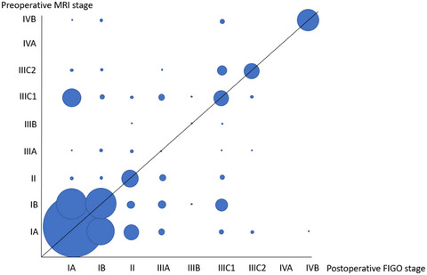
3.3 Correlations between MRI-based clinical stages and surgical stagesWe also evaluated the agreement between MRI-based clinical stages and surgical stages (Table 4). The top two highest rates of agreement were FIGO stage IVB (93.8%; 15/16) followed by IA (85.2%; 283/332). The lowest rate of agreement was found on FIGO stage IIIA (5.3%; 1/19). Understaging by preoperative MRI was most common for patients with FIGO stage IIIA disease (adnexal metastases) (68.4%; 13/19), and overstaging by preoperative MRI was most common for patients with FIGO stage IIIB disease (vaginal metastasis) (33.3%; 1/3).
TABLE 4. Correlations between clinical stages by preoperative magnetic resonance image and postoperative surgical stages FIGO stage IA IB II IIIA IIIB IIIC1 IIIC2 IVA IVB Total MRI stage IA 283 26 10 4 0 3 2 0 1 329 IB 31 40 5 5 1 9 0 0 0 91 II 2 2 11 4 0 3 0 0 0 22 IIIA 1 2 2 1 0 1 1 0 0 8 IIIB 0 0 1 0 1 1 0 0 0 3 IIIC1 12 3 2 4 1 10 2 0 0 34 IIIC2 2 2 0 1 0 6 9 0 0 20 IVA 0 0 0 0 0 0 0 0 0 0 IVB 1 2 0 0 0 2 0 0 15 20 Total 332 77 31 19 3 35 14 0 16 527 Bold means consistent numbers between MRI stage and FIGO stage. 3.4 Performance of preoperative MRI indetecting pathological parametersTable 5 shows the performance of preoperative MRI in detecting the pathological parameters. The sensitivity, specificity, PPV, and NPV for MI ≥50% on preoperative MRI were 60.8%, 88.5%, 68.4%, and 84.7%, respectively. The results for LR+ showed that, when the preoperative MRI revealed MI ≥50%, a patient would be 5.3-times as likely to have deep MI (≥50%) than if this parameter was not detected on MRI. In contrast, a negative result on MRI was likely 40% (LR−, 0.4) of the time in a patient with deep MI compared to a patient without it. The overall accuracy for deep MI was 80.5%, with good consistency between radiologists and pathologists. The degree of underestimating and overestimating deep MI on preoperative MRI was 11.4% and 8.2%, respectively. The overestimating rate was 15.3% (79/511) from stage IA to IIIC2.
TABLE 5. The measurements of the reliability of MRI in 527 EEC women MI ≥50% CI AM IAM PLNM PaLNM Sensitivity (%) 60.8 53.2 25.6 91.7 46.3 68.8 Specificity (%) 88.5 96.6 97.5 99.0
留言 (0)