
記住我
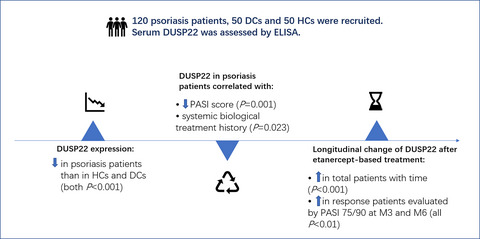
Psoriasis is a chronic and recurrent immune-mediated skin disease involving the rapid proliferation and abnormal differentiation of keratinocytes and activation of T cells, with variable morphology, distribution, and severity.1, 2 Clinically, psoriasis is marked by erythematous patches or plaques covered with slivery-white scales typically over the extensor surfaces, scalp, and lumbosacral regions, which is associated with increased risks of comorbidities (such as psoriatic arthritis, cardiovascular disease, and obesity).3-6 The primary treatment options include topic agents, phototherapy, systematic immunosuppressants, and biologics (such as tumor necrosis factor alpha (TNF-α) inhibitors, interleukin (IL)-12/23 inhibitors, and IL-17 inhibitors).7-10 Among the biologics, etanercept is one of the most frequently administered TNF-α inhibitors in China, which is effective in alleviating disease activity in psoriasis patients.11 However, etanercept is expensive compared to conventional treatments, meanwhile, a number of psoriasis patients fail to response to etanercept or discontinue treatment due to side effects over time.7, 12 To optimize the use of etanercept and individualize the treatment in psoriasis patients, the search of potential biomarkers for reflecting a clinical response to etanercept is vital.
MicroRNA (MiR)-146a and miR-146b have been reported to be mainly involved in the innate immune pathways by regulating Toll-like receptor signaling and ensuing cytokine response in immune-mediated diseases, including psoriasis.13-17 For instance, miR-146a dysregulation modulates IL-1R-associated kinase 1 (IRAK1), which is linked to the inflammation in psoriasis.13 Meanwhile, miR-146b modifies inflammation via downregulating the expression of atypical chemokine receptor (ACKR2) transcripts and protein in keratinocytes and lymphatic endothelial cells, which relates to the development of plaques in psoriasis.14 Clinically, plasma miR-146a is dysregulated in psoriasis patients compared with healthy control participates, and it displays the potential to predict clinical response to adalimumab (TNF-α inhibitors) in psoriasis patients.15 On the basis of the aforementioned evidence, it was hypothesized that plasma miR-146a and miR-146b might reflect the clinical response to other TNF-α inhibitors, such as etanercept treatment in psoriasis patients as well. However, no research has been published yet.
Therefore, this study aimed to investigate the correlations of miR-146a and miR-146b with disease risk and clinical features, as well as the linkages of their longitudinal changes with clinical response to etanercept treatment in psoriasis patients.
2 METHODS 2.1 ParticipantsThis study consecutively recruited 84 moderate-to-severe plaque psoriasis patients about to receive etanercept treatment in our hospital between January 2018 and February 2020. The inclusion criteria were as follows: (1) diagnosed as plaque psoriasis; (2) psoriatic body surface area (BSA)>10%, and psoriasis area and severity index (PASI)>8; (3) age between 18 and 80 years; (4) no use of biologics within 6 months; (5) about to receive etanercept treatment for at least 6 months; (6) able to be regularly followed up; (7) agreed with blood sample collection at each study visit. Following patients were excluded: (1) contraindications to etanercept; (2) known poor response to etanercept treatment; (3) patients at high risk of infection; (4) severe liver or renal dysfunction; (5) complicated with hematologic malignancies or tumors; (6) pregnant or lactating women. Besides, 80 disease controls and 80 healthy controls were enrolled from our hospital at the same period. The disease controls were selected from patients with dermatitis who treated in our hospital, and they were required to meet the criteria: (1) dermatitis patients, including contact dermatitis, atopic dermatitis, stasis dermatitis, and seborrheic dermatitis; (2) age and gender were matched with recruited psoriasis patients; (3) no history of psoriasis; (4) no history of autoimmune disease; (5) no history of hematologic malignancies or tumors. The health controls were screened from healthy people who came to our hospital for physical examination, and they were required to (1) have age and gender matched with recruited psoriasis patients, (2) no history of psoriasis or other inflammatory skin diseases, (3) no history of autoimmune disease or malignancies; (4) no obvious abnormality in main biochemical indexes.
2.2 Ethics statementThe current study was approved by Institutional Review Board and conducted according to the standards set by the International Conference on Harmonization and Good Clinical Practice. All participants provided the written informed consents.
2.3 Baseline data collection for psoriasis patientsClinical characteristics of psoriasis patients were collected at baseline (M0): age, gender, body mass index (BMI), disease duration, psoriatic BSA, PASI, treatment history, including topical therapy, phototherapy, systemic non-biologic treatment, and systemic biologic treatment.
2.4 Treatment for psoriasis patientsAll psoriasis patients received a subcutaneous injection of 25 mg etanercept twice a week, with intervals of 3–4 days, for a total of 6 months. Once the AE occurred, firstly, the according therapy would be applied to alleviate the symptom. After that, if their symptom was still not alleviated, then the dose of study drug would be reduced or withdrawn. Once the poor response occurred, the investigator would first judge the extent of poor response: if no response occurred, then the study drug would be withdrawn directly; and if poor response occurred, other treatment modalities would be used in combination, which included topical therapy, phototherapy, and systemic non-biologic therapy, while the combined treatment of patients was required to be documented in detail.
2.5 Response assessment for psoriasis patientsAfter initiation of etanercept treatment, PASI score of psoriasis patients was assessed at 1 month (M1), 3 months (M3), and 6 months (M6), which was used to calculate PASI75 response and PASI90 response. The PASI75 response was defined as a decrease of PASI score above 75% from baseline to each visit, and PASI90 response was defined as a decrease of PASI score above 90% from baseline to each visit. In addition, the patients who achieved PASI75 response at M6 were defined as PASI75 responders, correspondingly, the patients who did not achieve PASI75 response at M6 were defined as non-PASI75 responders in the analysis.
2.6 Blood sample collectionPeripheral blood (PB) samples of psoriasis patients were collected at M0, M1, M3, and M6. The PB samples of disease controls and healthy controls were collected after enrollment. All PB samples were collected using commercially available anticoagulant-treated tubes and immediately centrifuged at 1000 × g using a refrigerated centrifuge for 10 min, followed by centrifugation for 10 min at 2000 x g to isolate the plasma samples, which were immediately transferred into a clean polypropylene tube using a Pasteur pipette and stored at −80°C until the following detection.
2.7 MiR-146a and miR-146b detectionThe relative expressions of miR-146a and miR-146b in the plasma samples were determined by reverse transcription-quantitative polymerase chain reaction (RT-qPCR) assay. Briefly, total RNA was extracted from plasma samples by QIAamp RNA Blood Mini Kit (Qiagen, Duesseldorf), then complementary DNA was synthesized by RT-PCR Quick Master Mix (Toyobo). After that, qPCR was carried out by THUNDERBIRD® SYBR® qPCR Mix (Toyobo). The relative expressions of miR-146a and miR-146b were calculated using 2−△△Ct method and U6 was applied as the internal reference.18 The following are qPCR primers: miR-146a, Forward (5'->3’): ACACTCCAGCTGGGTGAGAACTGAATTCCATGGGT, Reverse (5'->3’): TGTCGTGGAGTCGGCAATTC; miR-146b, Forward (5'->3’): ACACTCCAGCTGGGTGAGAACTGAATTCCATAGGC, Reverse (5'->3’): TGTCGTGGAGTCGGCAATTC; U6, Forward (5'->3’): CTCGCTTCGGCAGCACATATACTA, Reverse (5'->3’): ACGAATTTGCGTGTCATCCTTGC.
2.8 Statistical analysisStatistical analysis was performed using SPSS 22.0 Software (IBM), and graph was plotted using GraphPad Prism 7.01 Software (GraphPad). Data were displayed as mean with standard deviation (SD), median with interquartile range (IQR), or number with percentage (No. (%)). Comparisons of miR-146a and miR-146b expressions among psoriasis patients, disease controls, and health controls were determined by Kruskal-Wallis test followed by Dunn's multiple comparisons test. Correlation of miR-146a with miR-146b, or correlations of miR-146a and miR-146b with continuous variables were determined by spearman's rank correlation test. Correlations of miR-146 and miR-146b with categorical variables were determined by Wilcoxon rank sum test. Variation tendency of miR-146a and miR-146b from M0 to M6 was determined by Friedman test. Receiver-operating characteristic (ROC) curve analysis and the derived area under the curve (AUC) were used to assess the performance of miR-146a and miR-146b in differentiating different subjects. Multivariate logistic regression analysis was also performed to exclude the potential cofounding factors. Once the patients had at least one-time measurement of the miR-146a/b level, then their data were included in the final analysis by using the last observation carry forward (LOCF) method. p value <0.05 was considered as significant.
3 RESULTS 3.1 Clinical features of psoriasis patientsIn this study, a total of nine patients were dropped out, including one patient who occurred an adverse event; five patients had poor responses to the study drug and three patients lost to follow-up. In psoriasis patients, the mean age was 52.2 ± 12.1 years, and there were 50 (59.5%) males and 34 (40.5%) females (Table 1). Furthermore, the mean BMI was 24.2 ± 3.5 kg/m2 and the median disease duration was 10.0 (4.0–15.8) years. Additionally, the median psoriatic BSA was 17.5 (14.0–22.0) %, and the median PASI score was 12.2 (9.4–18.9). The detailed information of treatment history and combined treatment is exhibited in Table 1.
TABLE 1. Clinical characteristics of psoriasis patients Items Psoriasis patients (N = 84) Age (years), mean ± SD 52.2 ± 12.1 Gender, No. (%) Male 50 (59.5) Female 34 (40.5) BMI (kg/m2), mean ± SD 24.2 ± 3.5 Disease duration (years), median (IQR) 10.0 (4.0–15.8) Psoriatic BSA (%), median (IQR) 17.5 (14.0–22.0) PASI score, median (IQR) 12.2 (9.4–18.9) Treatment history, No. (%) Topical therapy 77 (91.7) Phototherapy 69 (82.1) Systemic non-biologic treatment 54 (64.3) Systemic biologic treatment 12 (14.3) Combined treatment, No. (%) Topical therapy 64 (76.2) Phototherapy 43 (51.2) Systemic non-biologic treatment 33 (39.3) Abbreviations: BMI, Body mass index; BSA, Body surface area; IQR, Interquartile range; PASI, Psoriasis area and severity index; SD, Standard deviation. 3.2 MiR-146a and miR-146b in health controls, disease controls, and psoriasis patientsMiR-146a expression was decreased in psoriasis patients compared with disease controls (p < 0.001) and health controls (p < 0.001) (Figure 1A). Meanwhile, miR-146b expression was also lower in psoriasis patients compared with disease controls (p < 0.001) and health controls (p < 0.001) (Figure 1B).

MiR-146a and miR-146b expressions among health controls, disease controls, and psoriasis patients. Comparisons of miR-146a (A) and miR-146b (B) expressions among health controls, disease controls, and psoriasis patients. The performance of miR-146a in discriminating psoriasis patients from disease controls (C) and health controls (D), respectively. The performance of miR-146b in differentiating psoriasis patients from disease controls (E) and health controls (F), respectively
ROC curve analyses disclosed that miR-146a discriminated psoriasis patients from disease controls (AUC: 0.732, 95%CI: 0.655–0.809) (Figure 1C) and health controls (AUC: 0.888, 95%CI: 0.840–0.936) (Figure 1D). As to miR-146b, it could also distinguish psoriasis patients from disease controls (AUC: 0.702, 95%CI: 0.621–0.782) (Figure 1E) and health controls (AUC: 0.823, 95%CI: 0.757–0.889) (Figure 1F).
3.3 Correlation of miR-146a with miR-146b in psoriasis patients, disease controls, and health controlsMiR-146a positively correlated with miR-146b in psoriasis patients (p < 0.001, r = 0.519) (Figure 2A) and disease controls (p = 0.005, r = 0.310) (Figure 2B), while no correlation of miR-146a with miR-146b was observed in health controls (p = 0.062, r = 0.210) (Figure 2C).

MiR-146a correlated with miR-146b in psoriasis patients and disease controls but not in health controls. Correlation of miR-146a expression with miR-146b expression in psoriasis patients (A), disease controls (B), and health controls (C), respectively
3.4 Correlations of miR-146a and miR-146b with clinical features in psoriasis patientsAs for the correlations of miR-146a and miR-146b with continuous variables, miR-146a negatively correlated with psoriatic BSA (p = 0.011, r = −0.275) and PASI score (p = 0.003, r = −0.320), while it was not correlated with age, BMI, or disease duration; miR-146b negatively linked with PASI score (p = 0.020, r = −0.253), while it was not correlated with age, BMI, disease duration, or psoriatic BSA (Table 2). In terms of correlations of miR-146a and miR-146b with categorical variables, no correlation of miR-146a with gender, history of topic therapy, history of phototherapy, history of systemic non-biologic treatment, or history of systemic biologic treatment (all p > 0.05) was exhibited; meanwhile, miR-146b was also not correlated with these factors, either (all p > 0.05) (Table 3).
TABLE 2. Correlation between miR-146a/b and continuous variables in psoriasis patients Psoriasis patients (N = 84) MiR−146a expression MiR−146b expression r p value r p value Age 0.179 0.103 0.076 0.493 BMI −0.073 0.509 −0.051 0.647 Disease duration −0.076 0.493 −0.118 0.286 Psoriatic BSA −0.275 0.011 −0.174 0.113 PASI score −0.320 0.003 −0.253 0.020 Abbreviations: BMI, Body mass index; BSA, Body surface area; MiR, microRNA; PASI, Psoriasis area and severity index. TABLE 3. Correlation between miR-146a/b and categorical variables in psoriasis patients Psoriasis patients (N = 84) MiR−146a expression MiR−146b expression Median (IQR) p value Median (IQR) p value Gender 0.877 0.813 Male 0.337 (0.232–0.556) 0.441 (0.264–0.543) Female 0.360 (0.235–0.509) 0.428 (0.244–0.651) History of topical therapy 0.437 0.616 No 0.241 (0.192–0.651) 0.434 (0.241–0.496) Yes 0.362 (0.236–0.516) 0.434 (0.254–0.551) History of phototherapy 0.775 0.372 No 0.373 (0.224–0.450) 0.470 (0.241–1.124) Yes 0.343 (0.235–0.610) 0.425 (0.254–0.539) History of systemic non-biologic treatment 0.588 0.848 No 0.366 (0.252–0.561) 0.438 (0.255–0.537) Yes 0.348 (0.230–0.509) 0.434 (0.240–0.569) History of systemic biologic treatment 0.587 0.175 No 0.359 (0.240–0.454) 0.412 (0.248–0.543) Yes 0.239 (0.214–0.622) 0.481 (0.319–0.767) Abbreviations: BMI, body mass index; BSA, body surface area; MiR, microRNA; PASI, psoriasis area and severity index. 3.5 Correlations of longitudinal changes of miR-146a and miR-146b with clinical response to etanercept treatment in psoriasis patientsAfter etanercept treatment, the PASI75 response rate was 14.3%, 32.1%, and 69.0% at M1, M3, and M6 in psoriasis patients, respectively. Furthermore, the PASI90 response rate was 1.2%, 17.9%, and 36.9% at M1, M3, and M6 in psoriasis patients, respectively.
MiR-146a and miR-146b expressions were assessed at four time points (M0, M1, M3, and M6). Besides, total patients were divided into PASI75 responders and non-PASI75 responders based on the PASI75 response at M6. In total patients, miR-146a expression exhibited an increased trend throughout these four time points (p < 0.001) (Figure 3A). Further subgroup analyses disclosed that in PASI75 responders, miR-146a expression also showed an elevated trend throughout these four time points (p < 0.001) (Figure 3B); while in non-PASI75 responders, the increasing trend of miR-146a expression was not significant throughout these four time points (p = 0.124) (Figure 3C).

Change of miR-146a and miR-146b expressions at different time points. Comparisons of miR-146a expression at M0, M1, M3, and M6 in total patients (A), PASI75 responders (B) and non-PASI75 responders (C), respectively. Comparisons of miR-146b expression at M0, M1, M3, and M6 in total patients (D), PASI75 responders (E) and non-PASI75 responders (F), respectively
Meanwhile, in total patients, miR-146b expression also showed an increased trend throughout these four time points (p < 0.001) (Figure 3D). Further subgroup analyses revealed that in PASI75 responders, there was a raised trend of miR-146b expression through these four time points (p < 0.001) (Figure 3E); while in non-PASI75 responders, the increased trend of miR-146b expression was not significant (p = 0.357) (Figure 3F). These data indicated that longitude increment of miR-146a and miR-146b associated with clinical response to etanercept treatment in psoriasis patients.
To exclude the potential cofounding factors affecting the PASI75 response, the multivariate logistic regression analysis was also performed, which showed that the miR-146a at M0 (odds ratio (OR): 0.061, p = 0.033), disease duration (OR: 0.881, p = 0.018), history of systemic biologic treatment (OR: 0.045, p = 0.005) were independently correlated with the lower PASI75 response rate. However, history of topical therapy (OR: 14.521, p = 0.024) and combined with systemic non-biologic treatment (OR: 3.941, p = 0.041) were independent factors in predicting higher PASI75 response rate (Table S1).
4 DISCUSSIONPreceding investigations unravel that miR-146a and miR-146b are functionally linked to the immune and inflammatory responses of psoriasis.13-15 As an example, miR-146a is involved in the inflammation through regulating the expression of IRAK1 in psoriasis.13 Meanwhile, aberrant miR-146b downregulates ACKR2 and mediates the release of proinflammatory chemokines, which, in turn, devotes to the uncontrolled inflammation and the formation of plaque in psoriasis.14 Clinically, miR-146a is dysregulated in psoriasis patients compared with healthy controls, and it also positively correlates with PASI score in psoriasis patients; furthermore, miR-146a expression is markedly changed after 12 weeks of adalimumab (TNF-α inhibitor) treatment, and miR-146a expression after treatment inversely correlates with PASI reduction.13, 15 Under the context of aforementioned evidence, it was hypothesized that miR-146a and miR-146b might be implicated in the indication of disease activity and clinical response to other TNF-α inhibitors, such as etanercept in psoriasis patients, while the related information remains still unknown.
In this study, miR-146a and miR-146b expressions were initially detected in psoriasis patients, disease controls, and healthy controls. The results disclosed that both miR-146a and miR-146b expressions were decreased in psoriasis patients compared with disease controls and healthy controls, which could differentiate psoriasis patients from disease controls and healthy controls. The following is the possible explanation: miR-146a and miR-146b probably modified the production of inflammatory cytokines through regulating inflammatory pathways (e.g., NFκB) and anti-inflammatory proteins (e.g., ACKR2), which was linked to the skin inflammation, keratinocyte proliferation, neutrophil infiltration, and skin thickening erythema, then contributed to decreased risk of psoriasis.1, 15, 19 Besides, miR-146a was positively correlated with miR-146b in psoriasis patients and disease controls, while the correlation of miR-146a and miR-146b displayed a positive trend in health controls. These results could be explained by that: miR-146b probably functioned coordinately and assisted miR-146a in the regulation of keratinocyte proliferation and inflammatory responses in psoriasis; hence, miR-146a was positively correlated with miR-146b in psoriasis patients and disease controls.20, 21
Furthermore, this study analyzed the associations of miR-146a and miR-146b with disease features in psoriasis patients, which disclosed that miR-146a was negatively correlated with psoriatic BSA and PASI score, meanwhile, miR-146b was negatively linked with PASI score. These data indicated that miR-146a and miR-146b were related to less exacerbated disease activity in psoriasis patients. Herein, several possible reasons were suggested: (a) MiR-146a and miR-146b probably suppressed the immune and inflammatory response, which mediated less release of cytokines and chemokines, resulting in less severe inflammation and decreased disease activity in psoriasis patients1, 13, 14; (b) MiR-146a and miR-146b probably intercepted the proliferation and migration of keratinocytes but induced the apoptosis of keratinocytes in the lesioned skin, which devoted to the reduced disease activity in psoriasis patients, hence, miR-146a and miR-146b were correlated with less exacerbated disease activity in psoriasis patients.1, 20, 21
Additionally, all recruited psoriasis patients received etanercept treatment for 6 months, and their miR-146a and miR-146b expressions were examined at multiple points (at M0, M1, M3, and M6) after etanercept treatment. The results revealed that in general, miR-146a and miR-146b expressions were increased gradually with the treatment of etanercept over time. Further subgroup analyses exhibited that miR-146a and miR-146b expressions were also elevated gradually with the treatment of etanercept over time in PASI75 responders, while the increased trends of miR-146a and miR-146b expressions were not significant in non-PASI75 responders during the 6-month etanercept treatment period. These suggested that longitude increments of miR-146a and miR-146b were related to the clinical response to etanercept treatment in psoriasis patients. These results might be explained by that: MiR-146a and miR-146b probably modulate the downstream immunity-related and inflammation-related pathways to repress the secretion and release of cytokines (e.g., IL-12 and IL-23), which retarded the inflammation and attenuated disease activity in psoriasis patients; meanwhile, etanercept (TNF-α inhibitor) alleviated signs, symptoms, and disease activity in psoriasis patients; therefore, miR-146a and miR-146b expressions increased over time with etanercept treatment in psoriasis patients, and the increasing trend associated with PASI75 response.1, 8, 13, 14, 22
We notice that some studies disclose a pro-inflammation role of miR-146a/b,23, 24 while other studies show an anti-inflammation role of miR-146a/b in psoriasis patients.25, 26 Our study exhibited that lowly expressed plasma miR-146a and miR-146b correlated with exaggerated disease activity, and their increments during the treatment were closely linked with clinical response to etanercept in psoriasis patients which implied an anti-inflammation role of miR-146a/b in psoriasis patients. The possible reason for this finding might be that the source of the sample was different in separate studies. In detail, we detected the miR-146a/b in the PBMC of psoriasis patients, while other studies detecting the miR-146a/b expression in skin tissue disclosed a pro-inflammation role of miR-146a/b.
Nonetheless, some limitations should not be ignored in this study. First, the detailed mechanism regarding the effects of miR-146a and miR-146b on clinical response to etanercept was not investigated, thus, further experiments were needed. Second, the relatively small sample size (only 84 moderate-to-severe plaque psoriasis patients) probably reduced the statistic power of the analyses, and further validation studies were necessary. Third, since the original source of miR-146a/b in plasma was still not clear until now, we hypothesized that the miR-146a/b in plasma might derive from the inflammation-related cells, such as the macrophage, neutrophil, or Tregs. However, this hypothesis needed further exploration. Fourth, according to current literature, some non-coding RNA (such as: circular RNA RSF127 and long non-coding RNA SNHG1628) might regulate the miR-146a/b in other inflammation-related disease; thus, we hypothesized that these non-coding RNA might also be the upstream factors affecting miR-146a/b in psoriasis. However, this hypothesis needed further exploration. Lastly, all patients received etanercept for a relatively short duration (6 months), meanwhile, psoriasis is a disease with a high risk of recurrence; hence, the correlations of longitudinal changes of miR-146a and miR-146b with persistent clinical response and/or sustained remission to etanercept in psoriasis patients needed further exploration.
To conclude, lowly expressed plasma miR-146a and miR-146b correlate with exaggerated disease activity, and their increments during the treatment are closely linked with clinical response to etanercept in psoriasis patients. These data might offer insights into tailoring the use of etanercept and improving the treatment outcomes in these patients. However, more comprehensive studies are needed to validate.
CONFLICTS OF INTERESTThe authors declare that they have no conflicts of interest.
留言 (0)