
記住我


A 44-year-old Caucasian man presented to the Emergency Department with acute mechanical lower back pain, which occurred suddenly as he rotated his trunk while sitting. The patient denied any neurological, cauda equina or B symptoms (night sweats, fever or unintentional weight loss). His past medical history and family history were unremarkable.
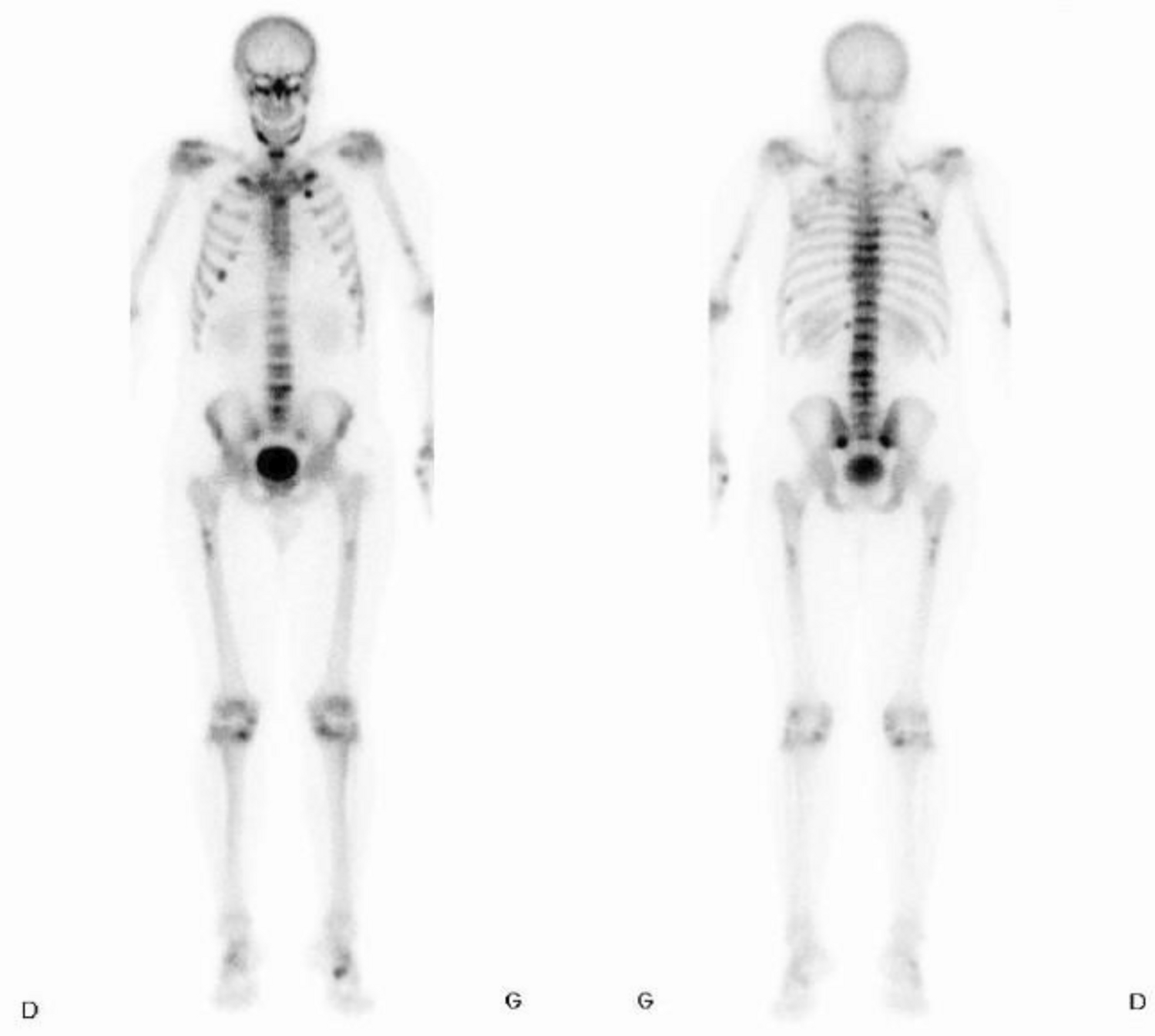
Apart from tenderness on percussion of the L4 and L5 vertebrae and the presence of a paravertebral muscle contracture, physical examination was unremarkable. Lumbar spine radiographs were reported as normal, but an MRI of the spine identified acute subchondral fractures of all the vertebrae from D2 to L5. Dual-Energy X-ray Absorptiometry (DEXA) scan found normal values for age at the hip, with Z-scores of − 1.2 SD at the femoral neck and − 1.0 DS at the total hip, but a low Z-score of − 2.8 SD at the lumbar spine, with an osteoporotic T-score value of − 3.0 SD at this site, though not really interpretable due to the lumbar fractures. A bone scan highlighted diffuse hyperfixation foci corresponding to non-consolidated fractures of the ribs, the sacrum and all the dorsal and lumbar vertebrae (Fig. 1).
Fig. 1
Bone scan showing diffuse bone hypefixation with several hyperfixating foci corresponding to non-consolidated fractures of the ribs, pelvis and vertebrae
Blood tests showed severe hypophosphatemia (0.33 mmol/l; normal range (NR): 0.8–1.45), mild hypocalcemia with corrected calcium levels at 2.18 mmol/l (NR: 2.20–2.50 mmol/l), raised alkaline phosphatase (209 U/l; NR: 25–102), slightly reduced 25-OH vitamin D (47 nmol/l; NR: 75–105) and 1.25-OH vitamin D (33 pmol/l; NR:40–140). Parathyroid hormone (PTH) levels were mildly elevated (7.10 pmol/l, NR: 1.1–6.8). Hypokalemia (3.4 mmol/l; NR: 3.6–4.6) and hypouricemia (235 µmol/l; NR: 286–518) were also present. Creatinine (72 μmol/l, NR: 62–106), arterial blood pH (7.43) and bicarbonate levels (24.5 mmol/l; NR: 22–27) were normal. Bone turnover markers were elevated with β-crosslaps and amino-terminal propeptide of type I collagen (P1NP), respectively, at 736 ng/l (NR: 158–442) and 69 μg/l (NR: 15.1–58.6). TSH, tryptase, anti-tissue transglutaminase, immunglobulin-A antibody and β2-microglobulin values were normal; HIV serology was negative. Serum electrophoresis and immunofixation did not detect any abnormal proteins, and levels of urinary free light chains were normal.
Urinalysis revealed microalbuminuria and normoglycemic glycosuria (83.2 mmol/l, normal (N) < 15). Tubular maximal phosphate reabsorption/glomerular rate (TmP/GFR) was very low at 0.27 mmol/GFR (NR: 0.80–1.35), consistent with renal phosphate wasting. Urinary glycine levels were also elevated (497 mmol/mol; N < 280).
Calcium and potassium levels normalized after oral supplementation, but phosphate levels remained low despite oral and intravenous administration, combined with calcitriol substitution.
Hypokalaemia, hypouricemia, glycosuria, microalbuminuria and aminoaciduria, together with tubular phosphate wasting, without renal failure or metabolic acidosis, suggested partial Fanconi syndrome. Drug history did not identify any culprit medication. The levels of heavy metals (cadmium, mercury and lead) came back as normal.
Due to the late onset of bone fragility, inherited causes of FS were unlikely. Ophthalmological examination did not find elements for cystinosis nor Kayser-Fleischer rings for Wilson’s disease. With the absence of hepatic or neurological involvement, and normal ceruloplasmin levels, this latter diagnosis was unlikely. A positron emission tomography and computed tomography (PET-CT 18-FDG) showed the recent vertebral and pelvic fractures, and it identified multiple recent rib fractures; moreover, it highlighted the presence of a 11 × 15 mm hypermetabolic subcutaneous lesion in the proximal left upper thigh, which was later described as an arteriovenous malformation on ultrasound (Fig. 2).
Fig. 2
The tumor on the PET-CT and at the ultrasound scan. A Nodular subcutaneous hypermetabolic (max SUV 3) lesion in the left thigh. B Subcutaneous hypoechogenous lesion at the ultrasound measuring 17 × 8 × 14mm, vascularized with arterial and venous flow with Doppler
Serum levels of fibroblast growth factor-23 (FGF23) finally came back elevated at 300 pg/mL (N < 50 pg/mL). Tumor-induced ostomalacia was therefore suspected. The patient underwent surgical resection of the lesion in the left thigh. Histopathological analyses showed a benign (19 × 18 × 11mm) mesenchymatous phosphaturic tumor. Molecular diagnosis identified a fibronectin 1- fibroblast growth factor receptor 1 (FN1-FGFR1) gene fusion, encountered in over 50% of cases of phosphaturic mesenchymal tumors, therefore confirming the diagnosis.
Hypophosphatemia and other dyselectrolytemia sustainably resolved 2 days after surgery, enabling us to stop both phosphate and potassium substitution. Glycosuria also disappeared after one week. Calcium and vitamin D supplementation was continued to support a hungry bone syndrome following surgery. The patient has not had any new fracture. The DEXA scan performed 2 years after surgery showed a 37% and 16% increase in bone mineral density at the lumbar and femoral sites, respectively, with complete normalisation of Z-scores.
留言 (0)