
記住我


Human cytomegalovirus (HCMV), is omnipresent, and infection is very common1. Following primary infection, the virus establishes latent infection within the host and may later reactivate in immunocompromised individuals and during pregnancy, where immunity is also altered, leading to an array of possibly severe clinical outcomes for the fetus1,2. Congenital HCMV infection (cCMV) is caused by in-utero mother-to-foetus transmission via the placenta or postnatally through breastfeeding3. The incidence of cCMV reported is 0.2–2.2 per cent of all live births worldwide, where India bears the highest burden of cCMV births (>1.5%)4,5. Sequalae reported in 15 per cent cCMV cases at birth include low birth weight, hepatosplenomegaly, microcephaly, jaundice, sensorineural hearing loss and later onset conditions such as developmental disability and impaired vision6. Israel and eight European countries routinely screen pregnant women for HCMV7,8. Whereas India, despite having the highest HCMV seroprevalence among women of reproductive age and the highest birth prevalence of cCMV, does not yet have a national maternal or neonatal screening programme5. Recent evidence has emerged to show that the dynamics of maternal HCMV infection could have an impact on pregnancy outcome and cCMV with accompanying sequalae9,10. Thus, screening for maternal infection could facilitate better management of pregnancy outcomes as well as neonatal interventions. We previously reported the importance of integrating both serology and PCR for HCMV screening in high-risk pregnancies11. This longitudinal study aimed to determine HCMV infection status across trimesters in women with and without BOH and the consequent impact on cCMV as well as pregnancy outcome.
Materials & MethodsThe present prospective cohort study was conducted at ICMR-National Institute for Research in Reproductive and Child Health (NIRRCH), Mumbai in collaboration with Nowrosjee Wadia Maternity Hospital (NWMH) and Bai Jerbai Wadia Hospital for Children (BJWHC), Mumbai, Maharashtra. This study was approved by the Institutional Ethics Committee of all participating sites (NIRRCH, NWMH, BJWHC). A signed informed consent was obtained from all the study participants, and assent was taken from parents of the newborns.
Study participantsEighty-three pregnant women aged 18–45 yr were recruited (2021-2023), either in their first (<12 wk) or second trimester (13-20 wk) from NWMH (Supplementary Table I); 38 had no history of previous or current pregnancy complications [without BOH (bad obstetric history) group] and 45 had either pregnancy complications diagnosed at recruitment or bad obstetric history (BOH group)11. Participants were followed until delivery, and newborns were recruited, and their birth weight, APGAR score, NICU (neonatal intensive care unit) admissions, and symptoms shown (if any) were noted. Pregnant women with gestational diabetes, autoimmune disorders, HIV, Hepatitis B, reproductive tract infections/sexually transmitted infections (RTIs/STIs) were excluded.
Sample collectionPeripheral blood and saliva were collected at the time of recruitment and once during each trimester: first or second trimester upto third trimester (>28 wk). Cord blood12 at delivery and saliva (<72 h of birth) of newborns were collected before or post-1 h of breastfeeding4,13 to detect cCMV.
TORCH serologyPeripheral and cord blood plasma was used for TORCH (Toxoplasma, Rubella, HCMV, HSV 1 & 2) IgM, IgG ELISAs and IgG avidity was measured as described previously11.
HCMV chemiluminescent microparticle immunoassay (CMIA)IgG antibody titers against HCMV were determined in plasma samples by CMIA using ARCHITECT™ Abbott™ kits (Abbott Diagnostics, USA) on Architect i1000SR immunoassay analyzer by Abbott Diagnostics as described previously14. Specimens with concentration values <6.0AU/ml are considered nonreactive and ≥6.0AU/ml are considered reactive to IgG antibodies and indicate past or acute infection.
HCMV nested-PCRHCMV nested-PCR was performed on saliva of participants and of their newborns and DNA extracted from PBMCs, plasma and whole blood to detect HCMV mtrII region as described previously11,15. Individuals having positivity in any two compartments, i.e. whole blood, plasma or saliva were considered HCMV infected and positivity only in PBMCs was considered as latent HCMV infection16.
Statistical analysisStatistical analysis was performed using GraphPad Prism version 10.0.1 (GraphPad Software, San Diego, California, USA). Data sets were compared using Fisher’s exact test, One-way ANOVA and P values <0.05 were considered significant.
Results Participant characteristicsEighty-three pregnant women, with or without BOH were recruited and followed up as shown in supplementary figure 1, and supplementary table I. Of the 45 BOH participants, majority had a history of spontaneous abortion (SA; n=30), followed by missed abortion (MA; n=17), neonatal death (NND; n=10), intrauterine foetal death (IUFD; n=10) and foetal growth restriction (FGR; n=2; Supplementary Fig. 2).
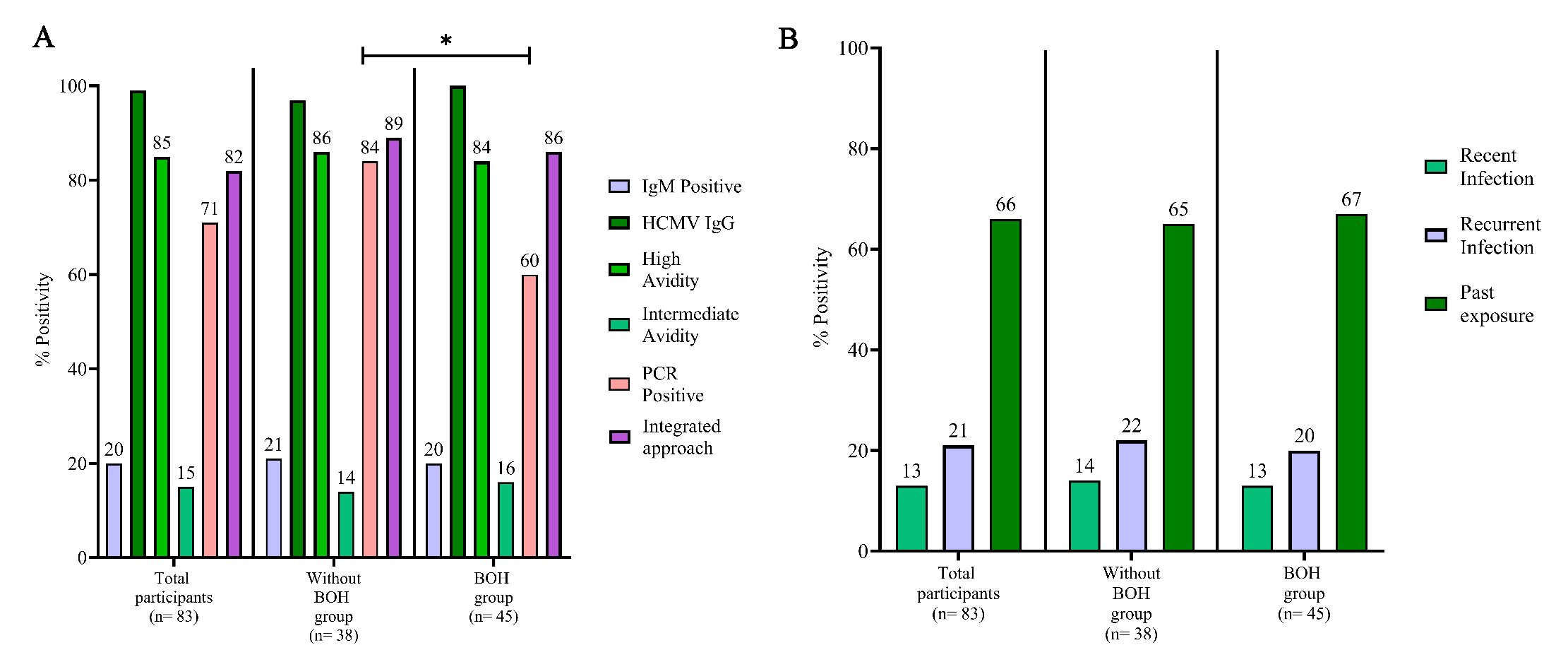
Integrated HCMV screeningHCMV-infected participants by serology were 34 per cent with 13 per cent having recent HCMV infection, 21 per cent recurrent HCMV infection and 66 per cent past exposure to HCMV (Fig. 1 and Supplementary Table II). Distribution of IgM positivity, high avidity and intermediate avidity was similar in both groups. HCMV PCR positivity revealed a much higher prevalence of infection overall (71%) with significantly higher prevalence (84%) in ‘without BOH’ group compared to the BOH group (60%; P=0.0171). Serological titers of anti-HCMV IgG were also estimated prospectively using CMIA which showed high concordance (95.65%) with IgG positivity as expected (Supplementary Table III). Integrated screening revealed a total prevalence of 82 per cent HCMV infection in the cohort.
Export to PPT
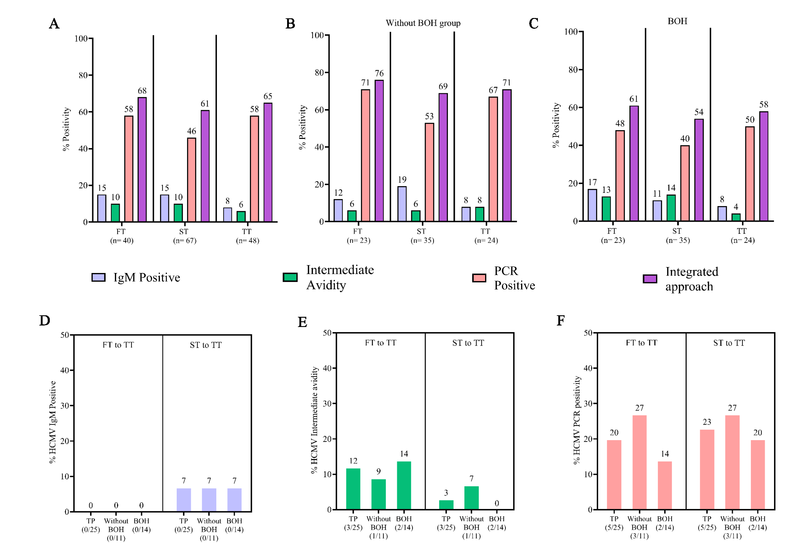
Prevalence and incidence across trimestersTrimester wise analysis (Fig. 2A-C) demonstrated that HCMV IgM positivity (15%) and intermediate avidity (10%) based prevalence was lower than that obtained by PCR (58%) and showed a gradual decline from first to third trimester. Increased prevalence of HCMV intermediate avidity IgG was observed in both the first and second trimesters for BOH group. HCMV PCR positivity also demonstrated a consistent dip in 2nd trimester, which was more pronounced in ‘without BOH’ group (Supplementary Table IV).
Export to PPT
As depicted in figure 2D-F, IgM screening alone detected a very limited incidence of infection, only from the second trimester onwards (7%). In the same cohort, however, concurrent avidity and PCR-based screening clearly highlighted higher incidence, 12 per cent and 20 per cent, respectively.
Adverse pregnancy outcomes: Pregnancy loss and association with HCMV infectionIn our cohort (Table IA), 14 per cent (n=10) were cases of pregnancy loss (PL), including IUFD (1.4%) and miscarriage (12.6%). Of these, a greater proportion, although not statistically significantly associated (P=0.1887), belonged to BOH group. Distribution of PL was marginally higher in first trimester compared to that in the second. In PL cases, the prevalence of BOH and concurrent HCMV infection was almost twice that observed in women with normal pregnancy outcomes (NPO; Table IB). Nine out of 10 cases with PL showed evidence of active HCMV infection, with only two scoring positives for HCMV IgM (Table IC). None of these individuals were IgM positive and showed only past exposure for other TORCH pathogens revealed through IgG positivity and high avidity (Table ID).
Table IA. Pregnancy loss and association with HCMV infection
Total participants (n=83) Without BOH group (n= 38) BOH group (n= 45) P value Recorded pregnancy outcome (n= 71) 36 35Pregnancy loss (PL): 14% (10/71)
IUFD: 1.4% (1/71)
Miscarriage: 12.6% (9/71)
8% (3/36) 20% (7/35)0.1887
RR: 2.4
SA 8.4% (6/71) MA 4% (3/71) First trimester 60% (6/10) Second trimester 40% (4/10)Table IB. Association of BOH and HCMV with PL
Pregnancy loss *Normal pregnancy outcome BOH + HCMV 60% (6/10) 35% (11/31) HCMV 30% (3/10) 48% (15/31) BOH 10% (1/10) 9.6% (3/31) No BOH + No HCMV (0/10) 6 (2/31)Table IC. HCMV infection status measured by different parameters in PL cases
Parameters HCMV infection PCR positive 60% (6/10) IA 30% (3/10) HCMV IgM positive 20% (2/10) HCMV exposed 10% (1/10)Table ID. Infection status of PL participants
Groups HCMV serology IgM/IA/PCR status Pregnancy outcome TRH IgM/IgG/Avidity Without BOH group RR IgM, P MA TRH, IgG, High PE P MA RI IA, P SA BOH PE P IUFD PE N SA PE P SA PE P MA RI IA SA RI IA SA RR IgM SA Adverse pregnancy outcomes: Symptomatic births and association with maternal HCMV infectionWe monitored 66 live births including two cases of twin births, 35 in ‘without BOH’ group and 31 in BOH group (Table II). In 14 cases of lost to follow up, pregnancy outcomes were recorded. HCMV infection associated symptoms were seen in 50 per cent of newborns with similar distribution in both groups (Table II). These included preterm birth (PT): 24 per cent, low birth weight (LBW) <2.5 kg: 32 per cent, very low birth weight (VLBW) <1.5 kg: 4.5 per cent and jaundice 17 per cent. BOH did not seem to be associated with these outcomes. When maternal HCMV infection status was analysed for these 33 neonates, 91 per cent of mothers were HCMV positive during first trimester, which declined to 48 per cent during second trimester and increased to 61 per cent in third trimester (Fig. 3A). The dynamics of infection in NPO group, remained unchanged across trimesters; 57 per cent, 60 per cent, and 60 per cent during first, second and third trimester, respectively. Co-occurrence of PT and LBW was high (30%), as expected (Fig. 3B; Supplementary Fig. 3).
Table II. Symptomatic births and association with congenital HCMV transmission
Total participants (n=83) Without BOH group (n= 38) BOH group (n= 45) Recorded pregnancy outcome (n=74) 36 38 Live births* (n=66) 35 31 Longitudinal sampling not available (n=14) 8 6 Complete sampling* (n=52) 27 25 APGAR score (1 min/5 min) 7-10 7-10 NICU admissions 5 4 a. Symptomatic births: 50% (33/66) 51% (18/35) 48% (15/31) Preterm births: 24% (16/66) 26% (9/35) 23% (7/31) Low birth weight (<2.5 kg): 32% (21/66) 37% (13/35) 26% (8/31) Very low birth weight (<1.5 kg): 4.5% (3/66) 3% (1/35) 6% (2/31) Jaundice: 17% (11/66) 17% (6/35) 16% (5/31) b. Congenital HCMV transmission (cCMV) Cord blood (n=32) 17 15 Neonatal Saliva (n=52) 27 25 1. Cord blood positive#: 34% (11/32) 47% (8/17) 20% (3/15) 2. Neonate saliva PCR positive: 23% (12/52) 26% (7/27) 20% (5/25) (1 or 2) cCMV births: 35% (18/52) 44% (12/27) 24% (6/25) Symptomatic births (SB) of complete sampling: 48% (25/52) 52% (14/27) 44% (11/25) SB within cCMV: 61% (11/18) 50% (6/12) 83% (5/6) c. Association of maternal HCMV infection status in second trimester with symptomatic births Recurrent HCMV infection and SB$ 33% (2/6) Recent HCMV infection and SB$ 100% (4/4)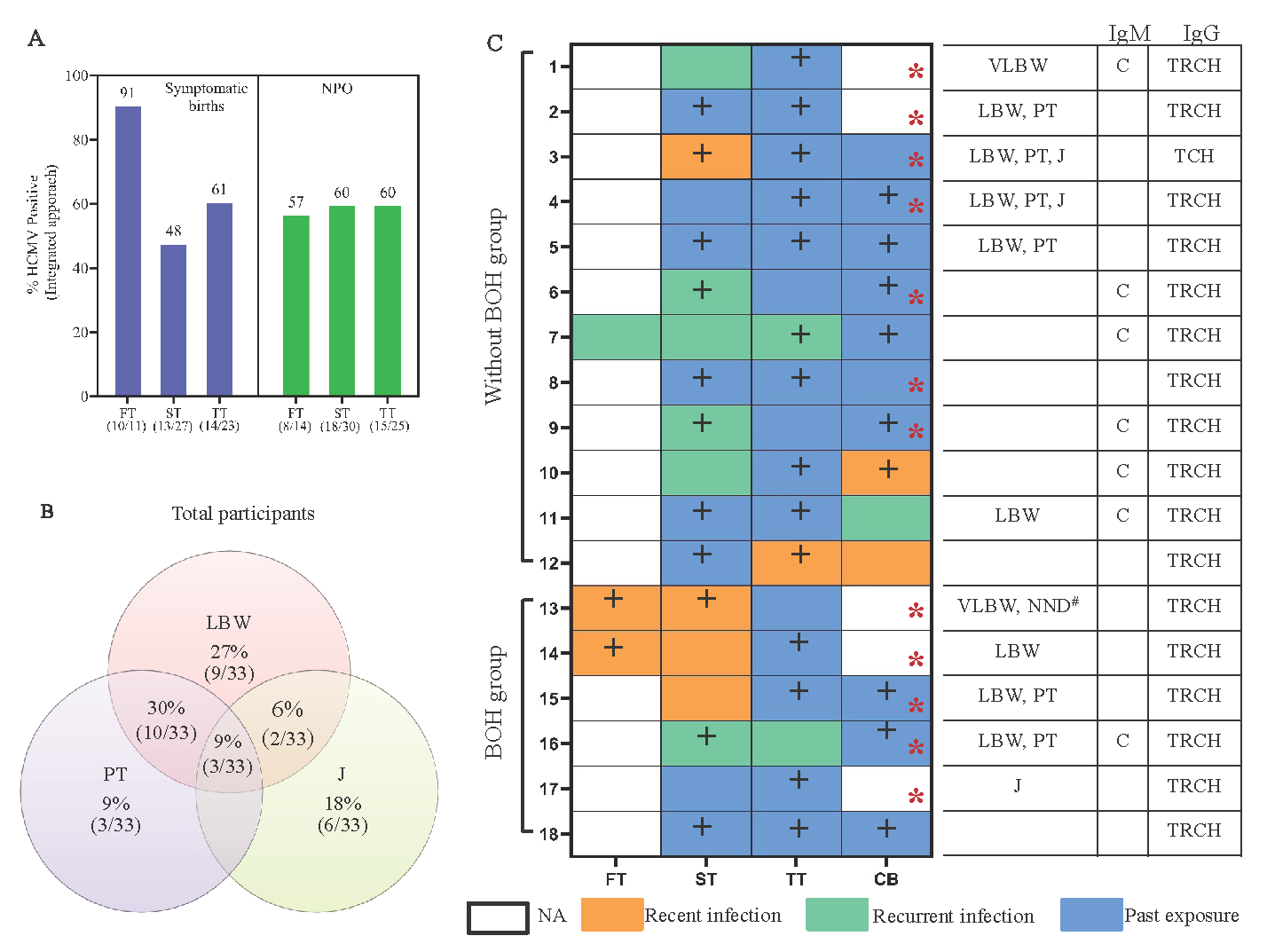
Export to PPT
Congenital HCMV infection: Association of maternal infection status with symptomatic birthMaternal and cognate neonate saliva screening was performed to assess cCMV in 52 mother-neonate dyads. Thirty-two cord blood samples were received (Table II). Overall rate of cCMV, detected either through cord blood (34%) or neonatal saliva positivity (23%) was 35 per cent with more frequent detection (44%) in ‘without BOH’ compared to the BOH group (24%), probably reflecting higher prevalence of HCMV PCR positivity observed overall in ‘without BOH’ group (Fig. 1A). When maternal infection status was assessed in 18 cCMV neonates (transmitter group; Fig. 3C), trimester-wise stratification of infection status showed, in BOH group, a greater proportion of recent infection (3/6) during second trimester compared to that in ‘without BOH’ group (1/12), a trend also apparent in limited data available from the first trimester. Conversely, without BOH group showed a preponderance of recurrent infection.
We evaluated the impact of these apparently distinct infection signatures on birth outcomes by integrating symptomatic births (SB) data with analysis of cCMV (Table II and Supplementary Fig. 4). SB occurred with a 48 per cent prevalence within the cCMV screening cohort. When distributed into cCMV cohort (n=18), the prevalence of SB was higher at 61 per cent. Groupwise distribution within transmission cohort showed much greater proportion of SB including the only case of NND in BOH (83%) compared to without BOH group (50%) that was associated with aforementioned unique signature of recent infection observed in BOH group. Furthermore (Fig. 3C and Table II), we observed higher prevalence of SB in women with recent infection (4/4) compared to recurrent infection (2/6; P=0.0762).
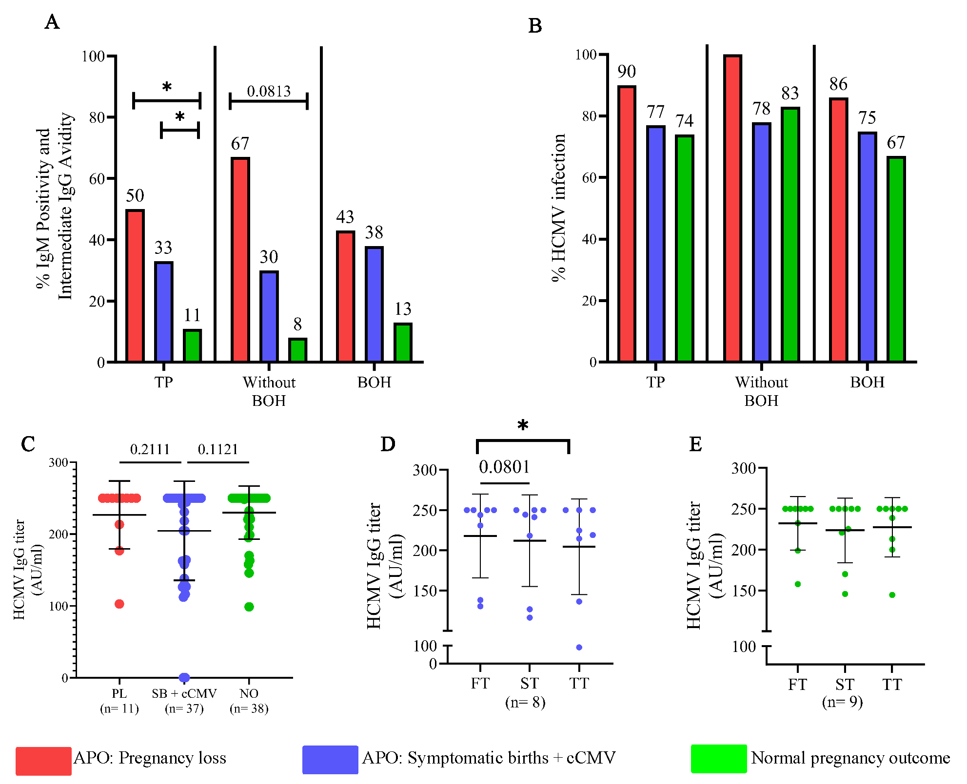
Distinct maternal HCMV infection signatures associated with pregnancy outcomesFurther, we wanted to ascertain if particular infection signatures (serological or molecular) were differentially associated with varying pregnancy outcomes (Supplementary Table V). For PL and SB+cCMV, significantly and incrementally higher frequency of detection by IgM and intermediate avidity IgG (Fig. 4A), but not PCR (Fig. 4B) was observed compared to women with NPO (P=0.0211, 0.0455). Also, within the ‘without BOH’ group, participants with PL showed the highest frequency of detection followed by those with SB+cCMV (P=0.0813) compared to women with NPO. However, in the BOH group, participants with either PL or SB+cCMV had a similar frequency of detection, which was higher than in women with NPO. Interestingly, analysis of anti-HCMV IgG titers by CMIA in these samples showed similar titers in PL and NPO which trended lower in the SB group (Fig. 4C). Further, prospective analysis of matched samples across trimesters showed a significant decline in IgG titers for SB group but not in NPO group (P=0.0801, 0.0455; Fig. 4D and E).
Export to PPT
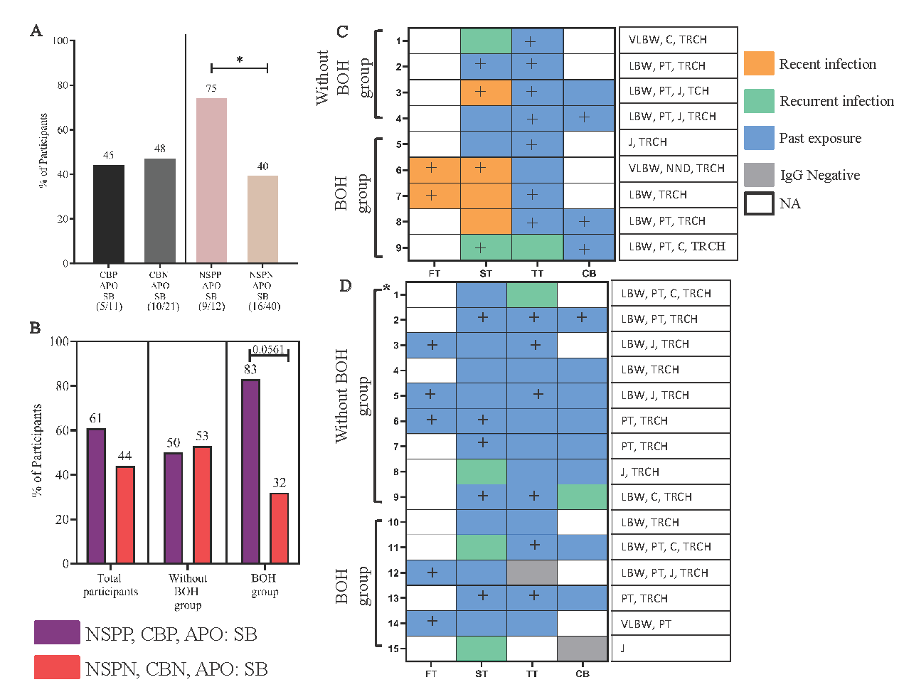
Adverse pregnancy outcomes: Symptomatic births and neonatal saliva HCMV infection statuscCMV was determined through screening of both cord blood and neonatal saliva detected in 18 neonates (Fig. 3C). We analysed the contribution of each screened compartment to the occurrence of SB (Fig. 5A). SB was much more frequent in saliva positive neonates (transmitters; 75%) compared to saliva negative (non-transmitters; 40%; P=0.0490). However, this discriminant signature was not observed in cord blood. Also, when positivity for both compartments was combined (Fig. 5B, Supplementary Table VI) a higher percentage of SB was observed in positive samples, mainly in BOH group (P=0.0561). Considering the aforementioned association of neonatal saliva positivity for HCMV with SB, we assessed maternal infection signatures (n=24) in the neonatal screening group (Table II) who had SB (Fig. 5C and D). A clearly disparate infection profile, comprising of early infection: intermediate HCMV IgG avidity in the first two trimesters, was apparent in transmitter compared to non-transmitters. Conversely, when the 27 mothers with NPO in the neonatal screening group were analysed, only three had neonates positive in saliva and did not show evidence of recent infection during pregnancy (Supplementary Fig. 5). In 24 non-transmitting mothers, only one in both groups showed evidence of recent infection. These results highlighted association of recent infection in early pregnancy with neonatal saliva positivity and SB. Consistent with our earlier observations, exposure to other TORCH pathogen was prevalent but IgM or intermediate avidity, when detected, was only for HCMV.
Export to PPT
DiscussionDespite India’s highest maternal HCMV seroprevalence and congenital transmission (cCMV) rates, basic prevalence data and incidence data on HCMV infection dynamics during pregnancy and cCMV-associated sequelae are lacking17,18. In our study, using an integrated approach: serology and PCR11, we longitudinally assessed maternal HCMV infection during pregnancy and cCMV with accompanying adverse pregnancy outcomes (APO) in an India setting. We clearly demonstrated the impact of BOH and concurrent HCMV infection on pregnancy loss (PL) and provided evidence for recent HCMV infection at an early stage of pregnancy leading to cCMV and APO.
While serology (IgM/IgG) is generally preferred for screening and diagnosis of HCMV infection, detection of low/intermediate avidity IgG antibodies indicative of early, class-switched and infection associated responses or PCR based screening is not generally performed19. Our results, incorporating serology, IgG avidity and PCR, greatly increased the sensitivity for detecting HCMV infection. We demonstrate utility and superiority of this approach, compared to IgM screening alone, in detecting the incidence of infection as early as in 2nd trimester. The study by Ebina et al20, which did not incorporate molecular screening, demonstrated that HCMV IgG avidity-based screening within 28 weeks of gestation, is useful in predicting cCMV20. Our incorporation of molecular screening highlights significant prevalence and incidence of early HCMV infection during pregnancy, including those without any previously reported BOH from India11,21.
Early HCMV infection/reactivation during pregnancy can have grave consequences on the foetus, resulting in PL22, including miscarriage and IUFD, significantly observed in our cohort. This prevalence was similar to that reported in 2019-2021 National Family Health Survey-5 for India23,24. This study also highlighted BOH as a risk factor for PL, which supports earlier cross-sectional data reported from India11. Interestingly, in our cohort, participants with PL majorly had BOH with concurrent HCMV infection compared to either BOH or HCMV infection alone. These results reveal a novel role for HCMV pathology in background of BOH and needs to be explored to evaluate possibly higher reactivation and re-infection occurring in these women.
Our use of sensitive nested-PCR delineated a unique signature of high positivity during the first trimester in women who had symptomatic births (SB), highlighting the importance of this approach in early pregnancy screening. Interestingly, when all adverse outcomes were evaluated till <20 wk, serological (including IgG avidity), but not PCR-based screening incrementally segregated participants in terms of outcome. Périllaud-Dubois et al19 and Faure-Bardon et al25, also underscored the importance of independently screening for HCMV IgG avidity in pregnancy19,25. Indeed, in our cohort, individuals with PL had high (similar to NPO group) HCMV IgG titers, which were mainly of intermediate avidity, thus underscoring the importance of viral IgG avidity screening in early pregnancy. Further, prospective CMIA analysis delineated a distinct profile of decreasing HCMV IgG titers observed in the SB, but not in NPO group, supporting an etiological role of cCMV. The apparent lack of resolution by integrating PCR for both trimesters may reflect sporadic replication of HCMV throughout pregnancy, which may be clinically relevant only when the immunogenic threshold is crossed, leading to the production of IgM (primary/recurrent infection) and low-intermediate avidity IgG (recent infection).
Our results correlating maternal HCMV infection signatures with cCMV, extended previously reported data supporting asymptomatic birth outcomes, known to be associated with recurrent maternal infection26,27. We also observed a unique maternal signature, mainly in the BOH group, of recent infection during the first and second trimesters that was associated strongly with SB including NND. Further, our results associating cCMV with SB underscored the importance of non-invasive screening of neonatal saliva4,13 using a sensitive, nested-PCR approach targeting HCMV mtrII region for the first time in a setting of cCMV28,29. Limitations of our study include limited sample size and further follow up of cCMV cases to evaluate post-natal sequelae such as sensory neural hearing loss.
The goal of maternal screening for HCMV infection is to mitigate possible APO and two recent, effective interventions are: passive immunization by administering anti-HCMV hyperimmunoglobulin and Valacyclovir as an effective treatment which can prevent cCMV, if infection is detected early in pregnancy30–33.
Our study highlights the importance of early maternal and neonatal HCMV screening in a high prevalence, resource-limited, public health setting, with potential to improve child health and limit resource intensive interventions required for many serious sequalae associated with cCMV.
留言 (0)