
記住我


Breast cancer is the most prevalent malignant form among women1,2 and by 2045, it is estimated that India may witness a rise of 448035 million new female breast cancers3. Despite the therapeutic advancements, primary ablative surgery is still the mainstay for breast cancer management.
Onco-anesthesiology prioritizes perioperative management for minimizing cancer recurrence and improving oncological outcomes4. The intravenous anesthetic propofol (2, 6-diisopropyl phenol)5, and the volatile anesthetic isoflurane (2-chloro-2-(difluoromethoxy)-1,1,1-trifluorethane)6 are commonly used during surgical resections with a favorable pharmacokinetic and pharmacodynamic profile7,8. Anesthetic agents have been indicated to deregulate immune and inflammatory responses either by regulating the stress response or impairing immune cells9. Studies have reported advantageous role of propofol over volatile anesthetics in terms of postoperative survival of patients10,11 and minimal immune suppression12. On the contrary, several recent clinical studies have observed no significant beneficial effect between propofol and other volatile anesthetics on cancer immunity and inflammatory responses13-16.
T lymphocytes play a vital role in the cell-mediated adaptive immune response. CD4+ helper T (Th) cells play a pivotal role in developing and sustaining effective anti-tumor immunity by stimulating other immune cells such as B cells, macrophages, and CD8+ cytotoxic T cells (Tc)17. The knowledge of the contradictory reports on the choice of anesthetic agent during surgery and the absence of any such reports from Indian background made it imperative to investigate the differential impact of propofol and isoflurane on the T cell immune responses, if any, among individuals of perioperative breast cancer patients.
Materials & Methods Study settingThis prospective study recruited individuals with breast cancer presenting to the Department of Surgical Oncology, Chittaranjan National Cancer Institute, Kolkata, who were advised for surgery [modified radical mastectomy (MRM)/breast conserving surgery (BCS)] according to the considered inclusion and exclusion criteria18. Recruitment of patients in this study was administered through informed consent form. Patients were block-randomized in two arms- volatile anesthestic isoflurane (n=50) in one arm and intravenous anaesthestic propofol (n=50) in the other. The study was approved by the Institutional Ethics Committee. The study adhered to the Indian Council of Medical Research’s ethical guidelines for biomedical research on human participants (2017)19 and was registered at Clinical Trials Registry-India (CTRI/2020/11/02886 dated Nov 3, 2020).
Protocol for anesthetic managementThe protocol for anesthetic management was same as reported before18. In the isoflurane group induction was achieved with thiopentone, 3-5 µg/kg and maintenance was done by using isoflurane 50 per cent in nitrous oxide to achieve minimum alveolar concentration of 1.0. For the propofol group induction was same but the maintenance was done using propofol with a target-controlled infusion pump, at an effect-site concentration of 2-3 µg/ml.
Collection of blood samplesPerioperative blood samples (preoperative, 1 day before surgery; intraoperative, 1 h after surgical incision and postoperative, 48 h after surgery) were collected from the study participants in both anesthetic arms18.
Typically, these anesthetic drugs are rapidly metabolized and eliminated from the body. The terminal elimination phase for propofol lasts for 1.5-31h20 whereas in case of isoflurane the elimination half-life of serum fluoride levels (metabolite of isoflurane) has been estimated to approximately 21 h21. Therefore, in this study, we have investigated the T cell immune responses for 1 h (intra) and 48 h (post) after incision.
Monitoring of anesthetic agents in serum samplespropofol was detected in serum samples using high-performance liquid chromatography (HPLC)22,23. Fluoride concentration during the pre, intra and postoperative periods after isoflurane anesthesia were measured directly from serum samples using fluoride ion selective electrode technique21 (Supplementary Material, Supplementary Fig. 1).
Flow cytometry for detection of lymphocyte subtypesThe immunophenotype of the lymphocytes was analyzed with flow cytometry24-26. Whole blood (100 µl) was incubated with a cocktail of fluorescent-tagged antibodies, in dark for 45 min at 4°C, suspended in PBS and was analyzed in a flow cytometer. The detailed protocol and gating strategy have been depicted in Supplementary Material (Supplementary Fig. 2 and 3).
Estimation of cytokinesELISA kits [Ray Biotech Peachtree Corners, GA, USA] were used to measure serum concentrations of interleukin (IL)-2, IL-12, IL-10, tumor necrosis factor (TNF)-α and interferon (IFN)-γ at 450 nm in a microplate reader18.
In silico analysesGrid-based Ligand Docking with Energetics (GLIDE, Schrödinger Release 2021-1: Maestro, Schrödinger, LLC, New York, NY, 2021) was used for docking analysis of isoflurane and propofol against CD4 and CD8 targeted proteins in extra precision mode. LigPlot+ ( https://www.ebi.ac.uk/thornton-srv/software/LigPlus/) and the ligand interactions module of Schrödinger were used to show the presence of intermolecular bonds between protein-drug complexes. Desmond program (Schrödinger Release 2021-1: Maestro, Schrödinger, LLC, New York, NY, 2022) was used for molecular dynamic simulations (MDS) of the Holo-1: CD4-isoflurane complex, Holo-2: CD4-propofol complex, Holo-3: CD8-isoflurane complex, and Holo-4: CD8-propofol complex, to understand the dynamic behaviour, mode of binding and inhibitor specificity for all the systems (details provided in Supplementary Material).
Statistical analysisTo compare the clinicopathological characteristics, Student’s t-test and Chi-square test were performed. The comparison of repeated measurement indicators at different observation time among the isoflurane and propofol group was performed using a linear mixed effect model. The model was used with patient as a random effect and with arm, timepoints and arm-by-timepoints as fixed effects. The arm-by-timepoints interaction indicates whether the change over time differed between the two anesthetic arms27. Post-hoc analyses were used with Bonferroni correction wherever applicable. The same linear mixed effect model was applied to investigate the effect of the anesthetic arm on Th cells with adjustment of confounding factors including age, height, weight, type of surgery, histopathology, grade, stage, molecular subtypes, ASA classification, duration of anesthesia and surgery, and pain score. Data were analyzed using Statistical Package for Social Sciences (IBM SPSS, ver. 25.0, Chicago, IL, USA). A value of P< 0.05 was considered significant.
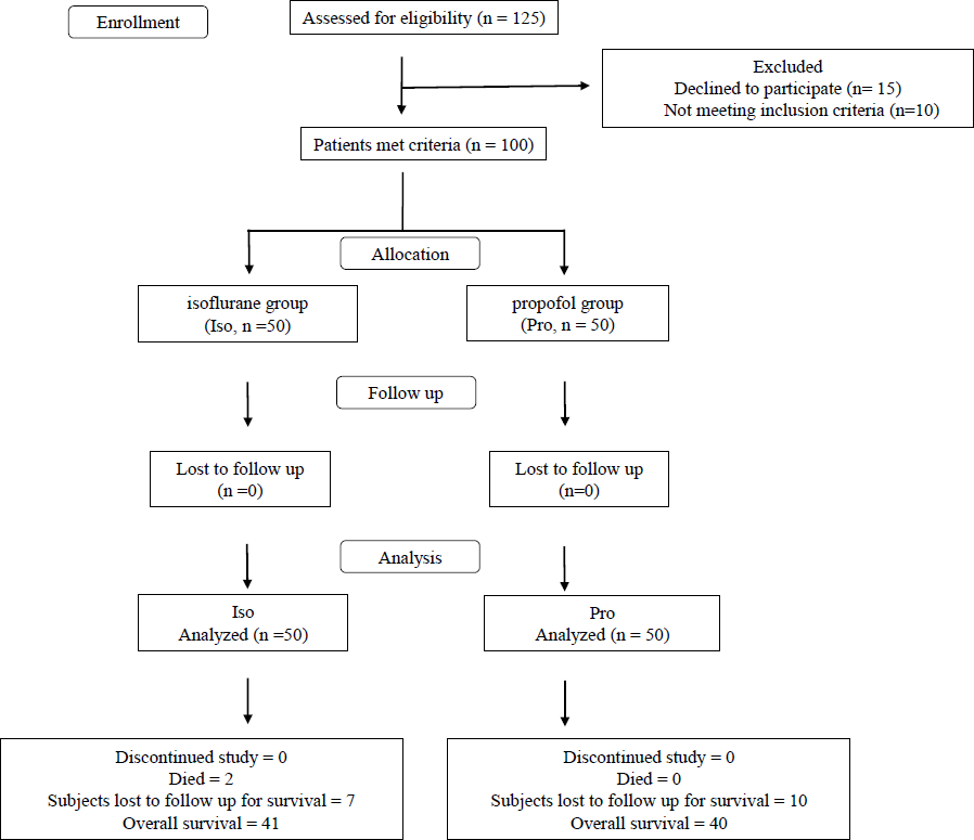
Results Participant clinicopathological characteristicsParticipants were recruited from December, 2020 to July, 2023 and followed up till August 2024 (Fig. 1). The clinicopathological characteristics of the enrolled participants with breast cancer (Table I) showed no significant difference between the two anesthetic groups in terms of age, weight, height, American Society of Anesthesiologists (ASA) classification, histopathology, estrogen receptor (ER)/ progesterone receptor (PR) / human epidermal growth factor receptor 2 (Her2) nu status, grade, stage, type of surgery, duration of surgery, duration of anesthesia and the post-operative numerical pain score. We also monitored the presence of anesthetic agents in serum samples. propofol and fluoride were detected only in the intraoperative serum samples (Supplementary Fig. 1).
Export to PPT
Table I. Clinicopathological characteristics of breast cancer patients recruited for surgical resection in two different anesthestic arms- isoflurane and propofol
Characters Character subtype Anesthetic agent P valueIsoflurane
(n=50)
Propofol
(n=50)
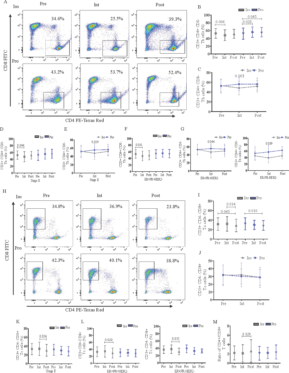
Age (yr) 52.36±11.35 53.10±11.75 0.749 Height (cm) 149.5±4.85 149.4±7.75 0.959 Weight (kg) 54.05±10.48 53.35±9.42 0.728 ASA classification; n (%) ASA I 20 (40) 21 (42) 0.839 ASA II 30 (60) 29 (58) Histopathology; n (%) Invasive carcinoma of NOS type 14 (28) 16 (32) 0.531 Invasive/ Infiltrating ductal carcinoma 28 (56) 30 (60) Ductal carcinoma in situ 6 (12) 2 (4) Mucinous carcinoma 2 (4) 2 (4) Molecular subtypes; n (%) ER+PR+HER2- 18 (36) 24 (48) 0.615 ER-PR-HER2+ 8 (16) 4 (8) ER-PR-HER2- 7 (14) 7 (14) ER+PR+HER2+ 6 (12) 7 (14) ER+PR-HER2- 6 (12) 6 (12) ER+PR-HER2+ 5 (10) 2 (4) Type of surgery; n (%) MRM 35 (70) 26 (52) 0.065 BCS 15 (30) 24 (48) Duration of surgery (min) 88.10±25.61 93.18±32.26 0.385 Duration of anesthesia (min) 110.5±27.93 115.8±30.36 0.365 Pain score (Number) 1.76±0.68 1.72±0.64 0.764 Grade; n (%) Grade I 2 (4) 5 (10) 0.212 Grade II 33 (66) 25 (50) Grade III 15 (30) 20 (40) Stage; n (%) Stage I 5 (10) 5 (10) 0.893 Stage II 34 (68) 32 (64) Stage III 11 (22) 13 (26) Impact of anesthetic agents on Th and Tc cellsisoflurane significantly suppressed the frequency of Th (CD3+CD8-CD4+) cells during the intraoperative than the preoperative period which was evident from the ‘intra-group’ analysis (Fig. 2; panels A and B). On the other hand, Th cells were significantly increased in postoperative compared to pre and intraoperative periods of propofol group (Fig. 2A and B). ‘Inter-group’ analysis showed the intraoperative Th cell depletion by isoflurane was significant compared to propofol (β-coefficient: -8.75; 95% CI: -13.00 to -4.51; Fig. 2C). Further subgroup analysis showed that the isoflurane-induced suppression of Th cell frequency during intraoperative period was prominent in stage II breast cancer as observed from ‘intra-group’ comparisons (Fig. 2D) and ‘inter-group’ comparisons (β: -8.51; 95% CI: -13.96 to -3.06; Fig. 2E). The intraoperative suppressive effect of isoflurane on Th cell frequency was also evident in the molecular subtype ER+PR+HER2- during the ‘intra-group’ comparison between intraoperative and preoperative period (Fig. 2F) and in the molecular subtype ER+PR+HER2- (β: -8.29; 95% CI: -14.37 to -2.21) as well as in ER-PR-HER2- (β: -11.10; 95% CI: -25.10 to 2.90) during the ‘inter-group’ comparison between isoflurane and propofol (Fig. 2G). According to the type III fixed effects of linear mixed model analysis (Supplementary Table I) it was further confirmed that Th cell frequency was significantly impacted by the interaction of the anesthetic arm with the timepoints. However, the age of the individuals also showed significant effect on the Th cells but not as an interaction of age with arm. The other confounding factors did not have any significant effect on the Th cell frequency of the individuals with breast cancer.
Export to PPT
Tc cells (CD3+CD4-CD8+) were also significantly reduced in count during the postoperative than preoperative period by both isoflurane and propofol (Fig. 2H and I) and the ‘inter-group’ comparison showed no differential effect of the anesthetics (Fig. 2J). However, subgroup analysis showed that isoflurane inflicted significant reduction in Tc cell frequency during the postoperative period compared to intraoperative period particularly in stage II cases but the same was not evident with propofol (Fig. 2K). Similarly, the decrease in Tc cells during postoperative period compared to intraoperative period by isoflurane was also observed across the molecular subtypes ER+PR+HER2- and ER+PR-HER2- whereas propofol did not induce any such changes (Fig. 2L). Additionally, isoflurane significantly increased the ratio of CD4+/CD8+ T cells postoperatively than intraoperative period, while the same ratio was maintained with propofol (Fig. 2M).
Effect of isoflurane/propofol on T cell activity markers, B cells and inflammatory cytokinesIn order to activate a Tc or a Th cell to proliferate and differentiate into an effector cell, an antigen presenting cell (APC) provides two signals – (i) through foreign peptide bound to major histocompatibility complex (MHC) on the surface of APC which signals through T cell receptor (TCR); and (ii) through co-stimulatory molecules - CD80 and CD86, which are identified by the co-receptor protein CD28 on the surface of the T cell28. Therefore, we checked the effect of isoflurane/propofol on the TCR and CD28 activity and did not observe any significant difference between the anesthetic groups (Supplementary Fig. 4).
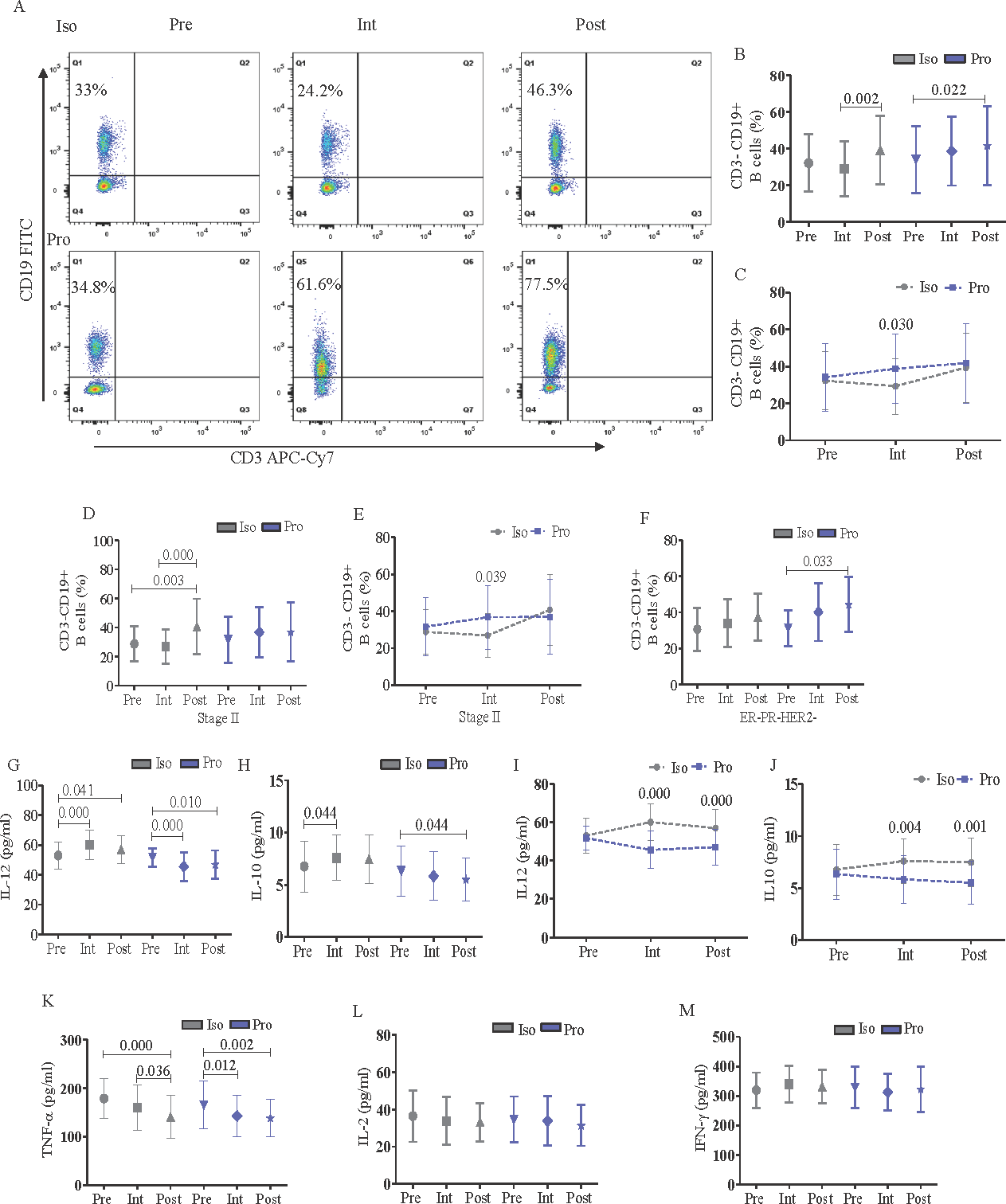
The Th cells inflict a direct anti-tumor response and help B lymphocytes to produce antibodies29. Therefore, we investigated the impact of the anesthetic agents on B cells. The ‘intra-group’ analysis revealed that the increase in B cell (CD3-CD19+) frequency was significant in the postoperative as compared to the intraoperative period in the isoflurane group whereas the same was higher in the postoperative than the preoperative period in the propofol group (Fig. 3A, B). However, ‘inter-group’ analysis showed significant decrease of B cell frequency by isoflurane compared to propofol during the intraoperative period (β: -7.51; 95% CI: -15.46 to 0.44; Fig. 3C). Subgroup analysis exhibited that isoflurane-induced increase in B cells during the postoperative period compared to pre and intraoperative period was significant only in breast cancer stage II cases (Fig. 3D). However, comparison between isoflurane and propofol showed that B cells were significantly reduced by isoflurane than propofol during the intraoperative period (β: -7.14; 95% CI: -16.47 to 2.18; Fig. 3E). The increment of B cell frequency by propofol during the postoperative than preoperative period was significant only in the ER-PR-HER2- breast cancer molecular subtype (Fig. 3F).
Export to PPT
A panel of cytokines including IL-12, IL-10, TNF-α, IL-2 and IFN-γ involved in innate and adaptive immune responses mediated through T and B cells were investigated in isoflurane/propofol groups at different timepoints. Both IL-12 (Fig. 3G) and IL-10 (Fig. 3I) were increased with isoflurane in the intra than preoperative periods and reduced with propofol in the post than preoperative period. However, inter-group analysis revealed significant increase in both IL-12 and IL-10 levels during intra (IL-12 β: 13.16; 95% CI: 8.73 to 17.60; IL-10 β: 1.33; 95% CI: 0.39 to 2.27) and postoperative periods (IL-12 β: 8.70; 95% CI: 4.26 to 13.13; IL-10 β: 1.53; 95% CI: 0.59 to 2.47) in the isoflurane group than propofol (Fig. 3I and J). TNF-α was significantly inhibited postoperatively in both the anesthetic groups (Fig. 3K) whereas IL-2 (Fig. 3L) and IFN-γ (Fig. 3M) did not show any significant alteration across isoflurane/propofol groups.
Molecular docking and trajectory analysis of isoflurane and propofol with CD4 and CD8The binding of the anesthetics with CD4 and CD8 can modulate their activity, altering immune responses. CD4 and CD8 co-receptors play crucial roles in T cell activation by binding to MHC and recruiting lymphocyte-specific protein tyrosine kinases (lck), which are essential for T cell signaling30. Any alteration in these interactions due to anesthetics can impact T cell function and the overall immune response. Therefore, next we investigated molecular docking and MDS of the anesthetics, isoflurane and propofol with CD4 and CD8. Molecular docking revealed that propofol elicited better binding energy against CD4/CD8 with higher binding scores than isoflurane (Table II). Propofol showed better binding affinity and higher scores compared to isoflurane against the said targets (Fig. 4; panels A and F). The 100 nanoseconds (ns) MDS trajectory revealed a stable root mean square deviation (RMSD) from 0 to 75 ns in Holo-1 but later exhibited higher deviation, which may be due to conformational changes. Holo-2 maintained a stable trajectory throughout the time frame. Holo-3 showed consistent deviations after 20 ns compared to Holo-4. Overall, propofol’s binding appeared to better stabilize CD4 and CD8 than isoflurane, as shown by lower RMSD values (Fig. 4B and G). Root mean square fluctuation (RMSF) analysis further elucidated the impact of ligand binding on residue mobility. The Apo state showed greater fluctuations, while specific residues in Holo states (notably in Holo-2 and Holo-4) exhibited reduced mobility, indicating constrained motions due to ligand interactions (Fig. 4C and H). Radius of gyration (rGyr) measurements indicated Holo-2 with more compactness compared to Holo-1. Similarly, Holo-3 and Holo-4 showed varying degrees of compactness, with Holo-4 being notably stable. These observations aligned with the RMSF results, highlighting the influence of ligand binding on the structural integrity of the proteins (Fig. 4D and I). Solvent accessible surface area (SASA) analysis during later simulation stages demonstrated that many residues transitioned from accessible to buried states upon ligand binding. In SASA, Holo-2 and Holo-4 depicted decreased values which signified its shift towards a buried state compared to Holo-1 and Holo-3 (Fig. 4E and J). Collectively, these results affirmed that propofol bonded more effectively to CD4 and CD8, contributing to their stability through reduced residue mobility, enhanced compactness, and significant changes in protein surface orientation. Additionally, post-MDS conferred that conventional H-bonds were broken down between CD4/CD8 and isoflurane but were retained with propofol (details provided in the Supplementary Material, Supplementary Fig. 5).
Table II. Binding energies and other interaction studies of CD4-isoflurane, CD4-propofol, CD8-isoflurane, and CD8-propofol complex.
S. No. Target PubChem CID Drug Binding energy (kcal/mol) No. of H-bonds H-Bond forming residues Average distance of H-bonds (Å) 1 CD4 3763 Isoflurane -3.80 1 Thr228 ∼2.556 2 CD4 4943 Propofol -4.85 1 Thr233 ∼1.715 3 CD8 3763 Isoflurane -2.74 1 Ser55 ∼2.504 4 CD8 4943 Propofol -3.70 1 Ser116 ∼1.985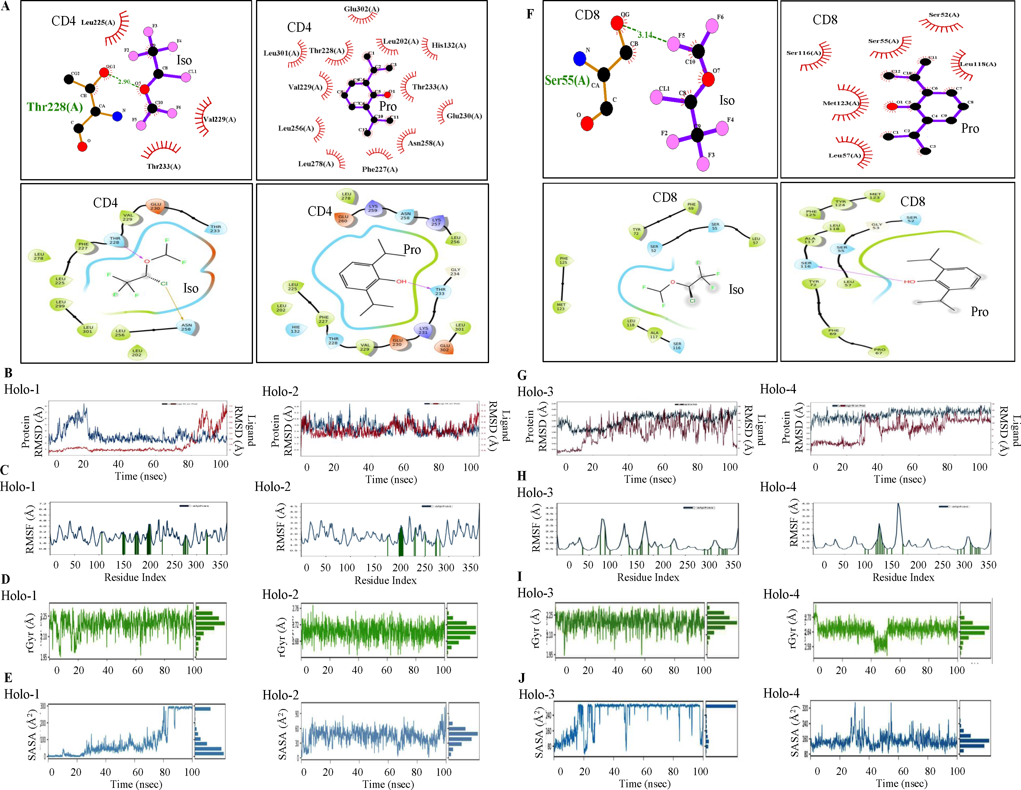
Export to PPT
DiscussionThe choice of intravenous anesthetics over volatile anesthetics has always been controversial regarding the perturbation of immune response during the perioperative period and consequential cancer recurrence12,14,31. The non-compliance of the beneficial11 or the non-beneficial32,33 role of propofol might have been due to variations in cancer type, grade, stage, hormone status, race, geographical location, and other socioeconomic factors. Therefore, in this study, we have considered only two specific types of surgery (MRM/BCS) with similar perioperative treatment regimens for both the groups. isoflurane in comparison to propofol induced a suppressive effect on Th and Tc cell frequency during the intra- and postoperative period respectively and this was prominent in stage II and some of the molecular subtypes of breast cancer. Small sample size in the stage and molecular subtype subgroups of breast cancer may have restricted their statistical significance.
The Th cells inflict a direct anti-tumor response and help B lymphocytes to produce antibodies as a part of humoral immunity17. In the current investigation, we observed that parallel to Th cells, B cell frequency decreased significantly with isoflurane than propofol during the intra-operative period. It might be corroborated that the reduction in the Th cell frequency by isoflurane might be one of the factors responsible for the depletion of the B cells in these patients.
Interestingly, during the intra- and postoperative period, IL-10 was predominantly upregulated by isoflurane than propofol. The reduction in the frequency of Th cells might have been partially regulated by the elevated levels of IL-10 during the intraoperative period of the isoflurane group. This finding conformed with a report where IL-10 secretion by Th2 cells inhibited Th cell differentiation and survival in an in vivo model34. In this study, T cell activity did not significantly differ between propofol and isoflurane as conferred by the expression of TCR, CD28, and cytokines IL-2, TNF-α, and IFN-γ. In concurrence with our findings another study reported that both propofol and desflurane triggered a similar beneficial immune response in terms of preservation of IL-2/IL-4 during the perioperative period of breast surgery13.
Studies have shown that the interaction of CD4 and CD8 co-receptors with p56lck may initiate tyrosine phosphorylation cascade leading to T-cell activation. The CD4- and CD8-p56lck complexes regulate several events in T cells including activation of transcription factors for gene expression, activation of integrin and intracellular calcium mobilization which are of prime importance in T-cell immunity related studies35. It has been reported that binding of glycoprotein 120 of human immunodeficiency virus (HIV) to CD4 on the T cells plays an important role in the induction of apoptosis36. The interaction of CD4 coreceptor with p56lck in its cytoplasmic tail is crucial in accelerating the HIV-induced apoptosis of CD4+ T cells37. Therefore, we were interested to check whether these coreceptors CD4 and CD8 have any interaction with the anesthetic drugs using in silico studies. Molecular docking exhibited that propofol strongly bonded with CD4 and CD8 as compared to isoflurane. MDS propagated that propofol formed a more compact and stabilized structure with CD4/CD8 than isoflurane. SASA analysis further portrayed that the binding of propofol with CD4/CD8 altered their surface chemistry and buried their accessible amino acid residues which might have prevented them from undergoing any form of interaction. In comparison, the binding of isoflurane kept the amino acid residues of CD4/CD8 more accessible for interaction. These interactions might play important role in anesthetic-induced T cell regulation including proliferation and apoptosis. However, these need further experimental validations.
The study had some limitations. Firstly, the effect of the anesthetic agents on the subtypes of Th and Tc cells and the interactions between the immune cells and surrounding factors were not studies. Secondly, the experimental validation of the functional molecular cues involved in the suppression of Th cells is yet to be deciphered.
Several studies have shown that these anesthetics have a significant impact on long term cancer outcome and perioperative immunoinflammatory profile. Immunomodulation during the intraoperative period may have a significant effect on the cancer metastasis and recurrence. Therefore, an anesthetic drug with minimal immunosuppressive effect during the intraoperative period may be beneficial for clinical practice12. In this study, propofol which minimally perturbed the T cell immune response and better controlled inflammatory mediators during intra or postoperative period might be indicated as a better anesthetic choice over isoflurane during surgical resection of breast cancer.
留言 (0)