
記住我
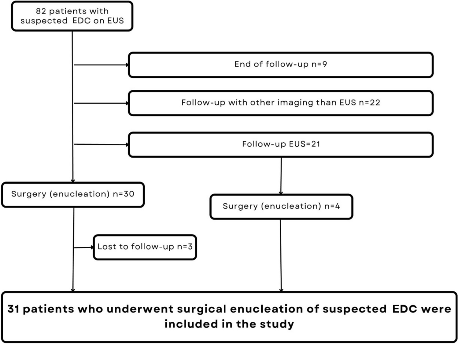
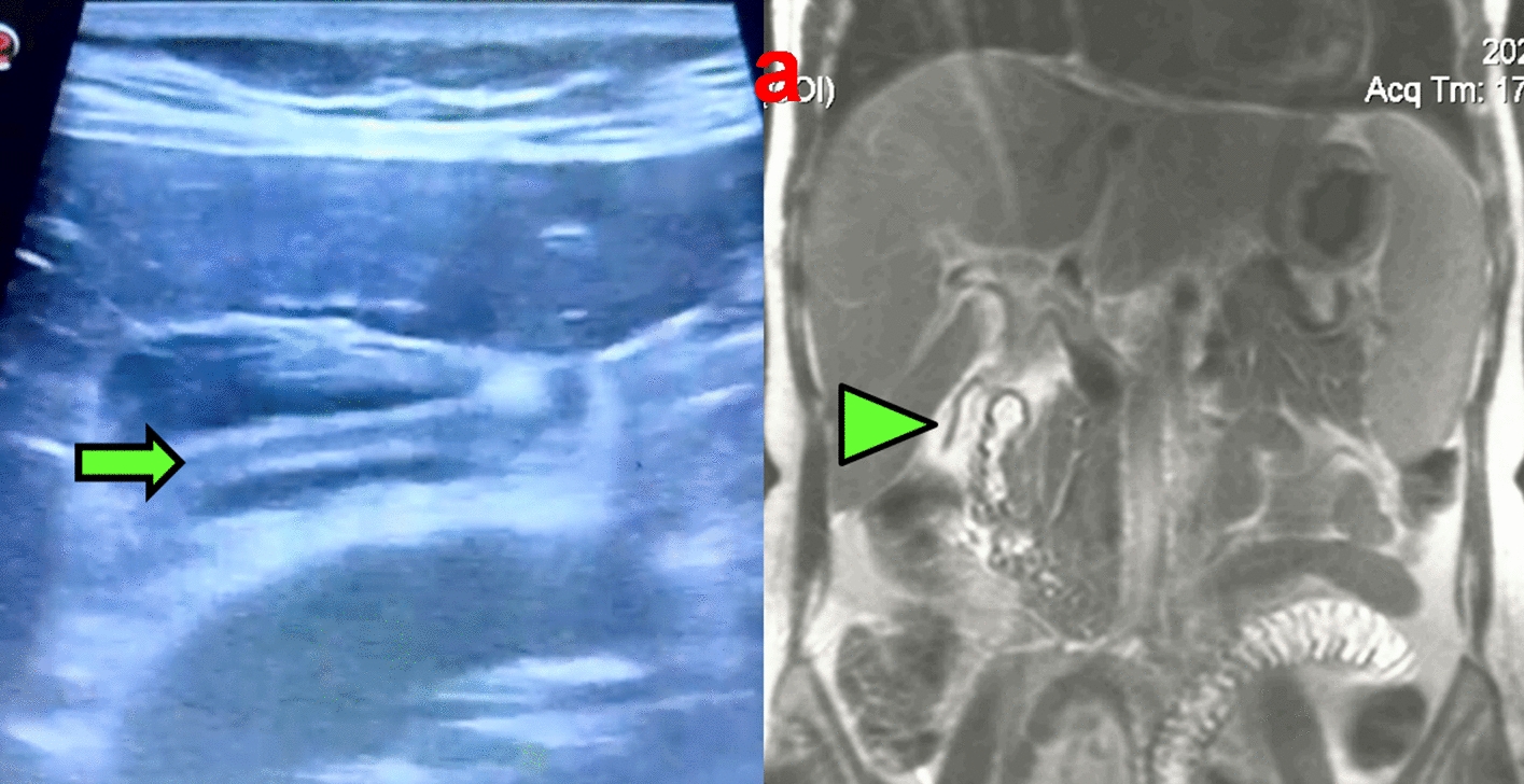

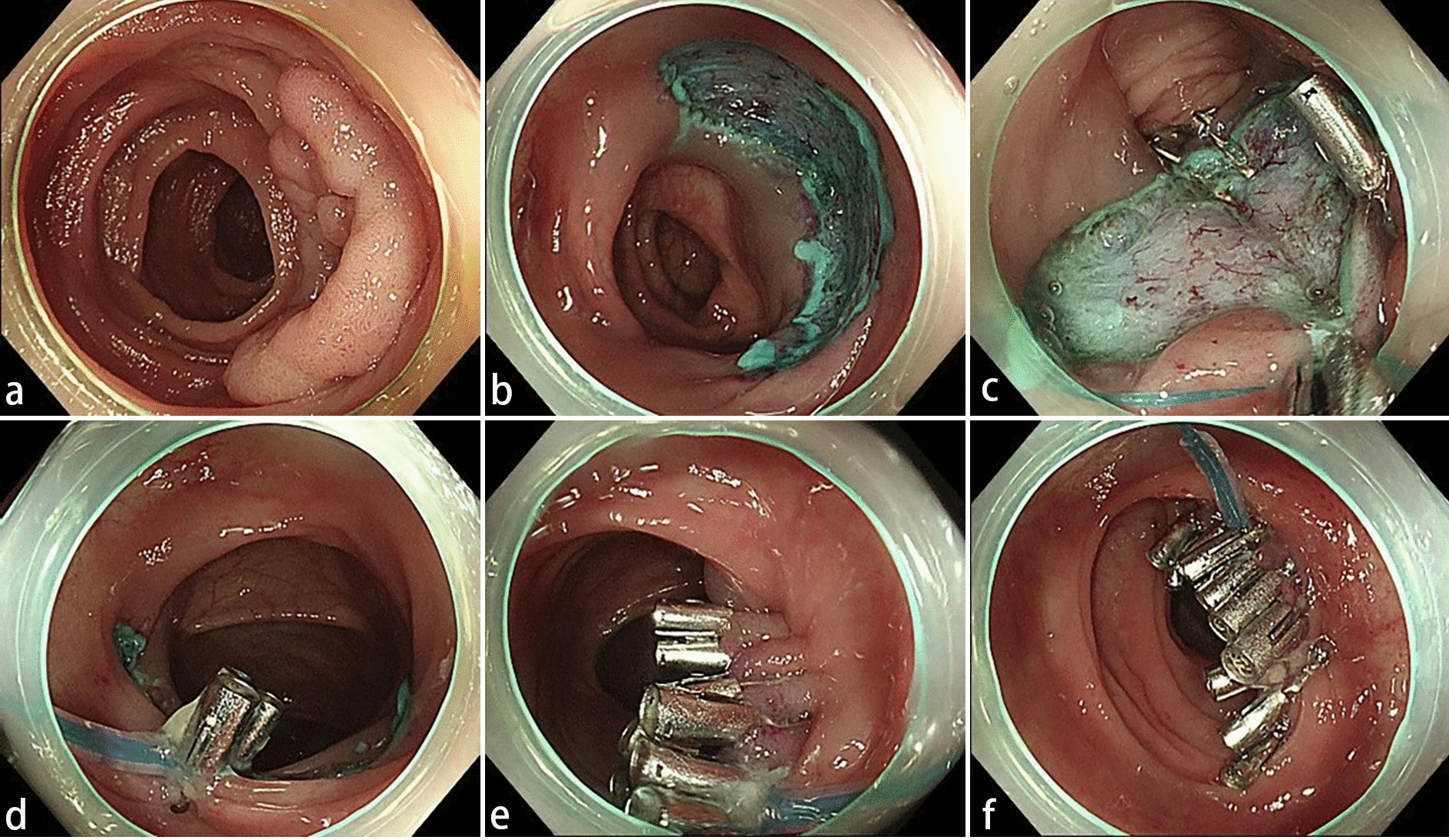
Table 1 presents the baseline characteristics of surgery and non-surgery patients.
Thirty-one patients out of 82 with suspected EDC (male: 15, 48%, median age 45 years, 25–75th percentiles: 36–57 years) underwent surgery. In the surgery group the lesion was an incidental finding in 18 (58%), but symptoms were present in 13 patients (42%); 2 had fever (6%), 1 had cough (3%), 4 had dysphagia (13%), and 6 had pain (19%). Gastroscopy was performed at diagnosis in 10 (32%); a submucosal lesion was discovered in 9 (90%). The surgically treated patients were younger and had larger cysts compared to the patients who did not have the suspected EDC surgically enucleated but remained in imaging follow-up (Table 1). There was no significant difference in the presence of enlarged lymph nodes between these two groups.
Features of Suspected Mediastinal EDCs in EUS and CT ScanOn endoscopic ultrasound the majority of suspected EDCs were located in the central (68%) and distal (26%) parts of the esophagus. Sixteen (52%) were intramural lesions. Fifteen (48%) were anechoic, two (6%) hypoechoic, and 13 (42%) had mixed echogenicity. Enlarged lymph nodes (> 10 mm) were present in only two patients (6%). The median size of the lesions was 49.5 mm, the size of the suspected EDC varied from 20 to 92 mm.
Thirty patients had a CT scan of the thoracic area available for re-evaluation. All but one of the re-evaluated CT scans were contrast enhanced. The density of the suspected EDC was reported as Hounsfield Units (HU) and 23 (77%) lesions had a density ≥ 20HU (soft tissue density). Only 6 (20%) had the density 0–19 HU as a cystic lesion containing simple fluid (i.e., transudate). One lesion had a density of -20HU (fat tissue) in CT scan and turned out to be a hamartoma in post-operative histology.
Table 2 shows comparison of clinical and imaging findings in patients with histologically documented lesions.
Table 2 Comparison of clinical and imaging findings in patients with histologically documented lesionsPatients are divided into two groups by histological diagnosis EDC and non-EDC. The histologically confirmed EDCs were all ≥ 2 cm, which is likely due to the inclusion criteria of the patients as larger cysts were referred to operation even when the appearance on EUS was consistent with EDC. Surprisingly majority as over 65% of the non-EDCs seemed intramural in both CT scan and EUS, which likely was partial reason for misdiagnosis. CT scan was performed on all 18 patients with EDC, interestingly showing that 16 (89%) as a majority of the duplication cysts had a density ≥ 20 HU and only two of the cysts (11%) density of cyst containing simple fluid. Figure 2 presents a CT scan and MRI image of a large histologically confirmed EDC.
Fig. 2
Large esophageal duplication cyst (9.1 cm) on the left originating from the upper esophageal region protruding into the left upper lobe of the lung. a Contrast-enhanced computed tomography image shows a high-attenuation mass with a density of 82 Hounsfield Units (HU). b Axial T2-weighted fat-saturated (Blade) magnetic resonance image reveals a lesion of increased signal intensity owing to liquid content. c Axial non-contrast-enhanced T1-weighted fat-saturated (Vibe) magnetic resonance image shows a lesion of increased signal intensity due to the high proteinous content of the liquid inside the duplication cyst
SurgeryOverall, 34 patients were referred to operative treatment for the suspected EDC, but three dropped out. In total, 31 patients underwent surgery. The indication for surgery was size ≥ 2 cm in 30 (97%), presence of symptoms in 13 (42%), and/or inconclusive appearance in imaging.
Post-operative complications were reported in eight patients (26%); three developed thoracic pain, two had cough, one had gastro-esophageal reflux disease, one had a phoniatric problem, and one suffered from post-operative hematoma.
Supplementary Table 1 shows surgical techniques used for enucleation of the lesions.
HistologyA histologic report was available for all 31 patients before the end of the study (Table 3), confirming an EDC in 18 (58%), a leiomyoma in 7 (23%), Müllerian duct cyst in 2 (6%), a mesenchymal tumor in 2 (6%), a paratracheal vascular malformation in 1 (3%), and a hamartoma in one (3%).
Table 3 Histological diagnosis compared to the diagnosis on EUS and CT scanOn EUS the diagnosis was correct in 58% (18/31) of the cases and on CT scan 57% (17/30). CT scan misdiagnosed three of the EDCs but found two leiomyomas correctly.
Imaging findings of histologically confirmed duplication cysts are presented in Table 3.
Interestingly, no malignancy was detected after a median follow-up of 3 years.
留言 (0)