Subjects
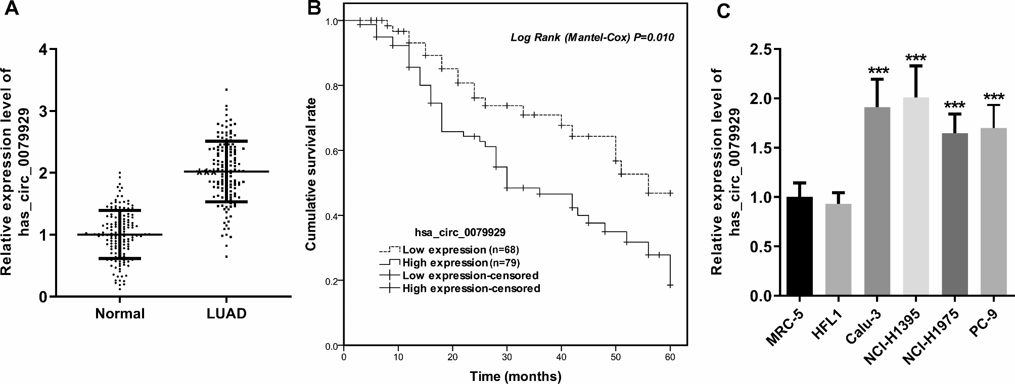
Clinical records of patients with lung cancer were retrospectively evaluated, to extract the visit and hospitalization information of patients who underwent curative resection by reason of primary lung adenocarcinoma, between 2011 and 2015 (n = 147). Patients who received any tumor-specific therapy, incomplete resection of other metastatic disease, or the presence of any nodules at the time of surgery were excluded from the study. Medical records of these patients were collected, recorded, and classified for age, sex, smoking status, TNM stage (the 7th edition of IASLC proposals), N status, distant metastasis, pleural invasion, vascular invasion, differentiation, and carcinoembryonic antigen (CEA) values. The follow-up information was collected, and the mean duration of clinical follow-up was 28 months (minimum/maximum: 3–60 months). This single-center retrospective study was with the approval of the ethical committee of Yuhuan Second People’s Hospital. All data and information derived from the tissues and clinical data was followed with written informed consent.
Cell lines and culture conditions
For investigation of human normal lung cell lines, MRC-5 and HFL1 were obtained from the Cell Bank of Chinese Academy of Sciences (Beijing, China), along with their specific culture mediums. For experiments of human lung adenocarcinoma cells, Calu-3, PC-9, NCI-H1975, and NCI-H1395, from the same institution as above, were cultured in RPMI 1640 (C11875500BT, GIBCO, Gaithersburg, USA), or MEM (41500034, GIBCO, Grand Island, USA), all supplanted with 10% FBS (GIBCO, Carlsbad, USA). The culture conditions for all cells were 37 °C with 5% CO2.
Overexpression and knockdown of hsa_circ_0079929
Small interfering RNA (siRNA) against hsa_circ_0079929 (siCIRC and siCIRC-2), the negative control scrambled siRNA (siCON), pCD25-ciR vector containing hsa_circ_0079929 (ovCIRC), and the negative pCD25-ciR vector (ovCON) were purchased from Geneseed Biotech (Guangzhou, China). Transfection of cells used Lipofectamine® 3000 (Life Technologies, Carlsbad, USA). Hsa_circ_0079929 overexpression and knockdown were confirmed using quantitative real-time PCR.
RNA isolation and quantitative real-time PCR
Samples for total RNA extraction were subjected to TRIzol Reagent (Introvigen, Carlsbad, USA) and RNase R (Epicentre, Madison, USA), following the manufacturer’s instructions. For miRNA, complementary DNAs were pre-amplified using the miScript II RT Kit (Qiagen, Hilden, Germany). Quantitative real-time PCR were performed, with primers for miR-1184 and the internal control U6, under the usage of miScript SYBR Green PCR Kit (Qiagen, Hilden, Germany). A SYBR Green qPCR system was used for circRNA expression analysis: an M-MLV First Strand Kit (Life Technologies, Grand Island, USA) and a Platinum SYBR Green qPCR Super Mix UDG Kit (Invitrogen, Carlsbad, USA). Transcript levels were quantified at the ABI 7500 FAST system (Life Technologies, Grand Island, USA). Relative levels were normalized to GAPDH or U6, using the 2−ΔΔCt formula.
Cell proliferation assay
Cell counting kit-8 (DOJINDO, Kumamoto, Japan) was taken for cell proliferation. After transfection, the cells were seeded at specific densities (Calu-3: 5000 cells/well; NCI-H1395: 8000 cells/well). 2-hour incubation post indicated points (0, 12, 24, 48, and 72 h), kit reagent was added and absorbance at 450 nm was measured on a plate reader (Bio-Rad, Hercules, USA).
Cell invasion and migration assays
Transfected and trypsinized Calu-3 and NCI-H1395 cells were resuspended and subjected to 24-hour starvation in their respective FBS-free media. Cell migration/invasion were determined using CytoSelect 24-well Cell Migration/Invasion Assays (8 μm, Cell Biolabs, San Diego, USA), with cell in serum-free medium in the top chamber and mediums containing 10% FBS in the bottom wells. After a 24-hour incubation, the migratory or invasive cells were dissociated and lyzed, following a measurement at 480 nm/520 nm (Bio-Rad Laboratories, Hercules, USA). The coefficient of variation was < 15%. The fluorescence was in ratio to that of the non-transfected cells.
Bioinformatics analysis
The downstream miRNAs of hsa_circ_0079929 were retrieved from Circular RNA Interactome, using the “miRNA Target Sites” module. MiR-1184 has been identified to inhibit lung adenocarcinoma previously [18].
Dual-luciferase reporter assay
The sequences of hsa_circ_0079929 including wild-type (WT-CIRC) or mutant miR-1184-binding sites (MUT-CIRC), were synthesized and inserted into luciferase reporter vectors by Genechem (Shanghai, China). Calu-3 cells were plated (10,000 cells per well) for a 24-hour incubation on 24-well plates. Then, cells were co-transfected with 100 ng of vector coding for either wildtype or mutant hsa_circ_0079929, 100 ng of either miR-1184 mimic or inhibitor (Geneseed Biotech, Guangzhou, China) using FuGENE HD Transfection Reagent (Promega, Madison, USA), for 50 h. After washing and lysing, dual luciferase assays were completed in the presence of Dual-Luciferase Reporter Assay Kit (Promega, Madison, USA). The values were displayed as Firefly luciferase values/Renilla luciferase values.
Statistical analysis
A P < 0.05, at two-tailed level, was considered significant. Associations between tumor expression of hsa_circ_0079929 and clinicopathologic characteristics were assessed using either the Chi-square tests, or Fisher’s exact test. Correlations between hsa_circ_0079929 and miR-1184 levels were analyzed using Pearson’s correlations. Overall survival time was estimated using the Kaplan-Meier method (the log-rank test). Multivariate Cox regression was utilized to examine the associations between relevant clinicopathologic features, along with hsa_circ_0079929 expression, with overall survival. Two-tailed Student’s t-test (or two-way analysis of variance test) was introduced to compare quantitative data.


留言 (0)