
記住我


Ethical compliance of this in vitro study was granted by the University Ethics Committee (18/24.09.2019). The sample size was calculated based on a previous study [25] with a similar methodology in the literature. As a result of the power analysis performed using G*Power 3.1 Software (Heinrich Heine University, Düsseldorf, Germany), following these input conditions: effect size = 0.58; α err = 0.05; power = 0.95, total sample size was determined to be at least 33. Forty freshly extracted human mandibular premolars with a single root and root canal, confirmed through radiographs, fully developed roots and no signs of root cracks or resorption, were selected for this study. The teeth were preserved in a saline solution at room temperature until they underwent the experimental procedure. Prior to root canal preparation teeth were decoronated at 16 mm from the apex for standardization.
Preparation and obturation of the root canalsA #10 K-file was introduced into the root canal until the tip of the file was visible from the apex; then, 1 mm was subtracted from this length to determine the working length for each specimen. Root canals were prepared using X1, X2 and X3 ProTaper Next rotary system files (Dentsply Maillefer, Balleigues, Switzerland) following the manufacturer’s recommendations. During the preparation, the root canals were irrigated with 5% NaOCl. Final irrigation was carried out using 17% ethylenediaminetetraacetic acid (EDTA), 5% NaOCl and distilled water. After the final irrigation procedure, the root canals were dried with paper points and filled with a bioceramic-based root canal sealer (BioRoot RCS; Septogon, Saint Maur Des Fosses, France) and #25/0.06 tapered ProTaper X3 gutta-percha (Dentsply Maillefer, Ballaigues, Switzerland) using the single-cone obturation technique. Coronal access to the root canal was sealed using temporary filling material (Cavit G; 3 M Espe, Seefeld, Germany). Specimens were stored at 37 °C under 100% humidity for 2 weeks to allow the sealer to completely set.
Retreatment of root canal filling materialsAfter removing the temporary filling material, the coronal 3 mm of the root canal filling was removed using a Gates-Glidden #2. Retreatment of the root canal filling materials was performed using D1, D2 and D3 ProTaper retreatment files until the working length was reached. The retreatment procedure was finalized using the ProTaper Next X4 (40/0.06). No solvent was used during retreatment procedure. Root canals were irrigated with NaOCl during the retreatment procedure. The specimens were randomly divided into four groups (n = 10) according to the final irrigation activation procedure.
Conventional needle irrigation (control)The root canals were irrigated with 17% EDTA and 5% NaOCl solutions for 1 min each. For irrigation, a 31-gauge closed end, two-sided vented irrigation needle (NaviTip Sideport; Ultradent Products. Inc., South Jordan, UT, USA) was placed in the canal 2 mm short of the working length and used in a back-and-forth motion.
SAThe root canals were irrigated with 17% EDTA and activated with a sonic irrigation activation device (EndoActivator, Dentsply Advanced Endodontics, Santa Barbara, CA, USA) using a 25/0.04 polymer tip placed 2 mm short of the working length. Activation was performed for 20 s at 10,000 cpm according to the manufacturer’s recommendations. This procedure was repeated three times for a total of activation time 1 min. The same procedure was repeated to activate the 5% NaOCl solution.
UIRoot canals were irrigated with 17% EDTA and then activated using an ultrasonic device (Newtron P5 XS BLED, Satelec/Acteon, Merignac, France) with an Irrisafe 20/0.01 tip (Satelec/Acteon, Merignac, France) placed 2 mm short of the working length. Activation was carried out for 20 s at a power setting of 6, following the manufacturer’s recommendations. This procedure was repeated three times for a total of activation time of 1 min. The same procedure was repeated to activate the 5% NaOCl solution.
XPRRoot canals were irrigated with 17% EDTA and then activated with the XP Endo Finisher R (30/0.00) retreatment file at 800 rpm and with a torque of 1 Ncm. Activation involved a 20-s up-and-down motion over a range of 7–8 mm, following the manufacturer’s recommendations. This procedure was repeated three times for a total of activation time of 1 min. The same procedure was repeated to activate the 5% NaOCl solution. Notably, the XPR file is activated at body temperature, and preparations for this group was carried out in a 37 °C water bath.
A 0.1% rhodamine fluorescent dye was added to the NaOCl solution before the irrigation procedure to enable the examination of solution penetration using CLSM. During the final irrigation procedure, each solution was applied at a volume of 2.5 ml for each specimen. At the conclusion of the final irrigation procedure, the root canals were irrigated with 2.5 ml of distilled water. All endodontic procedures were performed by an experienced endodontist.
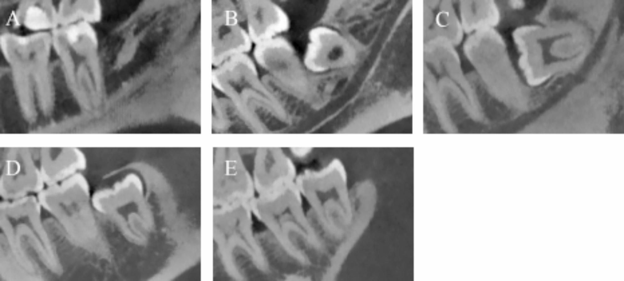
Assessment of penetration of NaOClFollowing the final irrigation procedure, the specimens were embedded in auto-polymerizing acrylic, and ∼ 1 mm thick horizontal sections were obtained at 3 mm (apical), 8 mm (middle) and 12 mm (coronal) from the apex using a diamond cutting disc (Isomed1000, Buehler, Lake Bluff, IL) under water cooling. The apical, middle and coronal thirds were scanned using CLSM at x5 magnification with a laser wave-length of 561 nm (LSM 800; Zeiss, Jena, Germany) (Fig. 1). Subsequently, after acquiring the images, the following three parameters were calculated for each third.
Fig. 1
Representative confocal laser scanning images of the coronal, middle and apical root thirds of each experimental group
1) Maximum penetration depth: This parameter was defined as the distance from the root canal wall to the deepest point of penetration at four standardized points with 90° angles. The total value of these four measurements was divided by 4 to calculate the mean maximum penetration depth (μm) [26] (Fig. 2a).
Fig. 2
(a) Measurement of maximum dentinal tubule penetration at four standardized points. (b) Measurement of sealer penetration (yellow lines) and circumference of the root canal wall (white line) to calculate percentage of sealer. (c) Measurement of the area of penetrations
2) Penetration percentage: The penetration percentage was calculated by dividing the length to which the irrigation solution penetrated the dentinal tubules along the root canal walls by the circumference of the root canal wall and multiplying this result by 100 (as %) [25] (Fig. 2b).
3) Area of penetration: To calculate the area of penetration, the total area covered by penetration of the irrigation solution was measured (μm2) [27] (Fig. 2c).
The investigator who performed the measurements on the CLSM images was blinded to the treatment groups. The images were analyzed using the Zeiss Zen software 2.3 (Carl Zeiss).
Statistical analysisThe study was conducted with an average of 10 replicates, following a 4 × 3 factorial experimental design. The analysis aimed to determine whether a statistical difference existed between the means of the activation methods (Control, SA, UI and XPR) and the root thirds (apical, middle and coronal). Statistical analysis was performed using the Anderson Darling test to assess normal distribution and Levene test to evaluate the homogeneity of group variances. However, these tests did not meet the prerequisites for parametric tests (P < 0.005). The Kruskal–Wallis test was employed to compare the medians of the activation methods in each root third, whereas the Friedman test was used for the median comparisons of the root third for each activation method. Statistical analyses were performed using R software (R Core Team, 2020). To determine which activation method and root third exhibited statistically significant differences between medians, the Bonferroni–Dunn multiple comparison test was applied at a significance level of 5%.
留言 (0)