Animals
All experimental protocols on animals were approved by the Animal Care and Use Committee at Shanghai Institute of Materia Medica, Chinese Academy of Sciences (IACUC number for C57BL/6J mice: 2023-07-XX-382). C57BL/6J mice were purchased from Slac Laboratory Animal (Shanghai, China) and maintained under a 12 h light/dark cycle with normal chow and free access to water.
Hormone secretion from isolated islets
Islets were isolated from anesthetized male C57BL/6J mice (8 wk old). 1 mg/mL Collagenase P (Roche, #11213873001) solution was injected into the pancreas via the bile duct, and the pancreas was digested at 37 °C for 15 min. The mixture was centrifuged at 800 rpm for 3 min to collect the precipitate, which was then washed three times with HBSS (5.4 mM KCl, 0.3 mM Na2HPO4, 0.4 mM KH2PO4, 4.2 mM NaHCO3, 1.3 mM CaCl2, 0.5 mM MgCl2, 0.6 mM MgSO4, 137 mM NaCl, 5.6 mM D-glucose, pH 7.4). Then, the precipitate was re-suspended in HBSS and filtered through a 70 μm filter to obtain islets. The detached islets were cultured overnight in RPMI 1640 supplemented with 10% fetal bovine serum (FBS). The islets were transferred to 48-well plates and starved with KRBB (118.5 mM NaCl, 2.54 mM CaCl2, 1.19 mM KH2PO4, 4.74 mM KCl, 25 mM NaHCO3, 1.19 mM MgSO4, 10 mM HEPES, pH 7.4) containing 0.5% BSA for 60 min. Subsequently, the islets were incubated with 0.1% dimethyl sulfoxide (DMSO) or compounds for a duration of 2 h at 37 °C in 1 mL KRBB containing 16.8 mM or 2.8 mM glucose. Supernatants were collected, and the insulin concentrations were measured using HTRF Insulin kit (Cisbio, #62INSPEC) and an Envision Plate Reader (PerkinElmer).
Cell culture and hormone secretion
The INS-1E cells were cultured in RPMI 1640 supplemented with 10% FBS, the αTC1-9 cells were cultured in low glucose DMEM supplemented with 10% FBS and the MIN6 cells were cultured in high glucose DMEM supplemented with 10% FBS. Cells were grown in 96-well plates at a density of 5 × 10⁴ cells (INS-1E and αTC1-9) or 2 × 10⁴ cells (MIN6) per well. After preincubation for 30 min at 37 °C in KRBB, the cells were incubated with either 0.1% DMSO or compounds for 1 h at 37 °C in KRBB containing either 16.8 mM or 2.8 mM glucose. Supernatants were collected and hormone concentrations were measured using HTRF Insulin Detection Kit (Revvity, #62IN1PEH) or HTRF Glucagon Detection Kit (Revvity, #62CGLPEH). For co-culturing of INS-1E and αTC1-9 cells, RPMI 1640 supplemented with 10% FBS was used, both cell densities were 2.5 × 10⁴ cells per well in 96-well plates.
Calcium assay
V1bR/HEK293 cells were seeded at a density of 4 × 10⁴ cells per well into 96-well culture plates and incubated for 24 h at 37 °C in 5% CO2. The cells were then incubated with 2 μM Fluo-4 AM in HBSS at 37 °C for 40 min. After thorough washing, 50 μL of HBSS was added. After addition of 25 μL antagonists and incubation for 10 min at room temperature, 25 μL agonists was dispensed into the well using a FlexStation III microplate reader (Molecular Devices), and the intracellular calcium change was recorded at an excitation wavelength of 485 nm and an emission wavelength of 525 nm.
cAMP accumulation assay
The cAMP assay was performed with GCGR/HEK293 or GLP-1R/HEK293 cell lines. Briefly, cells were harvested and resuspended in DMEM containing 500 μM IBMX at a density of 2 × 10⁵ cells/mL. Cells were then plated onto 384-well assay plates at 1000 cells/5 μL/well. DMEM (5 μL) containing different concentrations of antagonists were added to the cells and the incubation lasted for 15 min at 37 °C (this step was omitted in the agonist detection), then another 5 μL DMEM containing different concentrations of agonists were added to the cells and the incubation lasted for 30 min at 37 °C. Intracellular cAMP levels were detected with a LANCE Ultra cAMP kit (PerkinElmer, #TRF0264) and an Envision Plate Reader (PerkinElmer) according to the manufacturer’s instructions.
RNA sequencing of single mouse pancreatic islet cells
Pancreatic islets of male C57BL/6J mice were isolated as previously described and dispersed into single-cell suspension using nonenzymatic Cell Dissociation Solution (Sigma-Aldrich, #C5914) for 3 min at 37 °C. Single islet cells in RPMI 1640 medium (300 cells/μL) were mixed (3:2) with C1 Cell Suspension Reagent before loading onto C1 Integrated Fluidic Circuit (IFC). 20 μL LIVE/DEAD staining solution (2.5 μL ethidium homodimer-1 and 0.625 μL calcein AM in 1.25 mL C1 Cell Wash Buffer) was loaded onto the C1 IFC. Each capture site was carefully examined under microscope in bright field, GFP, and Texas Red channels for cell doublets and viability. Cell lysing, reverse transcription, and cDNA amplification were performed on the C1 Single-Cell Auto Prep IFC. Single cell cDNAs were sequenced by Berry Genomics. The sequencing data were standardized using the Z-score model, cluster analysis and t-SNE analysis were conducted.
Fluorescence-activated cell sorting and real-time qPCR assay
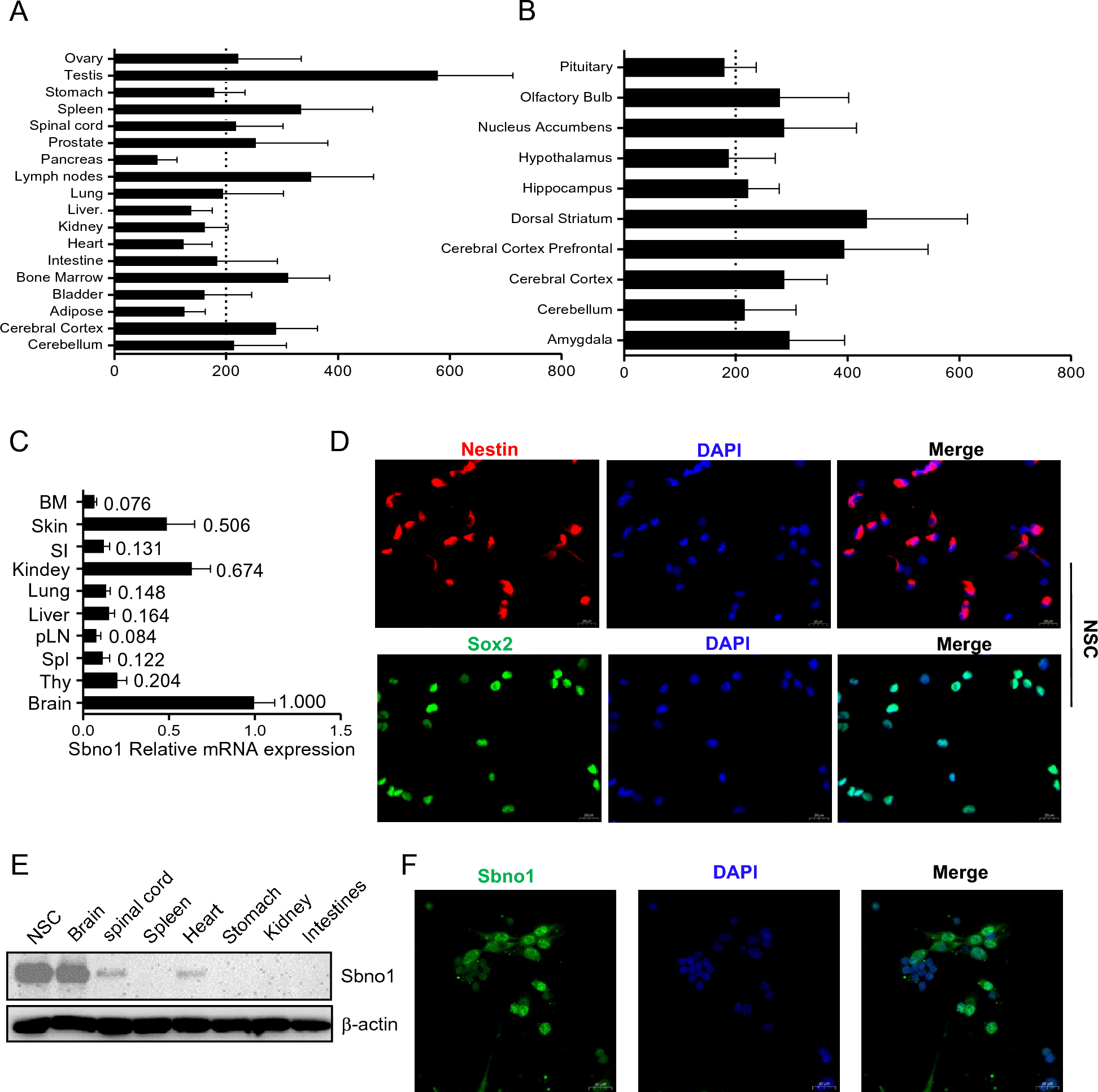
Pancreatic islets of male MIP-GFP mice (The Jackson Lab, #006864) were isolated and dispersed as previously described. FACS sorting was performed using an Influx cell sorter (BD Biosciences). Forward scatter (FSC) with parallel polarization and side scatter (SSC) were collected at 488 nm with each sample collected directly into Trizol to ensure immediate cell lysis and preservation of RNA integrity. RNA was isolated with guanidine thiocyanate and phenol method and RNA samples were reverse-transcribed using PrimeScript RT reagent Kit (TAKARA, #RR047Q). qPCR was conducted with Hieff qPCR SYBR Green Master Mix (Yeasen, #11202ES). The primer sequences used in qPCR are listed in Table 1.
Table 1 Summary of primer and shRNA sequencesshRNA transfection
Specific sequences of shRNAs (Table 1) targeting GCGR or GLP-1R mRNA were constructed into pLKO.1 puro lentiviral vector (Addgene, #8453). Lentiviral vectors and packaging vectors were transfected into HEK293T cells by FuGENE HD Transfection Reagent (Promega, #E2312) to produce virus particles. INS-1E cells were seeded onto 6-well plates at a density of 5 × 10⁵ cells per well and incubated with viruses and 5 μg/mL polybrene for 48 h. Transfected INS-1E cells were collected for real-time qPCR and co-culture experiments.
Western blot
MIN6 and αTC1-9 cells were lysed and sonicated in 1 × SDS buffer. Aliquots of proteins were fractionated by 10% SDS-PAGE and transferred to polyvinylidene difluoride membranes. The membranes were blocked with 5% nonfat milk for 30 min at room temperature and then incubated overnight at 4 °C in buffer containing anti-GAPDH (Cell Signaling Technology, #14C10 at 1:5000) or anti-V1bR (Abcam, #104365 at 1:900) antibodies. After through washing, the membranes were incubated with proper secondary antibodies for 1 h at room temperature. Immunostaining was visualized using Signal Fire™ ECL Reagent (Cell Signaling Technology, #6883) and images were taken with a ChemiDocXRS imaging system (Bio-Rad).
Oral glucose tolerance test and insulin detection
For oral glucose tolerance test (OGTT), mice were fasted overnight and then given either 0.1% DMSO in normal saline (vehicle) or 1 mg/kg AVP and 10 mg/kg Exendin (9–39) (n = 8 per treatment group) via intraperitoneal injection. A glucose bolus was delivered (1.5 g/kg orally) 15 min later. Blood was collected from a tail nick at designated time points, and plasma glucose levels were determined with a glucose meter. To detect insulin level, blood was collected from the retro-orbital plexus of mice 15 min after glucose administration (n = 4 per treatment group) and detected with an ELISA kit for mouse insulin (Crystal Chem, #90080).
Statistical analysis
Results are presented as mean ± SEM unless otherwise noted. Statistical significance was calculated using two-tailed Student’s t-test, P < 0.05 was considered significant. Statistical analysis was conducted using Graph-Pad Prism 8 (www.graphpad.com).


留言 (0)