
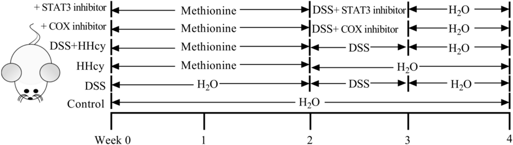
記住我
Levels of Hcy in serum and colon tissues of mice were measured to validate the hyperhomocysteinemia (HHcy) model. Compared to the control group, both serum and colon tissue exhibited elevated Hcy levels in the HHcy group [HHcy: 782.09 ± 182.39 vs. Control: 411.78 ± 72.17 ng/mL, P = 0.004; HHcy: 277.94 ± 50.58 vs. Control: 143.73 ± 24.07 ng/mg, P = 0.004] [Fig. 2A and B]. Additionally, in comparison to the DSS group, the HHcy + DSS group showed significantly higher Hcy concentrations in serum and Hcy content in colon tissues [DSS + HHcy: 755.55 ± 164.56 vs. DSS: 424.75 ± 58.68 ng/mL, P = 0.008; DSS + HHcy: 285.11 ± 57.30 vs. DSS: 149.79 ± 18.47 ng/mg, P = 0.008] (Fig. 2A and B).
Fig. 2
Homocysteine regulates intestinal inflammation in DSS mice. A Concentration of homocysteine in serum; B content of homocysteine in colon tissues; C body weight; D DAI score; E histological scores; F the activity of MPO in colon tissues; G colon histopathological features (HE X 200). *DSS vs. Control, P < 0.05; # DSS + HHcy vs. DSS, P < 0.05; ^DSS + HHcy + COX inhibitor vs. DSS + HHcy, P < 0.05; +DSS + HHcy + STAT3 inhibitor vs. DSS + HHcy, P < 0.05; ⊙HHcy vs. Control, P < 0.05
Following DSS treatment, mice exhibited weight loss from day 2 onward in comparison to blank controls (Fig. 2C). The Disease Activity Index (DAI) score increased progressively with DSS treatment duration (Fig. 2D). Evaluating the degree of inflammation in DSS-induced colitis, histological scores and MPO activity were employed. Histological scores were significantly higher in the DSS group than the control group [DSS: 1.78 ± 0.39 vs. Control: 0.22 ± 0.19, P < 0.001] (Fig. 2E), indicating evident inflammatory changes. Similarly, MPO activity was notably elevated in the DSS group compared to controls [DSS: 0.86 ± 0.08 vs. Control: 0.46 ± 0.06, P < 0.001] (Fig. 2F). Colon histology of the control group showed no signs of inflammation, whereas DSS treatment led to pathological changes, such as epithelial and cupular cell loss, inflammatory cell infiltration, and crypt damage (Fig. 2G).
Further examination of Hcy’s impact on colonic inflammation revealed more pronounced weight loss in the HHcy + DSS group compared to the DSS group (Fig. 2C). Additionally, the HHcy + DSS group displayed a significantly higher DAI score (Fig. 2D). Histological scores demonstrated more severe inflammatory infiltration and crypt damage in colon tissue induced by HHcy-mediated colitis [DSS + HHcy: 2.78 ± 0.39 vs. DSS: 1.78 ± 0.39, P = 0.004] (Fig. 2E and G). Furthermore, HHcy intensified MPO activity in mice within the DSS group [DSS + HHcy: 1.32 ± 0.11 vs. DSS: 0.86 ± 0.08, P < 0.001] (Fig. 2F).
In this investigation, mice received treatment with the COX inhibitor indomethacin and the STAT3 inhibitor cryptotanshinone. It was observed that the COX inhibitor reduced both colon histological score and MPO activity in the HHcy + DSS group [DSS + HHcy + COX inhibitor: 2.00 ± 0.33 vs. DSS + HHcy: 2.78 ± 0.39, p = 0.019; DSS + HHcy + COX inhibitor: 1.10 ± 0.07 vs. DSS + HHcy: 1.32 ± 0.11, P = 0.005] (Fig. 2E and F). Notably, this treatment ameliorated pathological changes such as inflammatory cell infiltration and crypt damage in colon tissue compared to the preceding conditions (Fig. 2G). Moreover, the addition of the STAT3 inhibitor also yielded reductions in colon histological score and MPO activity in the HHcy + DSS group [DSS + HHcy + STAT3 inhibitor: 2.78 ± 0.39 vs. DSS + HHcy: 1.89 ± 0.51, P < 0.001; DSS + HHcy + STAT3 inhibitor: 1.32 ± 0.11 vs. DSS + HHcy: 0.99 ± 0.09, P < 0.001] (Fig. 2E and F). This intervention corresponded with a mitigation of inflammatory cell infiltration in colon tissue.
Changes in Expression of PLA2, PGE2, cAMP, and STAT3 After Addition of COX and STAT3 InhibitorsProtein expressions of COX2 (Fig. 3A and C) and p-STAT3/STAT3 (Fig. 3A and F) were notably higher in the DSS group compared to the control group [DSS: 0.53 ± 0.13 vs. Control: 0.12 ± 0.07, P < 0.001; DSS: 0.30 ± 0.04 vs. Control: 0.10 ± 0.01, P < 0.001]. Further investigation into Hcy’s impact on intestine-related signaling molecules in DSS-induced mice revealed significantly elevated protein expressions of COX2 (Fig. 3A and C) and p-STAT3/STAT3 (Fig. 3A and F) in the Hcy + DSS group compared to the DSS group [DSS + HHcy: 1.03 ± 0.055 vs. 0.53 ± 0.13, P < 0.001; DSS + HHcy: 0.60 ± 0.06 vs. DSS: 0.29 ± 0.04, P < 0.001].
Fig. 3
Signaling pathway of homocysteine regulating intestinal inflammation in DSS mice. A Immunoblotting analysis of the cPLA2, phosphorylated-cPLA2, COX2, cAMP, STAT3, and phosphorylated-STAT3 protein in the colon of mice; B quantitative analysis of the protein level of p-cPLA2/cPLA2; C quantitative analysis of the protein level of COX2; D quantitative detection concentration of PGE2 in serum by ELISA; E quantitative analysis of the protein level of cAMP; D quantitative analysis of the protein level of p-STAT3/STAT3. *DSS vs. Control, P < 0.05; #DSS + HHcy vs. DSS, P < 0.05; ^DSS + HHcy + COX inhibitor vs. DSS + HHcy, P < 0.05; +DSS + HHcy + STAT3 inhibitor vs. DSS + HHcy, P < 0.05. a: Control group, b: DSS, c: HHcy group, d: DSS + HHcy group, e: DSS + HHcy + COX inhibitor group, f: DSS + HHcy + STAT3 inhibitor group
Continuing to explore Hcy’s promotion of intestinal inflammatory responses via the PGE2/COX2 signaling pathway, mice received ongoing treatment with the COX inhibitor indomethacin and the STAT3 inhibitor cryptotanshinone. Post-treatment, the protein level of COX2 (Fig. 3A and C) significantly decreased compared to DSS + HHcy [DSS + HHcy + COX inhibitor: 0.74 ± 0.06 vs. DSS + HHcy: 1.03 ± 0.055, P = 0.001]. Moreover, the addition of the STAT3 inhibitor notably reduced the protein level of p-STAT3/STAT3 [DSS + HHcy + STAT3 inhibitor: 0.42 ± 0.04 vs. DSS + HHcy: 0.60 ± 0.06, P < 0.001] (Fig. 3A and F).
Notably, the addition of the COX inhibitor did not significantly alter the protein expression of p-PLA2/PLA2 [DSS + HHcy + COX inhibitor: 0.74 ± 0.06 vs. DSS + HHcy: 0.81 ± 0.04, P = 0.105] (Fig. 3A and B), but led to significant decreases in serum PGE2 concentration (Fig. 3D), cAMP, and p-STAT3/STAT3 protein expression (Fig. 3A, E and F) [DSS + HHcy + COX inhibitor: 101.65 ± 8.77 pg/mL vs. DSS + HHcy: 131.36 ± 11.63 pg/mL, P = 0.001; DSS + HHcy + COX inhibitor: 0.46 ± 0.08 vs. DSS + HHcy: 0.70 ± 0.07, P = 0.002; DSS + HHcy + COX inhibitor: 0.46 ± 0.08 vs. DSS + HHcy: 0.70 ± 0.07, P = 0.002].
The protein expressions of p-PLA2/PLA2, COX2, and cAMP upstream of STAT3 inhibitor addition did not exhibit significant changes [DSS + HHcy + STAT3 inhibitor: 0.74 ± 0.04 vs. DSS + HHcy: 0.81 ± 0.04, P = 0.077; DSS + HHcy + STAT3 inhibitor: 0.96 ± 0.05 vs. DSS + HHcy: 1.03 ± 0.055, P = 0.329; DSS + HHcy + STAT3 inhibitor: 0.63 ± 0.10 vs. DSS + HHcy: 0.70 ± 0.07, P = 0.28] (Fig. 3A-C and E). However, serum PGE2 concentration significantly decreased (Fig. 3D) and p-STAT3/STAT3 protein expression was notably reduced (Fig. 3A and F) [DSS + HHcy + STAT3 inhibitor: 93.24 ± 6.77 pg/mL vs. DSS + HHcy: 131.36 ± 11.63 pg/mL, P < 0.001; DSS + HHcy + STAT3 inhibitor: 0.42 ± 0.04 vs. DSS + HHcy: 0.60 ± 0.06, P < 0.001].
The above findings confirm that PGE2/STAT3 is the signaling pathway that may be responsible for the exacerbation of intestinal inflammatory responses by homocysteine.
Changes in the Expression of Th17 Differentiation and Proliferation-related Molecules After the Addition of COX and STAT3 InhibitorsTh17 cells are central cells in the regulation of intestinal inflammation in IBD, and RORγt is not only a specific transcription factor in the differentiation of Th17 cells, but also a powerful target molecule for the regulation of the transcription factor STAT3 [15]. We investigated whether Hcy aggravates the intestinal inflammatory response by promoting the expression of IL-17A and its specific transcription factor RORγt in colonic tissue colon tissues through the PGE2/STAT3 signaling pathway.
Western blotting showed that the protein expression of IL-17A and RORγt in colon tissue was significantly higher in the Hcy + DSS group compared with the DSS group [Hcy + DSS: 0.87 ± 0.11 vs. DSS: 0.47 ± 0.03, P < 0.001; Hcy + DSS: 0.92 ± 0.08 vs. DSS: 0.40 ± 0.06, P < 0.001] (Fig. 4A and B). Flow cytometry revealed that the level of CD4+17A+T cell was higher in the Hcy + DSS group than in the DSS group [Hcy + DSS: 7.95 ± 0.20 vs. DSS: 5.78 ± 0.21, P < 0.001] (Fig. 4D and F). IL-23R plays a crucial role in Th17-mediated inflammation, maintains the Th17 phenotype and promotes its proliferation, and is essential for the terminal differentiation of Th17 [16]. The expression of IL-23R was significantly higher in the Hcy + DSS group compared with the DSS group by flow cytometry [Hcy + DSS: 13.87 ± 1.55 vs. DSS: 8.80 ± 0.68, P < 0.001] (Fig. 4E and G).
Fig. 4
Homocysteine promotes the differentiation of Th17 cells in DSS-induced colitis tissue through the PGE2/STAT3 signaling pathway. A Immunoblotting analysis of the IL-17A and RORγt protein in the colon of mice; B quantitative analysis of the protein level of IL-17A; C quantitative analysis of the protein level of RORγt; D and E IL-17A and IL-23R in CD4+T cells analyzed by flow cytometry; F and G quantitative analysis of the T cell level of CD4 + IL-17A + and IL-23R. *DSS vs. Control, P < 0.05; #DSS + HHcy vs. DSS, P < 0.05; ^DSS + HHcy + COX inhibitor vs. DSS + HHcy, P < 0.05; +DSS + HHcy + STAT3 inhibitor vs. DSS + HHcy, P < 0.05. a: Control group, b: DSS, c: HHcy group, d: DSS + HHcy group, e: DSS + HHcy + COX inhibitor group, f: DSS + HHcy + STAT3 inhibitor
To further investigate whether Hcy promotes Th17 cells differentiation in the colon tissue of DSS-induced colitis mice through the PGE2/STAT3 signaling pathway, we added COX inhibitor to the experimental mice and found that the protein expression of IL-17A and RORγt (Fig. 4A-C) and the level of CD4+IL-17A+T and IL-23R in CD4+T cells (Fig. 4D-G) were significantly reduced after the inhibitor added [DSS + HHcy + COX inhibitor: 0.52 ± 0.05 vs. DSS + HHcy: 0.87 ± 0.11, P < 0.001; DSS + HHcy + COX inhibitor: 0.52 ± 0.05 vs. DSS + HHcy 0.92 ± 0.08, P < 0.006; DSS + HHcy + COX inhibitor: 6.06 ± 0.34 vs. DSS + HHcy 7.95 ± 0.20, P < 0.001; DSS + HHcy + COX inhibitor: 10.17 ± 0.45 vs. DSS + HHcy: 13.87 ± 1.55, P < 0.001].
After the STAT3 inhibitor was added, we also observed the protein expression of IL-17A and RORγt in colon tissues (Fig. 4A–C), and the level of CD4+IL-17A+T cell and IL-23R in CD4+ T cells was also significantly decreased [DSS + HHcy + STAT3 inhibitor: 0.50 ± 0.02 vs. DSS + HHcy: 0.87 ± 0.11, P < 0.001; DSS + HHcy + STAT3 inhibitor: 0.50 ± 0.02 vs. DSS + HHcy: 0.92 ± 0.08, P < 0.001; DSS + HHcy + STAT3 inhibitor: 7.95 ± 0.20 vs. DSS + HHcy: 5.93 ± 0.27, P < 0.001; DSS + HHcy + STAT3 inhibitor: 13.87 ± 1.55 vs. DSS + HHcy: 9.65 ± 0.64, P < 0.001] (Fig. 4D–G).
These results suggest that Hcy may exacerbate intestinal inflammation by promoting the expression of IL-17A, IL-23R, and RORγt in colonic tissues via the PGE2/STAT3 signaling pathway.
留言 (0)