Human Tissue Samples and Cell Culture
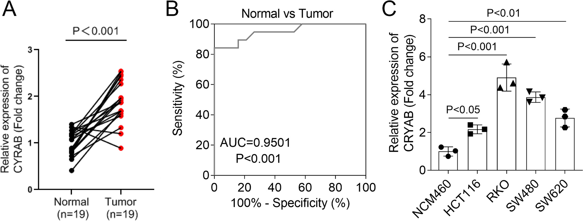
A total of 19 paired CRC tumor and adjacent noncancerous tissue samples were collected according to standard institutional protocols from Nanjing First Hospital. The study was reviewed and authorized by the Ethics Committee of our institution and was conducted in accordance with the Declaration of Helsinki.
All cell lines, including a human normal colonic epithelial cell (NCM460) and four CRC cell lines (HCT116, RKO, SW480, SW620), were purchased from the Shanghai Cell Bank of the Chinese Academy of Sciences (Shanghai, China). The above cell lines were maintained in commercially available DMEM medium (Corning, USA) supplemented with 10% FBS (Gibco-BRL) and 1% penicillin/streptomycin (Sigma, USA) at 37 °C and humidified with 5% CO2.
Plasmid and Cell Transfection
The shRNA targeting CRYAB was inserted into the pLKO.1-RFP vector to construct the PLKO.1-sh-CRYAB plasmid (labeled sh-CRYAB, TOP: CCGGGAAGAGCGCCAGGATGAACATCTCGAGATGTTCATCCTGGCGCTCTTCTTTTTG; Bottom: CTTCTCGCGGTCCTACTTGTAGAGCTCTACAAGTAGGACCGCGAGAAGAAAAACTTAA). His-tagged CRYAB plasmid was obtained from Sino Biological (Beijing, China). The corresponding overexpression plasmids (labeled TRIM6, TRIM24, TRIM47, TRIM52, and TRIM55) were constructed by cloning the coding sequences of CRYAB, TRIM6, TRIM24, TRIM47, TRIM52, and TRIM55 into a pcDNA3.0 expression vector. The TRIM55 sequence or β-catenin sequence was cloned into the expression vectors PCMV5-HA or pRK7-Flag, respectively, to generate the following plasmids: HA-TRIM55 and Flag-β-catenin. GST-tagged Ub wild type (GST-Ub WT) and mutant plasmids (GST-Ub K48R, GST-Ub K63R) were obtained from GeneWiz (Nanjing, China). The constructed plasmids were transfected with RKO or SW480 cells for 48 h with the help of Lipofectamine 3000 (Invitrogen) reagent.
Cell Viability Assay
Viability of log-phase RKO and SW480 cells was determined using the CCK-8 kit (Dojindo, Japan). Cells (3000 cells/well) were inoculated into 96-well plates containing culture medium and incubated for 24 h. Subsequently, CCK-8 reaction reagent (10 μL) was added to the cultures and maintained at 37 °C for 2 h. The cells were then incubated for 2 h at 37 °C. Finally, the absorbance at 450 nm of each well was measured using an automated enzyme marker (Molecular Device, USA) to calculate cell viability.
Determination of Intracellular Iron, Lipid Reactive Oxygen Species (ROS), Malondialdehyde (MDA), and Glutathione (GSH) Levels
Intracellular levels of unstable iron (Fe2+) were assayed using an iron assay kit purchased from Abcam. According to the standard experimental protocol, 1.5 × 10⁵ sh-NC, sh-CRYAB, Vector, CRYAB, CRYAB + vector, or CRYAB + TRIM55 group cells were homogenized in iron assay buffer and incubated with 5 µL of assay buffer for 30 min. Immediately after incubation of the above mixtures with 100 µL of iron probe for 60 min at 37 °C protected from light, the optical density (OD) of the reaction mixtures at 593 nm was evaluated on an enzyme labeling instrument.
Intracellular ROS levels were assessed relying on the C11-BODIPY 581/591 (50 ummol, Thermo Fisher) lipid peroxidation fluorescent probe and flow cytometry. Briefly, RKO and SW480 were transfected with specific plasmids and then stained with C11-BODIPY 581/591 as required for 1 h (protected from light, 37 °C). PBS solution was performed to remove excess dye and then changes in cellular fluorescence intensity were analyzed by flow cytometry (Accuri C6, BD Bioscience).
Malondialdehyde assay kit (Abcam, USA) and commercial GSH assay kit (Solarbio, China) were used to determine MDA and GSH content in CRC cell lysates, respectively.
Glutathione Peroxidase (GPx) Activity
GPx activity within cell samples was assessed using the GPx Activity Assay Kit (Solarbio, China). RKO and SW480 cell lysates, preheated GSH-PX assay working solution were sequentially added to a quartz cuvette and mixed, and A340 was read by spectrophotometer immediately after mixing. At this time, the initial value was recorded. After incubation for 3 min at 37 °C, A340 was read again. The blank tubes were operated in the same way as the sample tubes except that distilled water was used in place of the cell lysates. Finally, GPx activity in the cells was calculated according to the formula provided in the manual.
Western Blotting and Immunoprecipitation (Co-IP)
For western blotting, cultured CRC cells were lysed in 300 μL of RIPA lysis buffer (Thermo Scientific) mixed with protease inhibitors. After determining the protein concentration (BCA Protein Quantification Kit, Merck), an aliquot of denatured protein samples was loaded and separated on 15% SDS-PAGE and then transferred to a PVDF membrane (Millipore). Subsequently, the PVDF membrane was incubated with the target antibody (anti-β-catenin, 1:1000, ab224803, Abcam) overnight. After TBST washing, the membrane continued to be incubated with HRP-coupled secondary antibody (1:6500, ab205718, Abcam) for 2 h. Finally, the blot was visualized using an ECL chemiluminescence kit (Millipore).
For immunoprecipitation, lysates from CRC cells transfected with appropriate plasmids were extracted for IP. For IP of HA-tagged, Flag-tagged, or His-tagged proteins, lysates were incubated overnight with Anti-HA magnetic beads (Beyotime, P2121), Anti-Flag M2 magnetic beads (Sigma-Aldrich, M8823), or Anti-His magnetic beads (MedChenExpress, HY-K0209) at 4 °C. Beads were collected to release immunoprecipitated protein samples and used for subsequent western blotting analysis.
In Vivo Ubiquitination Assay
The GST-Ub construct and HA-TRIM55 and Flag-β-catenin were transfected into RKO and SW480 cells. After 24–48 h of transfection, cells were lysed in ubiquitination lysis buffer and sonicated. Immunoprecipitation was performed with anti-Flag antibody (Thermo Fisher), followed by western blot analysis of the precipitated complexes with anti-GST antibody (Merck).
Protein Stability and Degradation Analysis
RKO and SW480 cells transfected with TRIM55 overexpression plasmid or negative control plasmid were co-incubated with MG132 (10 μM, MCE), or incubated with CHX (50 μg/mL, MCE) alone for different times (0, 6, 12, 18, and 24 h) to obtain total cellular proteins and analyzed by western blotting using anti-β-catenin antibody.
RT-qPCR
RNA from CRC clinical tissues and cell lines was isolated by RNAiso Plus kit (TaKaRa, China). After quantification of RNA levels, complementary RNAs were generated using a reverse transcription kit (RevertAid First Strand cDNA Synthesis Kit, Thermo). RNAs for quantification of CRYAB and TRIMAB were isolated by using SYBR Green Master Mix (Vazyme, China) and an ABI Prism® Model 7500 Fluorescent Quantitative PCR System (Applied Biosystems, USA) was applied to perform PCR amplification for quantification of CRYAB, TRIM55, and GAPDH. The relative transcript levels of the target RNAs were calculated using the 2−ΔΔCt method and their expression levels were normalized to the expression of GAPDH. The primers used in this study were as follows: TRIM55, F, 5′-TGGTTTTGGGATAGACATGGGGGT-3′ and R, 5′-CTGGTGGGACTCCTGCTTGTA-3′. CRYAB, F, 5′-CAGCTGGTTTGACACTGGAC-3′ and R, 5′-TGGCGCTCTTCATGTTTTCC-3′.
Statistical Analysis
GraphPad Prism 8.0 software was applied to analyze the data in this study. All experiments used were conducted at least three times independently and measurements were reported as mean ± SD. Statistical differences between two consecutive groups of data were compared using Student’s t test, and comparisons of data between 3 and more groups were made utilizing one-way analysis of variance (ANOVA). A p-value of less than 0.05 was considered statistically significant.

留言 (0)