Chemicals and Food Preparation
VCD, 17 β-estradiol, progesterone, dimethyl sulfoxide (DMSO), and sesame oil were purchased from Sigma-Aldrich Corporation, St. Louis, MO. Three PBDE congeners [2,2’,4,4’-tetrabromodiphenyl ether (BDE-47), 2,2’,4,4’,6-pentabromodiphemyl ether (BDE-100), and 2, 2’,4,4′5,5’-hexabromodiphenyl ether (BDE-153)] were purchased from AccuStandard, Inc, New Haven, CT. Fulvestrant was purchased from AstraZeneca, Cambridge, UK.
Animals
Eight-week-old female C57BL/6 J mice were obtained from the Jackson Laboratory (BarHarbor, ME) and housed in AAALAC-accredited Animal Resources Center, City of Hope. Experimental procedures were approved by the Institutional Animal Care and Use Committee of City of Hope and performed according to the institutional and NIH guidelines for animal care and use. Mice were fed ad libitum a standard mouse chow diet and housed 5 per cage with free access to fresh water and kept on a 12-h light/dark cycle in a conventional clean room. Upon randomization and initiation of PBDE treatment, mice were moved to and housed in polypropylene cages with Sani-Chips beddings 2–3 per cage, and drinking water was filtered twice using reverse osmosis and carbon block system to avoid environmental exposure to potential endocrine-disrupting chemicals [2]. Food consumption was recorded weekly, and total oral exposure to PBDEs were calculated.
Pilot Evaluation
At 9 w after birth, VCD (130 mg/kg in sesame oil) was intraperitoneally administered 5 sequential days per week for 3 weeks, and treated animals were euthanized at 55, 75, and 130 d post VCD injection based on the previous study [10]. Intact mice received vehicle (sesame oil) injection and were euthanized at the same time points. Two mice per group were included in this pilot experiment. The ovaries, uterus, and mammary gland were collected and routinely processed.
Mammary Gland Characterization in the VCD-Treated Model
At 9 w after birth, mice were treated with a daily intraperitoneal injection of VCD (160 mg/kg in sesame oil) for 15 d in a row, according to the previous reports [11, 17, 18]. Control mice received vehicle (sesame oil) injection. During the entire experimental period, the animals were fed ad libitum with the control diet (described in the next section) for later comparisons with the PBDE experiments.
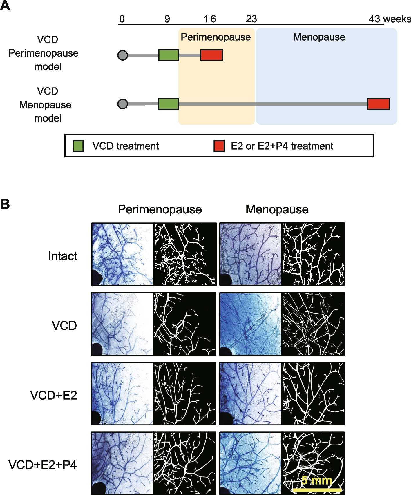
VCD Perimenopause model: In addition to the five vehicle-treated mice that were used as controls (Intact group), 30 VCD-treated mice were randomized into three groups of VCD, VCD + E2, and VCD + E2 + P4 (n = 10 each) at 7 w post VCD injection. The mice were treated with 17β-estradiol (E2) (1 μg/animal/day) and/or progesterone (P4) (1 mg/animal/day) for seven days as performed in our previous studies [1, 2]. The steroid hormones were dissolved in 100 µL sesame oil and administered intraperitoneally. Non-hormone groups received vehicle injections. Mice were euthanized 24 h after the last injection to collect ovaries, uterus, and mammary glands.
VCD Menopause model: 30 VCD-treated mice were randomized into groups of VCD, VCD + E2, and VCD + E2 + P4 (n = 10 each) at 14 w post VCD injection. After another 20 weeks, animals were treated with E2 and P4 for seven days and euthanized as in the Perimenopause model.
PBDE Exposure Experiments in the VCD Mouse Model
The VCD treatment was performed as described in the Mammary gland characterization in the VCD-treated model section. The experimental diet was prepared (Research Diets, Inc, New Brunswick, NJ) to achieve the relevant exposure with the ratio of the three congeners found in human blood (1 mg/kg/day, 0.056 mg/kg/day, and 0.126 mg/kg/day for BDE-47, -100, -153, respectively) [2, 8]. It was designated as the “high subacute” diet. Of note, the previous studies with the same “high subacute” diet resulted in the higher blood concentrations of the PBDE congeners by an order of magnitude compared to the human epidemiological study [1, 2, 8, 16]. In this investigation, we prepared another nutrient-matched diet with 1/20th of the PBDEs concentration (0.05 mg/kg/day, 0.0028 mg/kg/day, and 0.063 mg/kg/day for BDE-47, -100, -153, respectively). This food was designated as the “low chronic” diet. Control diet was made with a special nutrient-matched diet and vehicle (DMSO) (Research Diets, Inc).
1.Perimenopause High-Subacute PBDE model: 30 VCD-treated mice were randomized into VCD + PBDE, VCD + E2 + PBDE, VCD + E2 + P4 + PBDE (n = 10 each) at 7 w post VCD injection. The animals were treated with various combinations of the “high subacute” PBDEs treatment diet, 17β-estradiol (1 μg/animal/day), and/or progesterone (1 mg/animal/day) for seven days as performed in our previous studies [1, 2]. The special diet was fed ad libitum to mimic human environmental PBDE exposure via ingestion [19]. Mice were euthanized 24 h after the last injection to collect ovaries, uterus, and mammary glands.
2.Menopause High-Subacute PBDE model: 30 VCD-treated mice were randomized into three groups of VCD + PBDE, VCD + E2 + PBDE, and VCD + E2 + P4 + PBDE (n = 10 each) at 14 w post VCD injection. After another 20 weeks, animals were treated E2, P4, and/or “high subacute” PBDEs treatment diet for seven days and euthanized as in the Perimenopause-High PBDE model.
3.Menopause Low-Chronic PBDE model: 60 VCD-treated were randomized into three groups of VCD, VCD + PBDE, VCD + E2, VCD + E2 + PBDE, VCD + E2 + P4, and VCD + E2 + P4 + PBDE (n = 10 each) at 14 w post VCD injection. Feeding of “low chronic” PBDE diet was initiated for PBDE groups and continued for the subsequent 20 weeks. Non-PBDE groups were housed in the same special environment (described in the Animal section) as the PBDE groups and put on the control diet for these 20 weeks. During the last week on their special diets, animals received designated hormonal treatments for seven days as in the other models. Two mice from the VCD + E2 treatment group were treated with subcutaneous fulvestrant (FUL) injection (5 mg in 100 μL of sterile saline) one day prior to initiation of E2 treatment. These two mice formed an independent VCD + E2 + FUL group for single-cell transcriptomic analysis. Animals were euthanized 24 h after last injection, and the organs were harvested.
Whole-Mount Mammary Gland Scanning
The glands were fixed with 10% buffered formaldehyde and delipidated with toluene. After rehydration, the glands were stained with 0.025% toluidine blue, and permanent whole-mount staining slides were prepared. Then, the entire glands were captured using Cell3iMager Duos (SCREEN Holdings Co., Ltd., Kyoto, Japan) with 20 µm-intervals for the z-axis (Supplementary Fig. 1). Subsequently, the images were segmented with machine learning implementation using Cell3iMager Duos and the Model file CC8P06004V00 (SCREEN Holdings Co., Ltd.). The model file has been developed in our previous study [1], in which scanned images with manually labeled of ductal and end buds-like structures were fed into deep learning image analysis to construct an algorithm that automatically segments these structure in a whole-mount mammary gland image. The model file is currently distributed by SCREEN Holdings Co., Ltd. The segmented ductal structures were skeletonized and subjected to branching analysis using the ImageJ software and AnalyzeSkeleton plugin [20, 21], to calculate the total duct length and the number of branching points. The segmented end-buds were also counted using the ImageJ software.
For comparison, the data from our previous study was retrieved [1], in which the ovariectomized animals received E2 treatment (1 μg/animal/day) for seven days 20 weeks after menopause (ovariectomy), paralleling the menopause model in this study.
Mammary Gland Dissociation And Single-Cell RNA Sequencing
In short, the 4th mammary gland was dissociated with digestion buffer [1.5 mg/mL DNAse I (#10,104,159,001, Millipore Sigma, Burlington, MA), 0.4 mg/mL Collagenase IV (CLS-4, Lot: 47E17528A, Worthington Biochemical Corporation, Lakewood, NJ), 5% FBS, 10 mM HEPES in HBSS]. After dead cells were removed using Dead Cells Removal Microbeads (Miltenyl Biotec, Bergisch Gladbach, Germany), cells with high viability (> 80%) were loaded onto the Chromium Controller (10 × Genomics, Pleasanton, CA), targeting 2,000–5,000 cells per lane. The Chromium v3 single-cell 3′RNA-seq reagent kit (10 × Genomics) generated single-cell RNA-seq libraries. The NovaSeq 6000 system (Illumina, San Diego, CA) sequenced constructed libraries with a depth of 50 k-100 k reads per cell. Raw sequencing data were processed using the 10 × Genomics Cell Ranger pipeline (version 3.1.0) and then aligned to mm10 mouse genome. The datasets can be found in the NCBI GEO database under the accession GSE191219. They have been analyzed by our group for a perspective of fibroblast heterogeneity in the mammary gland, and the results have been published as an independent in-silico analysis [22].
Data Analysis
The downstream analysis of the scRNAseq analysis was performed using R (ver. 4.1.2) [23] and the Seurat R package (ver. 4.0.5) [24], unless otherwise specified. Raw data were mounted, and low-quality barcodes with < 500 gene count (nFeature_RNA) or > 10% proportion of mitochondrial genes (percent.mt) were filtered. The data from different samples were merged using merge function with a default setting. Then, normalization, scaling, variable feature identification, UMAP dimension reduction (dims = 30), and Louvain clustering (res = 0.1) were performed using a standard pipeline according to developer’s vignette. After a sensible clustering was obtained, the DEG analysis and cell cycle scoring were performed using FindAllMarkers and CellCycleScoring functions, respectively. Annotation of each cluster was determined based on marker gene expression and cell cycle indication.
Pseudo-RNA sequencing data was prepared using AverageExpression function. Subsequent clustering and principal component analyses were performed and visualized utilizing FactoMineR (ver. 2.4) [25], factoextra (ver. 1.0.7) [26], ape (ver. 5.5) [27], ggtree (ver. 3.2.1) [28] R packages. To unveil the impacts of “low dose” PBDE treatments, gene expression was compared between “VCD” and “VCD + LP”, “VCD + E2” and “VCD + E2 + LP”, “VCD + E2 + P4” and “VCD + E2 + P4 + LP”, individually. Then, commonly regulated genes were extracted from each pair. The same analysis was performed for “high dose” PBDE treatment (+ HP groups). The custom computer scripts and the relevant data are fully available in Zenodo (https://doi.org/10.5281/zenodo.7340869) [29]
The upregulated and downregulated genes in either LP or HP samples were further subjected to the Gene Ontology enrichment analysis using The Databese for Annotatrion, Visualization and Integrated Discovery (DAVID) (ver 2021) and the DAVID Knowledgebase (ver v2023q4) [30]. Briefly, the gene lists were uploaded, and the functional annotation charts for GOTERM_BP_DIRECT were retrieved with the default parameter by DAVID.
Statistical Analysis
All the statistical analyses were performed in R (ver. 4.1.2) and ggpubr R package (ver. 0.4.0). Wilcoxon rank-sum test was used to compare the distribution of two conditions of interest. p-value < 0.05 was considered as statistically significant. The number of samples can be found in the figure legends where applicable. The sample size of the study was determined based on the previous study [1, 2].


留言 (0)