
記住我
The GHR-KO mutation was introduced by CRISPR/Cas9 in cultured cells and GHR-KO founder pigs were generated by somatic cell nuclear transfer as described previously [14]. Pigs used in this study were generated by mating sows and boars heterozygous for the GHR gene mutation. The groups consisted of pigs homozygous for the GHR mutation (GHR-KO pigs) and wild-type (WT) littermate control animals. All animal experiments were performed in accordance with the German Animal Welfare Act and the Directive 2010/63/EU on the protection of animals used for scientific purposes. All animal procedures performed were approved by the responsible animal welfare authority (Regierung von Oberbayern, permissions ROB-55.2-1-54-2532-70-12, ROB-55.2Vet-2532.Vet_02-17-136 and ROB-55.2Vet-2532.Vet_02-22-92).
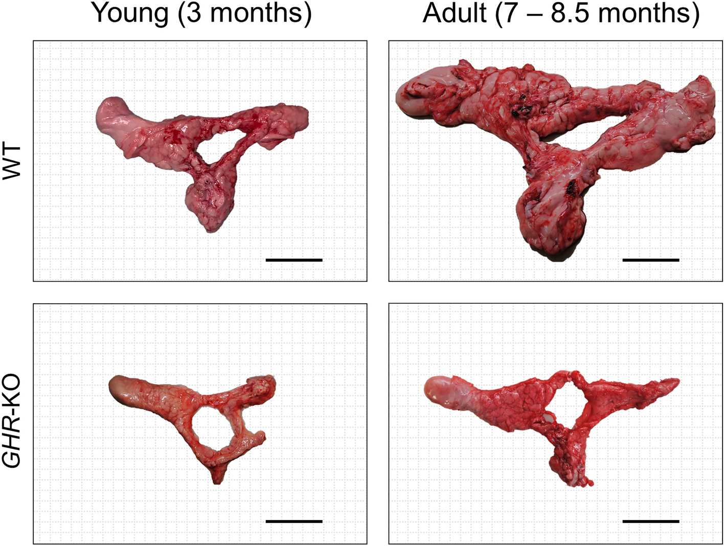
Necropsy, histology and pancreas samplingPancreata were obtained from GHR-KO (n = 12) and WT controls (n = 12) at either young (3 months; n = 5 vs. n = 5, all female) or adult age (7–8.5 months; n = 7 vs. n = 7, 3 males vs. 4 females each). Pigs were anesthetized by intravenous injection of ketamine (Ursotamin®, Serumwerk Bernburg) and xylazine (Xylazin 2%, Serumwerk Bernburg) followed by fentanyl (Fentadon®, Dechra) application. Subsequent to exsanguination, organs were sampled for further examination. After removal from the animal carcass and cleaning of connective tissue, the pancreas was weighed to the nearest g and photographed (Fig. 1). Representative samples of the pancreas were systematically and randomly sampled (as described in [17]). After cutting to size, tissue pieces were prefixed in 4% neutrally buffered formaldehyde solution for 24 h at room temperature, placed in embedding cassettes, routinely processed with a tissue processing system and embedded in paraffin. Microscopic sections (nominal thickness: 3 μm) were cut from paraffin blocks using a Microm HM 325 rotary microtome.
Fig. 1
Size comparison of fresh pancreata from GHR-KO and wild-type (WT) pigs at young and adult age. Bar = 5 cm
Immunohistochemical detection of insulinInsulin-positive cells in the pancreas were detected via immunohistochemistry according to Pilz et al. [18]. Briefly, after dewaxing, heat-induced antigen retrieval in citrate buffer (pH 6.0) for 15 min at sub boiling temperature, and endogenous peroxidase block by 1% H2O2, a mouse anti-insulin monoclonal antibody was used as the primary antibody (#I2018, Sigma; 1:3000) for overnight incubation at 4 °C, followed by biotinylated goat anti-mouse IgG (H+L) secondary antibody (#115-065-146, Jackson ImmunoResearch Laboratories, Inc.; 1:250 + 3% porcine serum) for 1 h at room temperature, and avidin–biotin complex (#PK-6100, Vector Laboratories) for 30 min at room temperature. A horseradish peroxidase DAB substrate kit (#SK-4100, Vector Laboratories) was used to detect bound antibodies. Nuclear counterstaining was performed with Meyer’s hemalum solution.
Unbiased quantitative stereological analysesThe volume density of immunohistochemically insulin-positive cells within the pancreas (Vv(β-cells/Pan)) was estimated by unbiased quantitative stereological analysis. An Olympus BX41 light microscope in combination with a connected camera (Olympus DP 72) and the newCAST™ stereology software (Visiopharm Integrator System, Visiopharm, version 3.6.2.0.) was used for the evaluation. Volume densities were determined using the point counting method. Points hitting the targeted structure’s sections were divided by those hitting the reference compartment within the same sections [19, 20]. The average number of systematically randomly sampled fields of view amounted to 233.7 ± 28.47 per case. On the average, 934.7 ± 113.9 points for pancreatic tissue and 23,367 ± 2846 points for β-cells were counted per case (at 200×magnification). Isolated β-cells were defined as individual or small clusters of insulin-positive cells (up to five nuclear profiles). The volume densities of β-cells (VV(β-cell/Pan)) and isolated β-cells (VV(isoβ-cell/Pan)) as well as the total volumes of β-cells (V(β-cell, Pan)) and of isolated β-cells (V(isoβ-cell, Pan)) in the pancreas were calculated according to [21, 22]. The terms “total volume” and “mass” are used synonymously. The specific weight of the pig pancreas (1.07 g/cm3) [21] was determined by the submersion method [23,24,25].
Intravenous glucose tolerance testsInsulin secretion capacity and glucose tolerance were assessed by intravenous glucose tolerance tests in GHR-KO pigs and WT controls at either age group (n = 3 per genotype and age). For glucose injection and blood withdrawal for insulin and glucose measurements, the pigs obtained central venous catheters (CareFlow™, Merit Medical®, size 2.5 or 3 French) into a marginal ear vein [26]. Pigs were fasted overnight (for 16 h) and afterwards attained a glucose bolus injection of 50% glucose solution (0.5 g/kg BW) though the central venous catheter. Blood samples were taken at the indicated time points before (− 10 and 0 min) and after glucose bolus injection (3, 5, 7, 10, 15, 20, 30, 40, 50, 60, 90 and 120 min).
Metabolite and hormone assaysSerum Insulin-like growth factor 1 (IGF1) concentrations were measured using the iSYS automated chemiluminescent IGF1 assay (Immunodiagnostic Systems) as described previously [27]. Blood glucose levels were immediately determined in duplicate from freshly collected full blood using a FreeStyle Freedom Lite blood glucose meter (Abbott) and FreeStyle Lite blood glucose test strips (Abbott) [28]. After clotting for 30 min at room temperature and centrifugation (1200×g, 20 min, 4 °C), aliquots of serum were stored at − 80 °C. Insulin levels were determined from serum with a sandwich chemiluminescence immunoassay (LIAISON® Insulin, DiaSorin) in combination with a fully automated immunoassay analyzer (LIAISON® XL, DiaSorin). Non-esterified free fatty acids (NEFAs) were analyzed from EDTA plasma using an AU480 clinical chemistry analyzer (Beckman Coulter) and adapted reagent kits from FUJIFILM Wako Chemicals GmbH as described previously [29].
StatisticsAll data are displayed as mean ± SEM. PROC GLM (General Linear Models, SAS 8.2) was used to analyze the body weight at the day of necropsy, the area under the curve (AUC) values for glucose and insulin and all assessed parameters concerning quantitative stereology of the pancreas. Effects of group (GHR-KO, WT), age (young, adult), sex (male and female adults) and the interaction group*age were taken into consideration. None of the investigated parameters was significantly affected by sex. Results of ivGTTs (glucose and insulin levels) were analyzed using PROC MIXED (Linear Mixed Models; SAS 8.2) considering effects of group (GHR-KO, WT), age (young, adult) as well as the interaction group*age. The Graph Pad Prism software (version 5.02 and 10.1.2) was used to generate figures and calculate AUC values, means and SEMs. P values < 0.05 were defined as significant.
留言 (0)