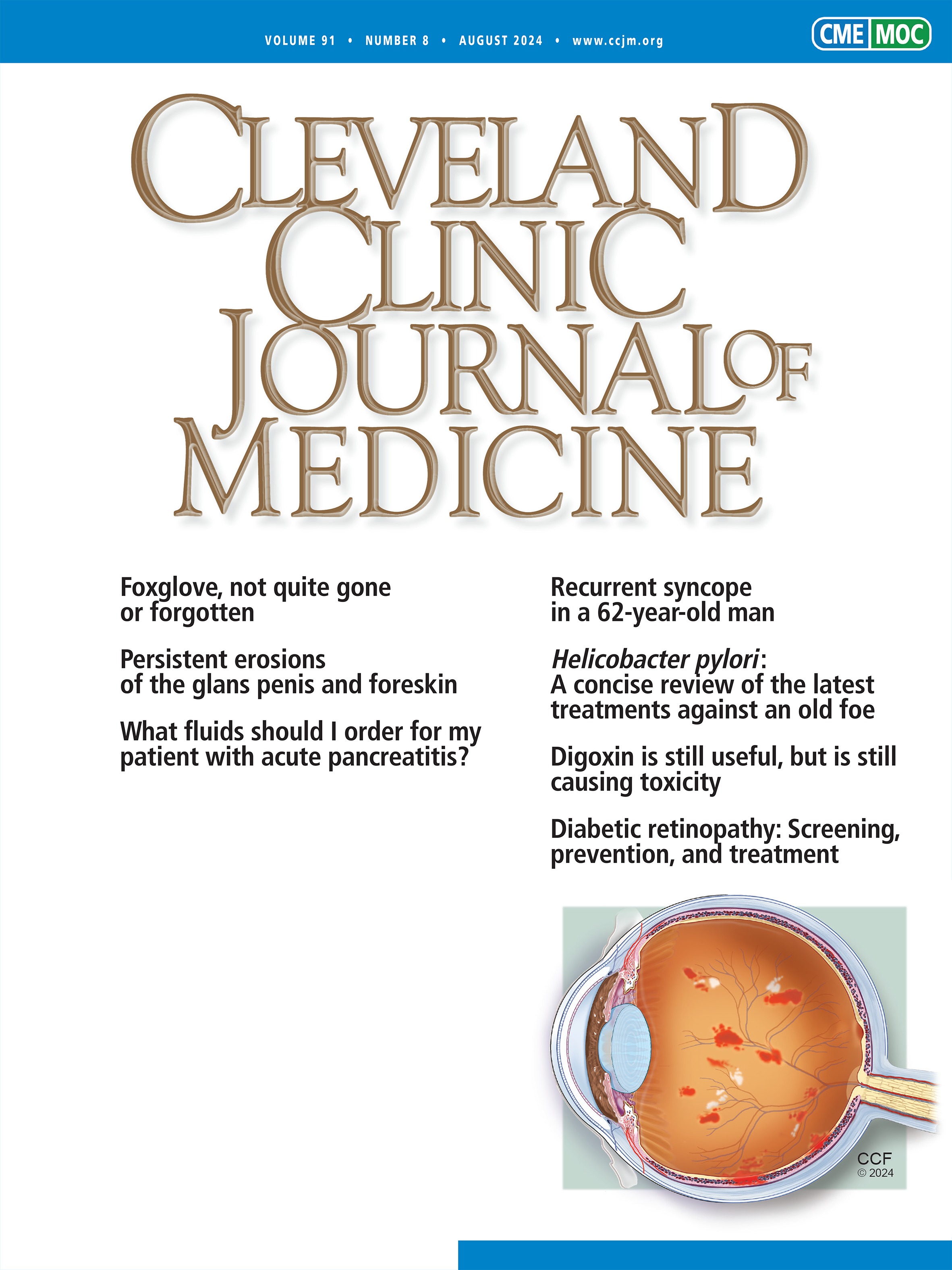
A 60-year-old man weighing 154 lb (70 kg) presented to his primary care physician with a 20-year history of slowly worsening prominent veins in both lower limbs (Figure 1). He described intense pruritis and swelling of the ankles and pretibial areas at the end of the day that improved with leg elevation overnight. He denied pain, history of venous thrombosis, leg trauma, or other medical problems. Family history was notable for varicose veins in his mother and 2 siblings. He had tried wearing compression stockings (20 to 30 mm Hg) but discontinued using them because he found them inconvenient to put on and uncomfortable. He no longer wore short pants in public due to embarrassment about the bulging veins as well as occasional comments from others asking him what was wrong with his legs.
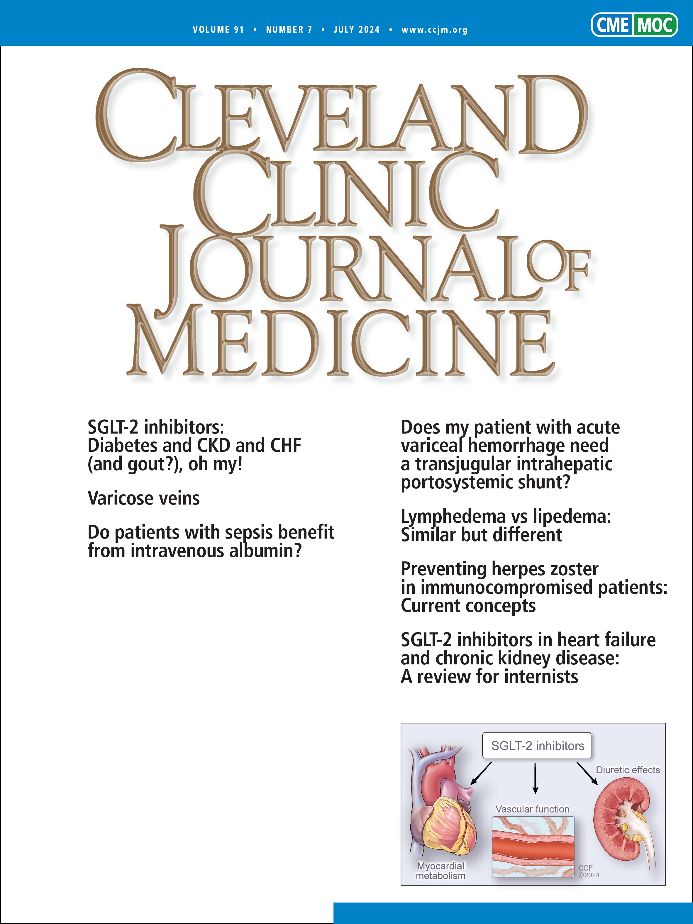
Due to cosmetic concerns, he was referred to a vascular surgeon. Venous duplex ultrasonography showed notable femoral and saphenous vein dilation and reflux without evidence of deep venous thrombosis. The surgeon performed bilateral radiofrequency ablation of the varicose veins. The patient had marked postoperative improvement (Figure 2). Follow-up Doppler ultrasonography 1 week after radiofrequency ablation did not show evidence of deep vein thrombosis.
Varicose veins are a common clinical manifestation of chronic venous disease. In the lower limbs this condition is characterized by subcutaneous dilated veins greater than or equal to 3 mm in diameter. It typically involves the great and small saphenous veins and their branches.1,2
Lower-limb varicosities are thought to result from elevated venous pressure in the extremities due to incompetent valves that allow reflux of blood backward, obstruction, or a combination of reflux and obstruction, thereby impairing adequate venous return.3 Risk factors for developing varicose veins include occupation requiring prolonged standing or walking and history of venous thrombosis, among others.4
Chronic venous disorders are graded using the Clinical, Etiologic, Anatomic, Pathophysiologic (CEAP) classification; in this system, the visible signs of chronic venous disease range from C0 (no visible or palpable disease) to C6 (venous ulcer), with varicose veins considered class C2.1
Evaluation
Patients with varicose veins can be asymptomatic or can present with clinical features including leg pain, heaviness, swelling, dryness, itching, skin changes, and ulceration.1 When examining patients with varicose veins, it is important to evaluate the varicosity pattern. In patients with symptomatic varicose veins and suprapubic or abdominal wall varicosities and in patients with leg heaviness, fullness, swelling, and claudication, evaluation for iliofemoral venous obstruction with Doppler ultrasonography is indicated to find the source of reflux.1 In those with medial thigh or vulvar varicosities and symptoms of pelvic venous congestion (eg, chronic pelvic pain, dysmenorrhea, dyspareunia, and urinary urgency), evaluation of pelvic venous pathology with Doppler ultrasonography is needed given the association between pelvic venous insufficiency and these varicosity patterns.1 Scrotal varicosities can suggest gonadal vein incompetence, nutcracker syndrome (left renal vein compression between the superior mesenteric artery and aorta), inferior vena cava lesions, or renal carcinoma.5
Color duplex ultrasound is the first diagnostic test recommended for patients presenting with varicose veins to confirm the absence of deep and superficial venous thrombosis.
Differential diagnoses to consider for chronic venous insufficiency include lymphedema, congestive heart failure, and renal disease.6
Treatment options
Untreated varicose veins can cause venous ulcers, pain, or, most commonly, aesthetic concerns, which is often why treatment is sought. Treatment of lower-extremity chronic venous disease depends on the CEAP classification and the severity of disease based on the Venous Clinical Severity score.1,6
Treatment of varicose veins often begins with conservative measures using moderate-pressure compression stockings (20–30 mm Hg) and lifestyle modifications such as weight loss and leg elevation.7 Compared with no stockings, compression stockings can improve symptoms, control edema, and improve orthostatic venous pressure.7 However, there is insufficient evidence to support their effectiveness as the primary treatment of varicose veins.1 When considering compression stockings, it is crucial to ensure that the patient does not have coexisting arterial insufficiency, which compression can worsen. Also, in practical terms, compression stockings have low compliance rates (as low as 37%) due to discomfort.1
Referral to vascular surgery for endovenous therapy (laser or radiofrequency ablation and sclerotherapy) or open venous surgical intervention (ligation or phlebectomy [or venous stripping]) may be indicated for patients with varicose veins refractory to conservative treatment or symptomatic varicose veins with axial reflux in the great or small saphenous vein.1 The type of surgical intervention is based on the size, location, and extent of venous involvement.4
Cosmetic improvement following radiofrequency ablation is around 70%, with optimal results occurring when patients wear compression stockings for 7 to 10 days after treatment8 and ambulate early. Complications of radiofrequency ablation include deep vein thrombosis, heat-induced thrombus extension, or, rarely, pulmonary embolism.1,7,8 Postprocedural duplex scanning within 1 week is routinely recommended.1


留言 (0)