Clinical blood samples
The inclusion criteria for patients were as follows: over 18 years of age and diagnosed with acute myocardial infarction. The diagnostic criteria for type 1 acute myocardial infarction were acute myocardial injury accompanied by clinical evidence of acute myocardial ischemia, at least one increase and/or decrease in cTn detected above the 99th percentile (the URL), and at least one of the following characteristics: symptoms of myocardial ischemia, new changes in the ischemic electrocardiogram, pathological Q-wave occurrence, or a time from onset to treatment of less than 6 h (Thygesen et al. 2018). The exclusion criteria for patients were as follows: under 18 years of age; lung diseases, such as inflammation, nodules or tumors; severe liver or kidney insufficiency; severe infection; rheumatic heart disease, primary cardiomyopathy, or other serious disease of the heart itself; recent ischemic stroke; severe immune system or blood system disease; long-term oral hormone use; and a history of malignant tumor. The study of human blood tissue was approved by the Ethics Committee of the Eighth Affiliated Hospital of Sun Yat-sen University (No. 202122-066-02) and complied with all relevant ethical guidelines. The participants with MI and healthy controls provided informed consent. There was no significant difference in basic information between the two groups (Supplementary Table 1).
Animals
Male C57BL/6J mice (6–8 weeks old, weighing 16–20 g, and SPF grade) were purchased from Zhuhai BesTest Bio-Tech Co., Ltd. (China; Animal Quality Certificate No. 44822700019760). All animals were housed in a controlled environment at 22 °C with a 12-h light-dark cycle and provided ad libitum access to food and purified water. All animal experiments were approved by the Animal Ethics Committee of the Affiliated Eighth Hospital of Sun Yat-sen University (2022-066-01).
Animal grouping and the MI model
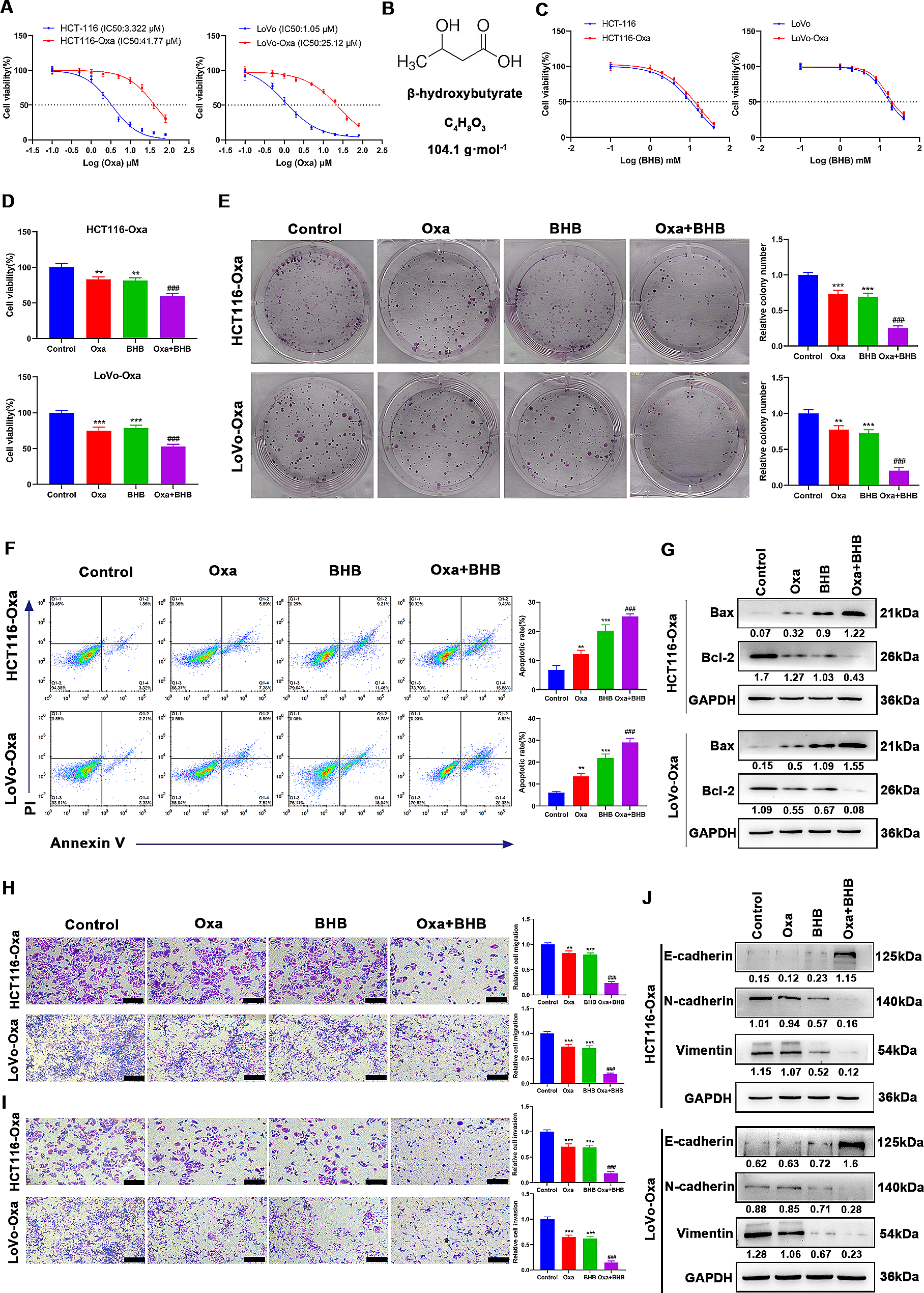
The experimental mice were randomly divided into the sham-operated group (sham), MI group (MI + Vehicle), MI with low-dose (2.5 mg/kg) AubipyOMe group (MI + L), and MI with high-dose (5 mg/kg) AubipyOMe group (MI + H), with six mice in each group. The MI mouse model was induced by ligation of the left anterior descending (LAD) coronary artery as previously described (Zheng et al. 2013). In brief, anesthesia was induced using a mixture of 2% isoflurane and oxygen, followed by disinfection. Subsequently, a 1–2 cm incision was made in the fourth intercostal space to expose the heart, which was then extruded from the thoracic cavity and secured with 6 − 0 sutures to ligate the LAD. Successful occlusion was confirmed by an electrocardiogram with elevation of the ST segment (Weng et al. 2018). Subsequently, the mouse heart was repositioned in the thoracic cavity, and air and blood were expelled before the chest was closed. The sham group underwent the same procedure without ligation of the LAD artery. On Days 12, 15, and 18 after sham operation or LAD ligation surgery, mice in the MI with AubipyOMe group received intraperitoneal injections of different doses of AubipyOMe (Sigma‒Aldrich, USA, #SML2210) (Hu et al. 2022). The MI group received injections of PBS and DMSO (solvent for AubipyOMe). Four weeks after MI surgery, cardiac function was evaluated by ultrasound imaging. Subsequently, the mice were euthanized, and the ventricular portion of the heart sample and serum were collected for subsequent experiments.
Echocardiography
The cardiac function of the mice was assessed using a high-resolution small animal ultrasound imaging system (Vevo2100, Visualsonics, Canada). Mice were initially anesthetized with 2% isoflurane using a mask. Measurements were taken for the following parameters: left ventricular (LV) internal diameter at end-systole (LVIDS) and end-diastole (LVIDD); interventricular septum thickness at end-diastole (IVSD) and end-systole (IVSS); and LV posterior wall thickness at end-diastole (LVPWD) and end-systole (LVPWS). Left ventricular fractional shortening (LVFS) and the left ventricular ejection fraction (LVEF) were calculated as described previously (Weng et al. 2018). The operators and analysts of the experiment were unaware of the group assignment.
Histological staining
Paraffin sections of mouse heart samples were obtained by rapid dissection, fixation, embedding, and slicing. The Sect. (4 μm) were stained using Masson’s trichrome staining kit (Servicebio, China, #G1006) and Sirius Red Stain Kit (Servicebio, China, #G1472) following the manufacturer’s instructions. The percentage of fibrotic tissue was determined by measuring collagen deposition in the sections using ImageJ software. For immunofluorescence staining, the heart sections were deparaffinized and blocked with 10% donkey serum. Primary and secondary antibody staining was performed accordingly. Subsequently, nuclear staining was performed using DAPI. Positive results were quantified using ImageJ software. For immunofluorescence staining, the heart sections were deparaffinized, followed by blocking with 10% donkey serum. Primary (1:200; Cell Signaling Technology, USA, #19,245) and secondary(1:300; Servicebio, China, #GB21303) antibody staining was performed accordingly. Subsequently, nuclear staining was performed using DAPI. Six images were collected for each heart and quantitative analysis was performed by Image-J software. Relative positive rate (%) : (positive area/total area) *100%.
Enzyme-linked immunosorbent assay (ELISA)
The Mouse ACP5 ELISA Kit was purchased from Signalway Antibody (USA, #EK12408), and the Human ACP5 ELISA Kit was purchased from BOSTER (China, #EK2138). ELISA kits were used to measure the levels of ACP5 in clinical patient samples and mouse blood samples according to the manufacturer’s instructions.
Cell culture and transfection
CFs and cardiac myocytes (CMs) were obtained from neonatal mice aged 1–3 days according to previous methods (Medzikovic et al. 2021). Briefly, the collected ventricles were cut to approximately 1 mm3 and digested at 4 °C for 12 h with 0.25% trypsin solution (without EDTA) (Beyotime, China, #C0205). The supernatant was discarded after centrifugation. The tissue samples were digested in a 37 °C water bath with 0.1% type II collagenase (Solarbio, China, #C8150) for 10 min at a time until all tissues were digested. After centrifugation, the cell suspension was resuspended in high-glucose Dulbecco’s modified Eagle’s medium (GIBCO, USA, #C11995500BT) supplemented with 10% fetal bovine serum (GIBCO, USA, #16000-044) and 1% penicillin‒streptomycin (HyClone, USA, #SV30010). Fibroblasts were obtained after two consecutive attachment times (approximately 45 min each). CMs in the culture medium were transferred to another culture plate and incubated at 37 °C and 5% CO2. The cells were used in experiments when they were passaged for 3–4 generations. Small interfering RNA (siRNA) targeting ACP5 (siRNA-ACP5) and a siRNA-ACP5 negative control (si-NC) were synthesized by Ruibo Biotechnology (China). The ACP5 adeno-associated virus (rACP5) and the negative control adeno-associated virus (rNC) were synthesized by Hanbio (China). Transfection was performed according to the manufacturer’s instructions using a transfection reagent. The cells were incubated with 1 µM Ang II (Sigma‒Aldrich, USA, #A9525) for 24 h to induce CF activation. The Ro 67-7476 ERK activator (MCE, USA, #HY-100403) was used to activate the relevant signaling pathway.
Immunofluorescence assay of cells
The cells were seeded into confocal dishes precoated with laminin (Sigma, USA, #L2020). After cell treatment, the cells were sequentially fixed, permeabilized, and blocked. After washing, the cells were incubated with primary antibody (ACP5, 1:200; Abcam; Britain, #ab191406) at 4 °C overnight. After washing, the cells were incubated with goat anti-rabbit IgG conjugated to Alexa 488 (1:500; Solarbio, China, #GB25303) for 45 min, and the cell nuclei were stained with DAPI at room temperature for 10 min. The cells were then observed with a confocal microscope (Primo Star, Zeiss Microsystems, Germany).
EdU assay and CCK8 assay
The proliferation of CFs was determined by the CCK-8 method and EdU fluorescence staining. After cell intervention, the cells were incubated with CCK-8 reagent (10 µl per well) (Tongren, Japan, #CK04) in the dark for 2 h, and the absorbance was then measured at 450 nm. EdU fluorescence staining was performed to evaluate the proliferation of CFs according to the manufacturer’s instructions (Beyotime, China, #C0075). Positive cell rate: The ratio of the number of EdU positive cells to the total number of cells.
Transwell assay and wound-healing assay
Cell migration ability was evaluated using wound-healing and Transwell assays according to previously described methods (Zhang et al. 2022). For the Transwell assay, a Transwell chamber (8-µm pore size) was placed in a 24-well plate containing culture medium. Fibroblasts (1.0 × 10^5 cells/mL) in serum-free medium were added to the Transwell chamber. After 24 h of incubation, the Transwell chambers were removed and gently washed with PBS, and the nonmigrated cells inside the chambers were removed by gently swabbing with a cotton swab. The chambers were then fixed with 4% paraformaldehyde for 15 min, washed with PBS, stained with crystal violet staining solution for 15 min, washed, and air-dried. Cell migration was observed under a microscope. In the wound-healing assay, when the density of CFs reached approximately 90% in a 6-well plate, a sterile pipette tip was used to scratch the cell layer. The cells were then gently washed with PBS to remove detached cells, and the medium was replaced with serum-free medium. Images of cell migration were captured at 0 and 24 h. The wound-healing and Transwell assays were analyzed by ImageJ software.
Quantitative reverse transcription-polymerase chain reaction (qRT‒PCR)
Total RNA was extracted using TRIzol reagent (Sigma, USA, #T9424) following the standard protocol, and cDNA synthesis was subsequently performed using a reverse transcription kit (Accurate Biology, China, #AG11706). The SYBR Green Premix Pro Taq HS qPCR Kit (Accurate Biology, China, #AG11718) was used for qRT‒PCR on the Roche Diagnostic Light Cycler 480 System. The relative mRNA levels were calculated using the 2^(-ΔΔCt) method. The amplification was expected to be about 18–30 bp and the annealing temperature was 60℃. The specificity was validated using the National Center for Biotechnology Information(NCBI) BLAST after the primer design. The qRT-PCR primer sequences were as follows:
ACP5(F): 5ʹ-GGAACTTCCCCAGCCCTTAC-3ʹ,
ACP5(R): AGGTCTCGAGGCATTTTGGG;
COL1A1(F): 5ʹ-GAACTGGACTGTCCCAACCC-3ʹ,
COL1A1(R): 5ʹ-TTGGGTCCCTCGACTCCTAC-3ʹ;
α-SMA(F): 5ʹ-TCCACGAAACCACCTATAACAGC-3ʹ,
α-SMA(R): 5ʹ-CCAGACAGAGTACTTGCGTTCT-3ʹ;
CoL3(F): 5ʹ-GAGGAATGGGTGGCTATCCG-3ʹ ,
CoL3(F): 5ʹ-TCGTCCAGGTCTTCCTGACT-3ʹ;
β-actin(F): 5ʹ-ATGTGGATCAGCAAGCAGGA-3ʹ,
β-actin(R): 5ʹ-AAGGGTGTAAAACGCAGCTCA-3ʹ;
Western blot analysis
CFs and cardiac tissue were lysed with RIPA lysis buffer (Beyotime, China, #P0013) containing phosphatase inhibitors (MCE, USA, #HY-K0021, #HY-K0022) and protease inhibitor cocktail (MCE, USA, #HY-K0010). The protein concentration was determined using a BCA Protein Assay Kit (CWBIO, China, #CW0014). The protein samples were mixed with SDS‒PAGE loading buffer (Beyotime, China, #P0015) and denatured at 100 °C for 10 min. After gel separation by SDS‒PAGE, the proteins were transferred onto PVDF membranes. Subsequently, the membranes were blocked with 5% skim milk at room temperature for 1 h. After washing, the membranes were incubated with the primary antibody overnight at 4 °C. On the following day, the washed membranes were incubated with an HRP-conjugated anti-rabbit IgG secondary antibody (1:5000; Cell Signaling Technology, USA, #7074). After washing, the chemiluminescent substrate was applied to the membranes, and the signal was detected using the ChemiDoc XRS + system (Bio-Rad). The grayscale values were analyzed using ImageJ software.
Statistical analysis
All analyses were performed using GraphPad Prism 8.0.2 software (GraphPad Software Inc., La Jolla, CA, USA). The experiments were repeated at least three times. The data are presented as the mean ± standard deviation (mean ± SD) when they followed a normal distribution with equal variances. Otherwise, the data are reported as the median and interquartile range. Student’s t test was used to compare differences between two groups. One-way analysis of variance (ANOVA) was used for comparisons among multiple groups. A significance level of P < 0.05 was considered statistically significant.


留言 (0)