PNS often appears as an abscess with or without sinuses in the middle of the natal cleft. It is a surgical-dermatological condition that is causing an increase in the frequency of surgical procedures, particularly among young adults [11, 12]. Although it is a common condition that can affect people of all ages, it is unusual in the extremities [13]. Males are more commonly affected than females, which may be associated with sex hormones and their more hirsute nature [1, 14]. The incidence of PNS has grown over the last 50 years, with the frequency in Germany increasing from 29/100,000 in 2000 to 48/100,000 in 2012 [12, 15]. In Asian countries, the occurrence of PNS is notably scarce, leading to a lack of epidemiological data on the disease. Despite this, a study noted an incidence rate of approximately 0.07% for PNS in 2017 [7]. Overall, the increasing incidence of pilonidal sinus over time may be multifactorial, influenced by a combination of improved diagnosis, changing lifestyles, environmental factors, genetic predisposition, population growth, and enhanced awareness.
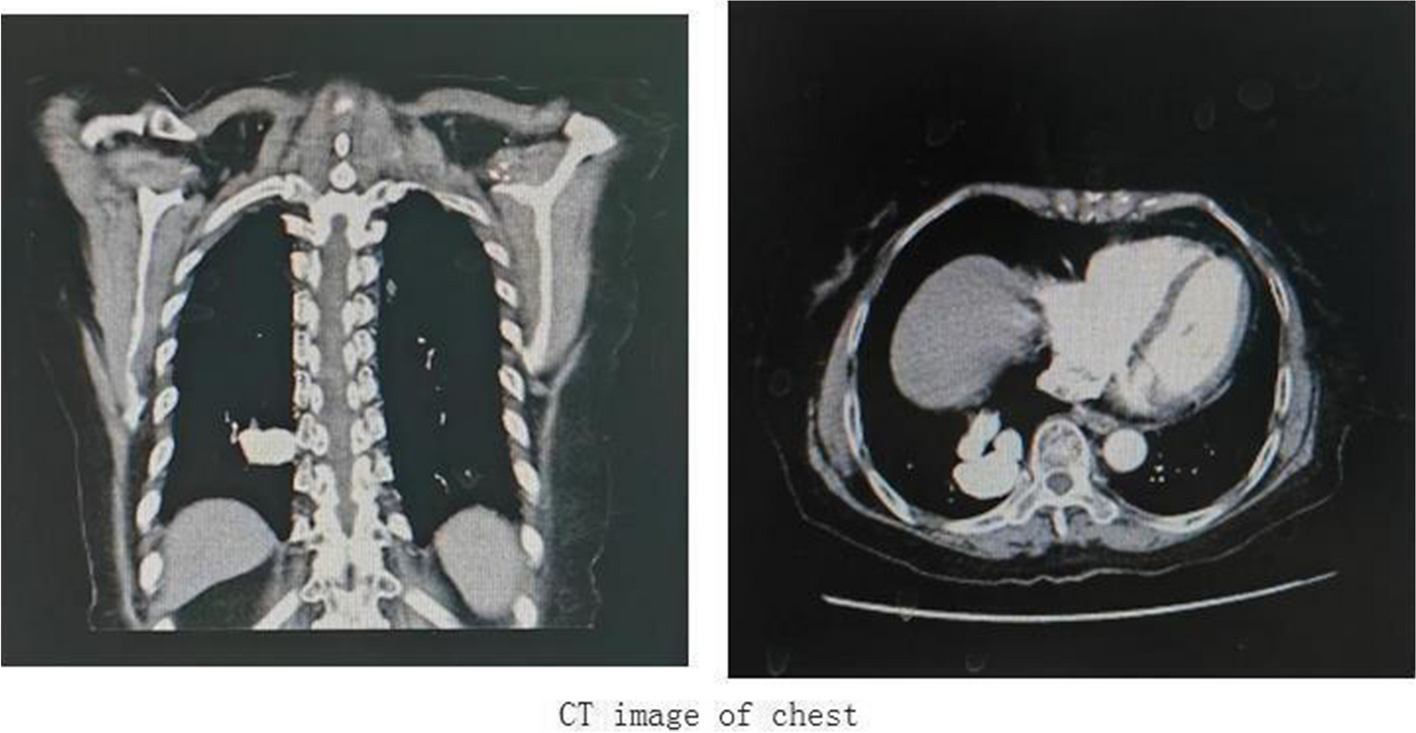
Although the PNS can occur in any other part of the body, its appearance on the posterior chest wall is an infrequently described presentation of this well-known condition [5, 7, 9]. Melanocytic nevi are benign lesions caused by the clonal proliferation of a melanocyte. It is expected that this change would occur either spontaneously or in response to extrinsic stimuli (for example, sun exposure), resulting in histologically symmetric, essentially uniform cell proliferation with homogenous alterations affecting the majority of component cells [16]. A hairy nevus can become irritated and give rise to an inflammatory reaction. This reaction may occur due to various factors such as friction, trauma, or exposure to certain irritants. Additionally, hair follicles within the nevus can become ingrown, leading to inflammation and irritation. However, it’s essential to note that not all hairy nevi will necessarily cause irritation or inflammation, and the severity of the reaction can vary depending on individual factors and the specific characteristics of the nevus [17].
The etiology of PNS is unknown; however, it is believed to be caused by an accumulation of hair and skin debris in the natal cleft, which can lead to infection and the creation of a cyst or sinus tract [1, 3, 16]. Some potential risk factors have been identified, including male gender, positive family history, particularly first-degree relatives, obesity, prolonged sitting periods exceeding 6 h daily, poor personal hygiene (bathing frequencies of 2 or fewer times per week), an inactive lifestyle, excess body hair, and a deep natal cleft [1, 5, 7, 18, 19]. Recurrence rates of 7–42% have been reported after primary closure [1, 19].
.
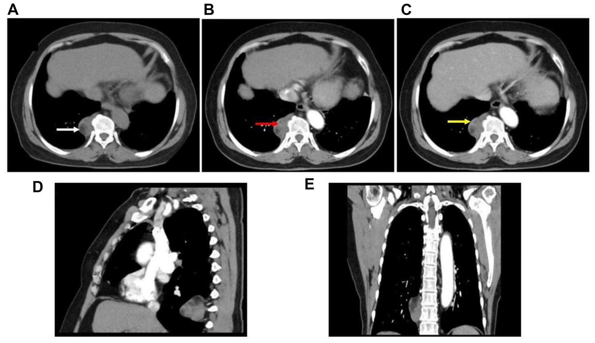
Clinical presentation is the primary method for diagnosing PNS. Ultrasonography can also detect hair strands within the lesion, while fine-needle aspiration biopsy offers another diagnostic option [20]. PNS may be asymptomatic or present with pain, redness, swelling, tenderness, abscess formation, or drainage of pus or blood [1, 3]. Nevus typically manifests as a nodular growth in young adulthood with limited progression, though some may develop into melanoma [20]. Nevus diagnosis can involve various methods, including clinical examination, dermoscopy, and sometimes biopsy. Clinical examination involves visual inspection of the skin lesion, noting its color, size, shape, and other characteristics. Dermoscopy, a non-invasive technique, allows for a closer examination of the lesion’s surface features and structures. In cases where the diagnosis is uncertain or further information is needed, a histological examination considered as a gold standard diagnosis [21]. The clinical diagnosis of the current case can be challenging, especially when encountering unusual presentations, pain, or tenderness, emphasizing the need for vigilance among healthcare professionals.
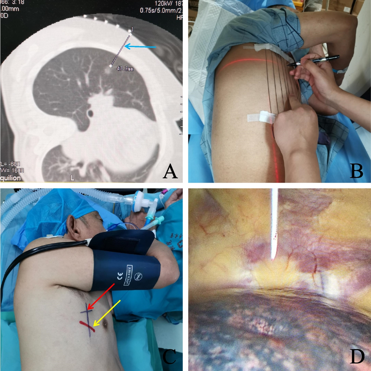
Eradication of the sinus tract, complete healing, and prevention of recurrence are the main three principles in the management of PNS [1]. The ideal treatment should be effective, safe, low cost, and minimize discomfort, hospital stay, and recurrence rate [1, 22]. Surgical excision of the PNS is the primary treatment, but special care must be taken during excision to avoid compromising the blue nevus. Regular follow-up is crucial to monitor both conditions and detect any potential complications or recurrences. The risk of wound infection following surgery is significant, particularly from anaerobic bacteria, which causes delayed wound healing [13, 23]. The current case was diagnosed clinically as a blue nevus and managed with surgical excision under local anesthesia.
The association between PNS and blue nevus is not fully understood, but it may involve congenital skin weaknesses or changes in the local environment, possibly influenced by shared genetic factors or embryological origins [10]. This rarity presents unique challenges for diagnosis and treatment, often leading to delays and potential mismanagement. Comprehensive documentation and analysis of such cases are needed to improve medical understanding and patient care. This study contributes valuable insights into this rare association, aiding clinicians in diagnosis and management.
In conclusion, the posterior chest wall PNS is another type of atypical PNS that is extremely rare. The association between PNS and blue nevus is a fascinating medical finding that deserves further investigation. Although rare, it underscores the complexity of dermatological and surgical interactions. Continued research into the underlying mechanisms of this association can help improve our understanding of both conditions and guide optimal management strategies for affected patients.

留言 (0)