
記住我


We retrospectively collected the clinical data of 120 patients who underwent lung nodule localization and resection surgery at the Department of Thoracic Surgery, First Affiliated Hospital of Bengbu Medical College, from January 2020 to January 2022. These patients were randomly selected according to our inclusion criteria for this study. Among them, 30 patients underwent CT-assisted body surface localization combined with intraoperative stereotactic anatomical localization, 30 patients underwent only CT-assisted body surface localization, 30 patients underwent only intraoperative stereotactic anatomical localization, and 30 patients underwent CT-guided percutaneous microcoil localization. All clinical information was from the hospital medical record. The success rates (The success rate of localization was defined by the rate of successful one-time localization, where a distance ≤ 1.5 cm between the nodule and the marking was considered successful, while a distance > 1.5 cm was considered failed localization. Regardless of the method of lung nodule localization, a localization failure is still recognized during surgery if the lung nodule is not found in the lung tissue that has been cut down, and then the operator re-searches for the nodule, and eventually the nodule is found in the lung tissue that has been cut down a second time.), complication rates, and localization times (CT-Assisted Body Surface Localization (Method for Group B) 's time consists of two parts, the first part is started when the patient is positioned on the CT examination table and stopped when the puncture point is drawn on the body surface. The second part is on the operating table when the operator holds the puncture needle, and stops the timing at the end of the puncture. Intraoperative Stereotactic Anatomical Location (Method for Group C)'s time: start the timer when the surgeon observes the patient's thoracic cavity during surgery, and stop the timer when the surgeon leaves a cautery mark on the lung surface with the electrocoagulation knife. CT-Assisted Body Surface Localization Combined with Intraoperative Stereotactic Anatomical Localization (Method for Group A)'s time is equal to CT-Assisted Body Surface Localization (Method for Group B)'s time plus Intraoperative Stereotactic Anatomical Location (Method for Group C)'s time. Time used for Preoperative CT-Guided Percutaneous Insertion of a Microcoil for Localization (Method for Group D): start the timer when the patient is positioned on the CT examination bed and stop the timer when the micro-spring coil has been placed and the puncture site is covered with gauze on the body surface). of the four lung nodule localization methods were statistically analysed.
Inclusion and exclusion criteriaThe inclusion criteria were as follows: 1. The maximum diameter of the pulmonary nodule was ≤ 30 mm. 2. The nodule was located in the outer 1/3 region of the lung field. 3. Patient age was ≤ 70 years. 4. The same pulmonary nodule localization method was used by the same person. 5. Prior to surgery the patient had already undergone a chest CT at our hospital. 6. The patient had not undergone a lung puncture biopsy or a thoracentesis.
The exclusion criteria were as follows: 1. A pleural indentation sign was present at the nodule site. 2. The solid component of the nodule was ≥ 50%. 3. Patients exhibited extensive pleural adhesion. 4. Presence of emphysema in the patient. 5. Patients had a history of radiotherapy or chemotherapy. 6. Surgical excision yielded ≥ 2 lesions. 7. Patients who underwent direct lobectomy. 8. Patients with enlarged hilar or mediastinal lymph nodes. 9. The patient had no pleural effusion.
Technical methodsCT-Assisted body surface localization combined with intraoperative stereotactic anatomical localization (Method for group A)Given the complexity of intraoperative localization and the potential possibility for anatomical markers to move during surgery, a two-pronged approach was employed. Initially, the approximate region of the nodule was determined using CT-assisted body surface localization. This approach reduces the scope of intraoperative anatomical localization, decreasing the difficulty of the process. Subsequent anatomical localization was more precise. When discrepancies arise between CT-assisted body surface localization and intraoperative stereotactic anatomical localization, two scenarios should be considered: If the patient is an elderly individual with loose skin or a female with the surface marker of the nodule near the breast area, the results of the intraoperative localization should be prioritized. Conversely, if the development of the lung fissure is suboptimal, the findings from CT-assisted body surface localization should be given precedence.
Preoperative CT-Assisted body surface localization (Method for group B)Patients were initially positioned in the same posture as was used during the surgical procedure. A localization assistant device composed of evenly arranged metal bars was placed on the patient's chest. After the patient took a deep breath and held it, a CT scan was performed. Upon identifying the most visible level of the lung nodule, the CT machine was retracted to that level. A line, termed the "X-line", was drawn on the patient's skin along the infrared line projected by the CT machine. Another line, parallel to the body's longitudinal axis and termed the "Y-line," was drawn along the metal bar corresponding to the surface projection point of the lung nodule. The intersection of the X and Y lines marks the surface projection point of the lung nodule. The distance from this point to the nodule and the thickness of the chest wall were measured along a line termed the "Z-line". After successful induction of general anaesthesia, the patient was repositioned as during the CT scan. A puncture needle was inserted vertically into the skin at the marked point, reaching a depth 1 cm greater than the chest wall thickness measured preoperatively on the CT scan to ensure penetration into the lung tissue. The anaesthetist then inflated the patient's lung before withdrawing the puncture needle. Following localization, a chest support pad was placed beneath the patient. Video-assisted thoracic surgery was initiated. During the procedure, haemorrhagic spots left by the puncture needle on the lung surface, indicating the location of the nodule, were visualized and marked using an electrosurgical knife. The operation procedure is shown in Fig. 1.
Fig. 1
Preoperative CT-Assisted Body Surface Localization (Method for Group B). Panel a shows the patient's chest CT scan at the level of the nodule, with the blue arrow indicating the distance from the nodule centre to the surface. Panel b shows the results obtained by the clinician marking the lung nodule's projection on the skin with the assistance of the CT infrared line and metal bars. Panel c shows the patient after general anaesthesia and intubation in the same position as during the CT scan; the red arrow points to the lung nodule's puncture location, and the yellow arrow points to the surgical incision site. Panel d illustrates the insertion of the locating puncture needle into the thoracic cavity
Intraoperative stereotactic anatomical location (Method for group C)Upon successful induction of anaesthesia, the patient was positioned in the lateral decubitus position, and a chest support pad was placed beneath the patient. Video-assisted thoracic surgery was commenced. During surgery, inherent anatomical landmarks of the thorax, such as the lung apex, lung margin, lung fissures, aortic arch, pulmonary artery, pulmonary vein, azygos vein arch, superior vena cava, oesophageal-tracheal groove, azygos vein notch and paravertebral line, were sought. Preoperatively, through meticulous examination of the patient's thin-slice chest CT images, the relationship between the nodule and related anatomical landmarks was determined. The surgeon compared intraoperatively identified landmarks with those from preoperative CT images to localize the nodule. Notably, due to deformations between the expanded and collapsed states of the lung, proportional relationships between landmarks were consulted. The identified location was then marked using an electrosurgical knife. The operation procedure is shown in Fig. 2.
Fig. 2
Intraoperative Stereotactic Anatomical Location (Method for Group C). The figure displays common reference markers for intraoperative anatomical localization. Panel a depicts the lung apex; Panel b displays the lung fissures; Panel c shows the azygos vein arch; Panel d represents the aorta
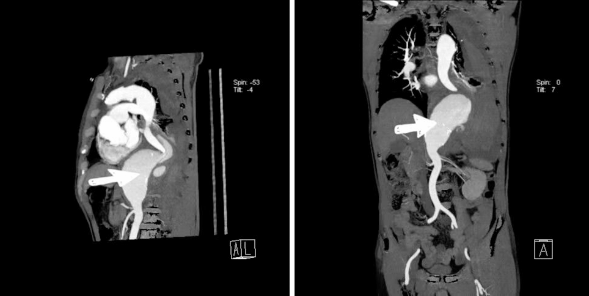
Preoperative CT-Guided percutaneous insertion of a microcoil for localization (Method for group D)After the localization assistance device, composed of adjacent evenly arranged metal bars, was placed on the patient's chest, a chest CT scan was performed. The puncture point was determined and marked on the skin, and the depth and angle of needle insertion were established. Following aseptic preparation and local anaesthesia, the patient was instructed to hold their breath and the puncture needle was inserted into the subpleural space of the lung. This procedure was followed by another CT scan, adjustments to the needle tip position, and further CT confirmation. Once the location was identified, the microcoil was deployed, anchoring near the nodule. After the placement, another CT scan was conducted to ensure proper coil localization. The patient was monitored for potential complications, such as haemorrhage and pneumothorax. The operation procedure is shown in Fig. 3.
Fig. 3
Preoperative CT-Guided Percutaneous Insertion of a Microcoil for Localization (Method for Group D). Panel a displays the lung nodule's projection on the skin under CT guidance. Panel b illustrates how the clinician inserted the puncture needle before deploying the microcoil. Panel c shows the CT image taken during the localization process, with the yellow arrow pointing to the puncture needle and the red arrow to the lung nodule. Panel d provides an intraoperative image showing the deployment of the microcoil on the lung surface
Procedure for lung nodule resectionThe area of lung tissue containing the marker point of the pulmonary nodule was gently elevated by the surgeon using atraumatic forceps. Subsequently, to ensure an adequate surgical margin, wedge resection of the lung tissue encompassing the nodule was performed using an endoscopic linear cutter. When the lung tissue was resected, we used a lancet to dissect the lung tissue in a direction perpendicular to the pleural surface right up to the lung nodule, and the distance between the dissection and the locator marking point on the pleura was measured with a straightedge and recorded in the operative record. If the nodule was not identified in the initial excised lung tissue, the resection range was expanded. The resected lung tissue was subjected to rapid frozen section pathology. When rapid frozen section pathology revealed that the nodule was malignant, we performed a lobectomy. For adenocarcinoma in situ or minimally invasive adenocarcinoma, the surgical margins have been more than 2 cm.
Statistical analysisCorrelations were analysed using SPSS 23.0 software. Quantitative data are presented as the mean ± standard deviation and One-way ANOVA was applied for analysis among different groups with Turkey post hoc test. Numerical data are presented as frequencies and percentages and were compared using chi-square tests. The level of significance was set at P < 0.05.
留言 (0)